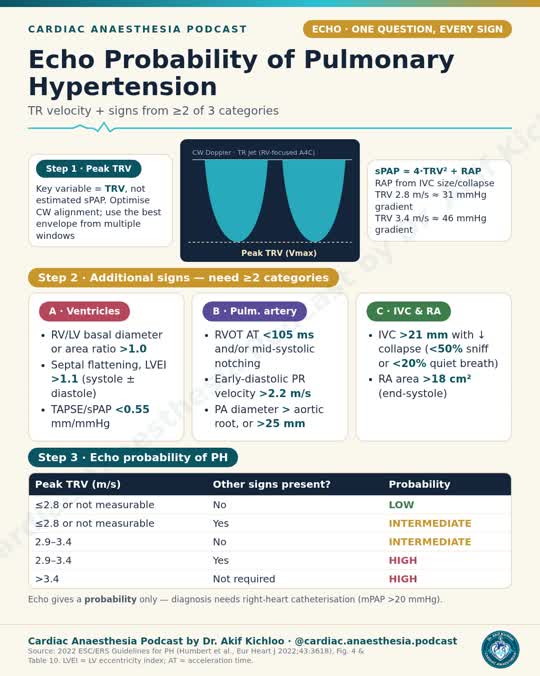
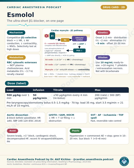
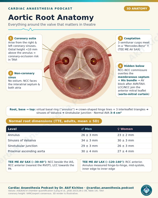
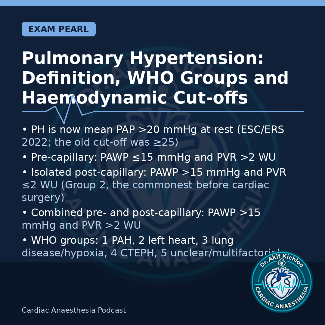
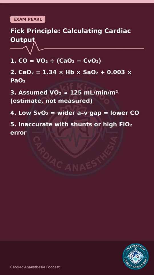
Cardiac Anaesthesia Podcast

Welcome to the reference library

Join the newsletter for more from Cardiac Anaesthesia. Subscription is optional.

Open the signup page in a new tab if the form does not load.

Cardiac Anaesthesia Podcast

Reference library

Dr. Akif Kichloo · @cardiac.anaesthesia.podcast

Cardiac Anaesthesia Podcast Homepage

Posts

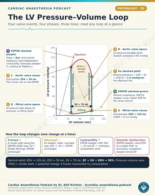
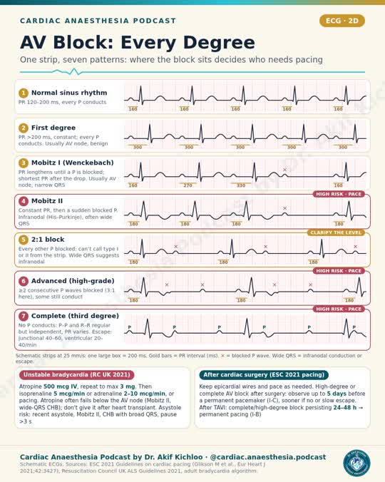
LV Normals: Men vs Women (ASE/EACVI 2015)Norepinephrine: what this U.S. label saysCO₂ flushing: the bypass circuit and the operative fieldTransfusion on CPB: haematocrit and fresh frozen plasmaSuccinylcholine: doses and contraindicationsClevidipine: Dosing and CautionsRV Function on Echo (ASE 2025)Milrinone: Doses and Cautions (US Label)Tetralogy of Fallot: spells, repair and the stiff right ventricleSeptic shock: fluids, MAP, vasopressors and inotropes (SSC 2026)Anaesthesia during bypassAbdominal Aortic Aneurysm: When to Repair (ESC 2024)Ketamine: Doses and Cautions (US Label)Diastolic function in 2 steps: the ASE 2025 algorithmPlanning Delivery in Heart Disease (ESC 2025)MAP, pump flow, pulsatility and prime on bypassVentilator Check: AARC 2024Flumazenil: reversing benzodiazepines safelySeptal flattening: pressure or volume overload?Procoagulants after cardiac surgeryPregnancy heart risk: mWHO 2.0 classes II–III to IVCardiac output monitors at a glanceProsthetic valves 5/5: intraoperative TEE after replacementProsthetic valves, part 4: thrombosis and perioperative VKAProsthetic valves 3/5: valve choice and anticoagulationProsthetic valves 2/5: grading dysfunction on echoProsthetic valves 1/5: types and pathophysiologyCyanide poisoning: hydroxocobalaminPeripartum cardiomyopathy: ESC 2025 essentialsM-TEER: What TEE ChecksEndotracheal Suctioning in Ventilated AdultsIV BP Drugs: Half-Life and Clinical OffsetAortic Root and Ascending Aorta: When to OperateMethylene Blue for Vasoplegia: The Serotonin TrapHyperkalaemia in Adults: Protect, Shift, RemovePressure Half-Time at a GlanceHeparin Reversal with Protamine After CPBAcid–Base in 4 QuestionsFlow–Time Curve: 6 CluesPulmonary Valve 5/5: TEE Views and Bypass PearlsPulmonary Valve 4/5: Anaesthetic Goals and HaemodynamicsPulmonary Valve 3/5: Intervention Criteria in Adult Congenital Heart DiseasePulmonary Valve 2/5: Grading PS and PR on EchoPulmonary Valve 1/5: Aetiology and PathophysiologyCalcium channel blocker poisoning: rescue and current caveatsHHS: slow correction, close monitoringAccidental hypothermia: stage it, then triageAdrenal crisis: hydrocortisone first, tests laterHeart transplantation: episode previewCalcium emergencies in adultsFloating a pulmonary artery catheter: read the waveformOne-lung ventilation: hypoxaemia rescueMassive transfusion: ratios, calcium and targetsLocal Anaesthetic Systemic Toxicity: Recognise and TreatMajor Burns: Fluids, Airway and the First HoursSteroid potency: what the numbers meanROTEM at a glanceMalignant hyperthermia: interpreting the crisis protocolProne positioning in severe ARDSSugammadex: dose by the depth of blockTEG: The Machine Doesn't Dictate the Bag, Part 2 of 2TEG at a Glance: Part 1 of 2IABP Timing Errors at a GlanceTricuspid regurgitation, part 5 of 5: intra-op TEE and bypass pearlsTricuspid regurgitation, part 4 of 5: anaesthetic goals and haemodynamicsTricuspid regurgitation, part 3: when to interveneTricuspid regurgitation, part 2 of 5: grading severity on echoTricuspid Regurgitation 1/5: Aetiology and PathophysiologyParacetamol Overdose: Nomogram, NAC and Transplant CriteriaWhich arterial-line trace is this? Understanding dampingHigh-Risk Pulmonary Embolism: Who Gets Thrombolysis?Unanticipated Difficult Intubation: DAS Plans A to DReversal Before Urgent SurgeryNeuraxial Blocks and AnticoagulantsDKA: Diagnosis and the First HoursTension pneumothorax: decompress firstReference library announcementHyponatraemia: Safe Correction LimitsStatus Epilepticus: The Timed Treatment LadderVasoactives by Shock State12 Vasoactives in 60 SecondsMitral Regurgitation 5/5: Intra-op TEE and Bypass PearlsMitral Regurgitation 4/5: Anaesthetic Goals and HaemodynamicsMitral Regurgitation 3/5: Intervention Criteria (ESC 2021 + ACC/AHA 2020)Mitral Regurgitation 2/5: Grading Severity on EchoMitral Regurgitation 1/5: Aetiology and PathophysiologyTricyclic Overdose: ECG Signs and Sodium BicarbonateSgarbossa Criteria: MI in LBBB or Paced RhythmAnaphylaxis: Adrenaline Doses That MatterARDS: Berlin Definition and Lung-Protective VentilationVasopressors and Inotropes at a Glance (infographic)CVP Waveforms at a Glance (infographic)Etomidate for Induction in Cardiac PatientsDigoxin Toxicity: ECG Clues and TreatmentPulsus Paradoxus: Mechanism and CausesCABG Conduits: LIMA, Radial and VeinElectrolytes on the ECG (infographic)The LV Pressure–Volume Loop (infographic)Pre-op Anaemia Before Cardiac SurgeryHeart Murmurs and Bedside ManoeuvresJones Criteria for Acute Rheumatic Fever (AHA 2015)Cell Salvage in Cardiac SurgeryAV Block: Every Degree (infographic)Echo Probability of Pulmonary Hypertension (infographic)Mitral Stenosis 5/5: Intra-op TEE and Bypass PearlsMitral Stenosis 4/5: Anaesthetic Goals and HaemodynamicsMitral Stenosis 3/5: Intervention Criteria (ESC 2021 + ACC/AHA 2020)Mitral Stenosis 2/5: Grading Severity on EchoMitral Stenosis 1/5: Aetiology and PathophysiologyPerioperative Beta-Blockers: Continue, Don’t StartEndocarditis Prophylaxis: Who Needs It?Prostaglandin E1 for Duct-Dependent LesionsCHA2DS2-VASc and HAS-BLED ScoresThe Bypass Circuit: Every Component (infographic)Cardiac Action Potential: Every Phase (infographic)Reversing Anticoagulants Before Urgent SurgeryNYHA Class vs ACC/AHA Heart Failure StageHumour: Muscle relaxants ranked by how long they stay at the partyShock Types: Haemodynamic ProfilesPA Catheter Normal ValuesRV Systolic Function on Echo: Every Method (infographic)LV Normals: Men vs Women (infographic)Aortic Regurgitation 5/5: Intra-op TEE and Bypass PearlsAortic Regurgitation 4/5: Anaesthetic Goals and HaemodynamicsAortic Regurgitation 3/5: Intervention Criteria (ESC 2021 + ACC/AHA 2020)Aortic Regurgitation 2/5: Grading Severity on EchoAortic Regurgitation 1/5: Aetiology and PathophysiologyCoronary Anatomy: Dominance and TerritoriesLAST After Chest Wall BlocksOxyhaemoglobin Dissociation Curve ShiftsWiggers Diagram: Key Cardiac Cycle EventsStopping DOACs Before Cardiac SurgeryOxygen content and delivery on one page9 arterial line waveforms to read at a glanceAortic Dissection: Stanford vs DeBakeyModified Duke Criteria for EndocarditisType 5 MI After CABG: Fourth Universal DefinitionHeart Failure by Ejection FractionWhich ECG leads should be on your screen?A1 to P3 on one pageMagnesium in Cardiac SurgeryAdenosine: Doses and CautionsAmiodarone in Cardiac SurgeryVasoactive Drugs: Receptor TableWhy inhaled NO drops PVR without dropping SVRFour CCS classes, exact wording — the pre-op and viva stapleAortic Stenosis 5/5: Intra-op TEE and Post-bypass PearlsAortic Stenosis 4/5: Anaesthetic Goals and HaemodynamicsAortic Stenosis 3/5: Intervention Criteria (ESC 2021 + ACC/AHA 2020)Aortic Stenosis 2/5: Grading Severity on EchoAortic Stenosis 1/5: Aetiology and PathophysiologySVR and PVR: Formulas and Normal ValuesVentricular InterdependenceFluid Responsiveness: PPV and SVV Cut-offsMyocardial O2 Supply vs DemandCoronary Blood Flow: LV vs RV TimingEsmolol: Mechanism, Doses and CautionsAortic Root AnatomyPulmonary Hypertension: Definition, WHO Groups and Haemodynamic Cut-offsFick Principle: Calculating Cardiac OutputSevere Chronic AR: Anaesthetic Goals and Bypass Pitfalls

For clinician education and exam study. Not patient-specific advice. Consult current guidelines and local protocols when making clinical decisions.

← Back to posts

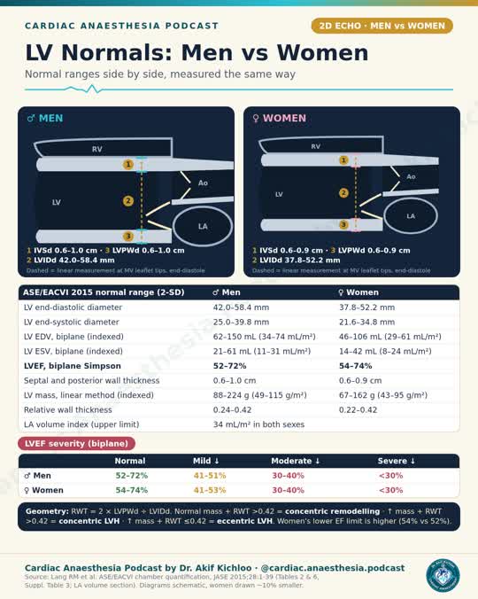
LV Normals: Men vs Women (ASE/EACVI 2015)

These are adult 2D transthoracic echo reference ranges. Use the method that goes with each number. Comparable LV dimensions and volumes can use the same limits on transoesophageal echo, but do not transfer this whole set to intraoperative views. The reel’s drawings show relative scale, not where to place the calipers.

Linear size and biplane volumes

In the parasternal long-axis view, measure at or just below the mitral leaflet tips, perpendicular to the left ventricular (LV) long axis. Measure wall thickness and internal diameter at end-diastole, then internal diameter again at end-systole. The diastolic diameter range is 42.0–58.4 mm in men and 37.8–52.2 mm in women. The systolic range is 25.0–39.8 mm and 21.6–34.8 mm, respectively. For both the septum and posterior wall, the diastolic ranges are 0.6–1.0 cm in men and 0.6–0.9 cm in women.

Use the biplane method of discs on non-foreshortened apical four- and two-chamber views. In men, end-diastolic volume is 62–150 mL (34–74 mL/m² indexed to body surface area) and end-systolic volume is 21–61 mL (11–31 mL/m²). In women, the respective ranges are 46–106 mL (29–61 mL/m²) and 14–42 mL (8–24 mL/m²).

Ejection fraction

The normal biplane LV ejection fraction (LVEF) range is 52–72% in men and 54–74% in women. Table 4 grades a low LVEF as mildly reduced at 41–51% in men or 41–53% in women, moderately reduced at 30–40% in either, and severely reduced below 30% in either.

These are echo reference bands, not heart-failure diagnoses. The 2026 ESC heart failure guideline no longer uses a separate mildly reduced EF category. Its heart-failure categories use clinical criteria as well as EF. A value of 50% is not within either ASE/EACVI biplane normal range.

Mass and geometry

With the 2D-guided linear method, normal LV mass is 88–224 g in men and 67–162 g in women. The indexed ranges are 49–115 g/m² and 43–95 g/m², respectively. Do not use these mass limits for the separate area-based 2D methods. The upper limit for maximum left atrial volume indexed to body surface area is 34 mL/m² in both.

Relative wall thickness (RWT) is twice the diastolic posterior wall thickness divided by the diastolic LV internal diameter. It has no units; reference ranges are 0.24–0.42 in men and 0.22–0.42 in women. With normal indexed mass, RWT ≤0.42 is normal geometry and RWT >0.42 is concentric remodelling. With raised indexed mass (>115 g/m² in men or >95 g/m² in women), RWT ≤0.42 is eccentric hypertrophy and RWT >0.42 is concentric hypertrophy.

References

Lang et al. ASE/EACVI adult chamber quantification. J Am Soc Echocardiogr. 2015;28:1–39. Tables 2, 4 and 6, Supplemental Table 3, Figure 6 and section 9.3.

2026 ESC Guidelines for the management of heart failure.

← Back to posts
← Back to posts

Norepinephrine: what this U.S. label says

The Sagent ready-to-use bag in this reel contains the equivalent of 16 mg of norepinephrine in 250 mL of 5% dextrose (64 mcg/mL). It needs no further dilution. That instruction is for this bag, not for a concentrate vial or a different product. The U.S. label is for raising blood pressure in adults with severe, acute hypotension. Sagent prescribing information, sections 1–3

Start, titrate and reassess

The labelled IV starting dose is 8–12 mcg/min. Titrate to a pressure that supports vital-organ perfusion. The 2–4 mcg/min figure describes typical maintenance, not a target, ceiling or weight-based dose. The label calls for blood-pressure checks every 2 minutes until the desired effect, then every 5 minutes for the rest of the infusion. The label says to address hypovolaemia before starting and to suspect occult hypovolaemia if the pressure does not respond. Separately, in unstable septic shock, the 2026 sepsis guideline allows immediate IV crystalloid and vasopressors together on a case-by-case basis. Sagent label, sections 2.1–2.2 and 5.1 · Surviving Sepsis Campaign 2026, recommendation 11 · Septic-shock reference

Protect the infusion site

The label calls for a large vein, with frequent checks for free flow and extravasation. It advises avoiding leg veins in older patients and in patients with occlusive leg-vessel disease. If extravasation is suspected, act immediately according to local protocol. The adult label describes infiltrating the affected area as soon as possible with 5–10 mg phentolamine mesylate in 10–15 mL of 0.9% saline. The label describes a local response when infiltration is done within 12 hours. Do not wait. In adult septic shock, the 2026 sepsis guideline suggests starting peripherally rather than waiting for central access, a conditional recommendation based on very low-certainty evidence. It does not specify a peripheral dose, site, line size or duration, and did not consider midlines. The guideline does not endorse this bag strength for peripheral use. Follow your local concentration and access protocol. Sagent label, sections 2.1 and 5.1 · Surviving Sepsis Campaign 2026, recommendation 12

Taper rather than stop abruptly. The label warns of arrhythmias, particularly with hypoxia or hypercarbia, and advises avoiding use in mesenteric or peripheral vascular thrombosis because ischaemia may worsen. Give whole blood or plasma separately if needed to expand blood volume. Do not use this bag if the solution is pinkish, darker than slightly yellow or contains a precipitate. The label lists no contraindications. These warnings still apply. Sagent label, sections 2.1, 2.3, 4 and 5

← Back to posts
← Back to posts

CO₂ flushing: the bypass circuit and the operative field

CO₂ can help in two different places during adult cardiac surgery. Before priming, it displaces air in the empty cardiopulmonary bypass circuit. In the operative field, it can displace air from open cardiac cavities. CO₂ dissolves in blood more readily than nitrogen, so bubbles containing CO₂ can dissipate more quickly. EACTS/EACTAIC/EBCP adult CPB guideline, section 4.4.2 and Recommendation Table 5

Before you prime the circuit

The guideline recommends flushing the empty bypass circuit with CO₂ before priming as standard practice to reduce gaseous microemboli (Class I, Level B). The evidence here concerns bubbles in the circuit and prime. It does not establish a reduction in postoperative stroke or cognitive complications. Recommendation Table 5

When the heart is open

The guideline says you may consider CO₂ insufflation of the operative field to reduce emboli (Class IIb, Level B). Keep the usual de-airing steps and monitoring in place. If you use field CO₂, adjust gas flow to avoid hypercapnic acidosis (Class I, Level C). This recommendation does not specify a single flow rate for every case. Section 4.4.2 and Recommendation Table 5

Several studies found fewer gaseous microemboli with field CO₂. The reviews cited by the guideline did not establish a postoperative neurocognitive benefit. Fewer detected bubbles and better cognition are different outcomes. A newer multicentre study in open left-sided valve surgery was designed to assess acute ischaemic brain injury after field CO₂ plus standard de-airing. The public 2026 meeting abstract does not give an effect estimate, so it does not settle the question here. CO₂ Study protocol · AATS 2026 abstract listing

← Back to posts
← Back to posts

Transfusion on CPB: haematocrit and fresh frozen plasma

These recommendations apply during adult cardiopulmonary bypass. When you read the haematocrit on the pump, ask whether the patient is receiving enough oxygen at the current flow. The haematocrit gives you only part of that answer. EACTS/EACTAIC/EBCP adult CPB guideline, Recommendation Table 45

Packed red cells during bypass

HCT below 18%: The guideline recommends packed red cells at this level, which corresponds to a haemoglobin of about 6 g/dL (Class I, Level C).

HCT 18–24%: You may consider red cells after assessing tissue oxygenation (Class IIb, Level B). The footnote describes indexed oxygen delivery above 273 mL/min/m² with satisfactory cerebral oximetry. A value below 273 does not, on its own, tell you to transfuse. Look at pump flow, arterial oxygen content and the wider perfusion picture.

HCT above 24%: The guideline advises against red-cell transfusion when oxygen delivery and extraction are acceptable (Class III, Level C). If either is inadequate, reassess the cause. The recommendation does not make transfusion automatic in that situation. Recommendation Table 45 and footnote d

The same guideline gives a separate goal-directed perfusion recommendation. During normothermic CPB, it says to consider a lower indexed oxygen-delivery threshold in the 280–300 mL/min/m² range (Class IIa, Level B). That range is a perfusion target, not another haemoglobin or haematocrit trigger. Recommendation Table 43

When plasma fits

If antithrombin deficiency is impairing the response to heparin, the guideline recommends antithrombin concentrate first (Class I, Level B). Consider fresh frozen plasma if the concentrate is unavailable (Class IIa, Level C). This advice is specific to antithrombin deficiency. The guideline advises against prophylactic plasma during bypass to reduce perioperative blood loss (Class III, Level B). Recommendation Table 45

Active coagulopathic bleeding after bypass is a different decision. FARES-II compared four-factor prothrombin complex concentrate with frozen plasma in adults who needed clotting-factor replacement after separation from bypass. Among the 420 patients in the primary analysis, 77.9% in the concentrate group and 60.4% in the plasma group needed no further haemostatic intervention between one and 24 hours after treatment started. The trial was unblinded. It did not test prophylactic plasma on bypass or treatment of antithrombin deficiency. FARES-II randomised trial, 2025

← Back to posts
← Back to posts

Succinylcholine: doses and contraindications

The adult dose in this reel comes from the August 2026 US Quelicin label for short surgical procedures. For that setting, the label gives an average of 0.6 mg/kg IV (individual range 0.3–1.1 mg/kg). Block usually starts in about a minute. After roughly two minutes of maximum block, the label describes recovery over the next four to six minutes. Do not rely on that timing as a rescue plan for a failed airway. Quelicin prescribing information, §§2.1–2.2

Children and the boxed warning

These are emergency airway doses, not routine paediatric induction doses. The US label lists 2 mg/kg IV for infants and smaller children and 1 mg/kg IV for older children and adolescents. If no suitable vein is accessible, the label gives up to 3–4 mg/kg IM, with a maximum total of 150 mg and an onset of about two to three minutes. The boxed warning matters even if a child looks well: succinylcholine can precipitate rhabdomyolysis and severe hyperkalaemia in an undiagnosed myopathy such as Duchenne muscular dystrophy. Ventricular arrhythmia, cardiac arrest and death have been reported. Reserve paediatric use for emergency intubation, an airway that must be secured immediately, or IM use when a suitable vein is inaccessible. Quelicin boxed warning, §§2.3–2.4 and 5.1

What to check before giving it

The US label contraindicates it in skeletal-muscle myopathy, known hypersensitivity to succinylcholine, known or suspected genetic susceptibility to malignant hyperthermia, and after the acute phase of major burns, multiple trauma, extensive skeletal-muscle denervation or upper motor neurone injury. In the last group, severe hyperkalaemia may lead to cardiac arrest. The label does not define a reliable start or end to that risk period. Quelicin §§4 and 5.2–5.5

Bradycardia can occur after the first dose in children and may progress to asystole. The risk rises after a repeat dose in both children and adults. Atropine pretreatment may reduce bradyarrhythmias; the label does not make it routine. Known low plasma cholinesterase activity is a separate reason to avoid succinylcholine because paralysis can last much longer. Quelicin §§5.6 and 5.9

Have oxygenation, ventilation and intubation support ready before giving the drug. Succinylcholine is a depolarising neuromuscular blocker. It paralyses without sedating. In routine use, induce unconsciousness first. The label recognises that an emergency can make this sequence impracticable. Quelicin §§2.1 and 5.14

Adult RSI is a different setting

Do not use the US short-procedure range as a universal adult RSI regimen. The 2025 Difficult Airway Society guideline gives 1.5 mg/kg suxamethonium or 1.2 mg/kg rocuronium as examples for achieving rapid-onset block in adults undergoing RSI. For critically ill adults, the Society of Critical Care Medicine suggests either agent when there are no known contraindications to succinylcholine. Use your local protocol and choose for the patient in front of you. DAS 2025, Table 1 · SCCM 2023 adult RSI guideline

← Back to posts
← Back to posts

Clevidipine: Dosing and Cautions

Clevidipine is a short-acting IV arterial vasodilator. This is the adult dosing approach in the US Cleviprex label when oral treatment is not feasible or desirable. Set the blood pressure goal for the patient and the indication before titrating.

Titrate to the response

Start at 1–2 mg/h. Early in the infusion, you can double the rate at 90-second intervals if the response calls for it. As the pressure approaches the goal, use smaller increases and wait 5–10 minutes between changes. Most patients respond at 4–6 mg/h. The label estimates a further systolic fall of 2–4 mmHg for a 1–2 mg/h increase, but the measured response matters more than that estimate.

Most patients in the label’s clinical experience received no more than 16 mg/h. Experience at 32 mg/h is limited to short-term use. At 0.5 mg/mL, the label recommends no more than 1,000 mL in 24 hours, equivalent to about 21 mg/h averaged over that day, because of the lipid load. Count other lipid sources as well. Experience beyond 72 hours is limited. The 2025 AHA/ACC high blood pressure guideline lists 21 mg/h in its hypertensive emergency table. Do not read the label’s limited experience at 32 mg/h as a routine maximum.

When to withhold it and what to monitor

Severe aortic stenosis, allergy to soy or eggs or their products, and defective lipid metabolism are contraindications. Examples of the last group include pathological hyperlipidaemia, lipoid nephrosis, and acute pancreatitis with hyperlipidaemia. Watch patients with heart failure closely because negative inotropy can worsen it.

Monitor blood pressure and heart rate throughout the infusion and until vital signs stabilise. If rapid titration causes hypotension or reflex tachycardia, reduce the infusion rate. The label advises against adding a beta-blocker specifically to treat clevidipine-induced tachycardia. That is separate from its warning that clevidipine does not protect against abrupt withdrawal of a beta-blocker.

In most patients, blood pressure recovers within 5–15 minutes of stopping. If a prolonged infusion is stopped without a transition to another antihypertensive, watch for rebound hypertension for at least 8 hours. The emulsion provides about 0.2 g lipid/mL. Use aseptic technique, discard any remainder within 12 hours of puncturing the stopper, and do not administer other medicines through the same line.

What the cardiac surgery trials tested

In ESCAPE-2, treatment failure after cardiac surgery occurred in 5 of 61 clevidipine patients versus 39 of 49 placebo patients. Failure meant not lowering systolic pressure by at least 15% or stopping study treatment within 30 minutes. ECLIPSE found better control within its prespecified pressure ranges than with nitroglycerin or sodium nitroprusside, and similar control to nicardipine within the prespecified range; clevidipine had fewer excursions when that range was narrowed. These trials describe blood pressure control in selected patients, not a universal postoperative target. If you are managing a patient having noncardiac surgery, the 2026 AHA/ACC guideline reaffirms that perioperative blood pressure management should be individualised.

Sources

← Back to posts
← Back to posts

RV Function on Echo (ASE 2025)

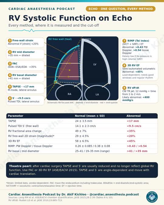
These are adult transthoracic echo reference limits, not cutoffs to apply unchanged to intraoperative transoesophageal echo. Start with an RV-focused apical four-chamber view. Tricuspid annular plane systolic excursion (TAPSE) measures lateral annular displacement. Tissue Doppler S′ measures peak systolic velocity at the same site. Read both alongside RV size and other measures of global systolic function.

Systolic function and size

The adult reference limits in ASE Table 1 are TAPSE >1.7 cm, S′ >9.5 cm/s, fractional area change (FAC) >35% and 3D RV ejection fraction >45%. FAC is the fall in RV area from end-diastole to end-systole divided by end-diastolic area, expressed as a percentage. The strain limits are magnitudes: >20% for RV free-wall longitudinal strain and >17% for six-segment RV global longitudinal strain.

For TAPSE, mild reduction is >1.3 to ≤1.7 cm, moderate is >1.0 to ≤1.3 cm and severe is ≤1.0 cm. For S′, the corresponding bands are >7.2 to ≤9.5 cm/s, >5.0 to ≤7.2 cm/s and ≤5.0 cm/s.

For FAC, those bands are >29 to ≤35%, >22 to ≤29% and ≤22%. For 3D RV ejection fraction, they are >39 to ≤45%, ≥32 to ≤39% and <32%. Exactly 32% falls in the moderate band.

On the RV-focused view, ASE Table 1 lists a basal diameter <4.1 cm, mid diameter <3.5 cm, longitudinal diameter <8.2 cm and end-diastolic area <25 cm². On the apical four-chamber view, the RV normally appears no more than about two-thirds the size of the left ventricle. That visual comparison does not replace measurements.

When the numbers disagree

A poorly aligned cursor or beam can make TAPSE or S′ read low. TAPSE may also read low with constrictive pericarditis or interference from a pacemaker lead or catheter. It can underestimate RV function after cardiothoracic surgery; the guideline specifically notes that S′ may do so in the immediate period after pericardiectomy. Look for corroborating measures before labelling global dysfunction.

TAPSE divided by pulmonary artery systolic pressure (PASP) is an echo estimate of RV adaptation to afterload. Studies of healthy people generally report 0.5–0.7 mm/mmHg. In pulmonary hypertension, values around 0.3–0.4 mm/mmHg have been associated with RV–pulmonary artery uncoupling and higher mortality. This is a prognostic association, not a stand-alone diagnostic cutoff.

References

Mukherjee et al. ASE guideline on the right heart in adults. J Am Soc Echocardiogr. 2025;38:141–186. Table 1 and text.

Journal correction. J Am Soc Echocardiogr. 2025;38:641.

Instagram reel

← Back to posts
← Back to posts

Milrinone: Doses and Cautions (US Label)

The Hikma US prescribing information, revised May 2025, covers short-term intravenous milrinone for acute decompensated heart failure. These adult doses are not a cardiac surgery or cardiogenic shock protocol.

Loading and maintenance

The label gives a loading dose of 50 mcg/kg IV over 10 minutes from the 1 mg/mL vial. Dilute drug drawn from the vial before giving a maintenance infusion. The 200 mcg/mL premix in 5% dextrose is ready for infusion but is not for loading.

The labelled maintenance range is 0.375–0.75 mcg/kg/min, with 0.50 mcg/kg/min listed as standard. Adjust the rate to haemodynamic response and renal function. The labelled maximum is 1.13 mg/kg over 24 hours, including the loading dose in the first 24 hours.

Renal impairment

Milrinone clears more slowly in renal impairment, so a lower maintenance rate may be needed. At creatinine clearances of 5, 10, 20, 30, 40 and 50 mL/min/1.73 m², the corresponding labelled rates are 0.20, 0.23, 0.28, 0.33, 0.38 and 0.43 mcg/kg/min, respectively.

Monitoring and cautions

Use continuous ECG monitoring and check blood pressure, heart rate, electrolytes and renal function. Have immediate treatment available for life-threatening ventricular arrhythmias. Correct hypokalaemia and slow or stop the infusion if blood pressure falls excessively. The label reports ventricular arrhythmias in 12.1% of phase II/III trial patients, supraventricular arrhythmias in 3.8%, and hypotension in 2.9%. Safety and efficacy beyond 48 hours of heart-failure treatment have not been established.

The label does not recommend use in the acute phase after myocardial infarction. Do not use milrinone in place of surgical relief of severe aortic or pulmonary valve obstruction; it may worsen outflow obstruction in hypertrophic subaortic stenosis. It can accelerate the ventricular response in atrial flutter or fibrillation that is not adequately rate-controlled. Do not give furosemide through an IV line containing milrinone: the drugs can precipitate on contact.

Hikma vial label · Hikma 5% dextrose premix label · Instagram reel

← Back to posts
← Back to posts

Tetralogy of Fallot: spells, repair and the stiff right ventricle

In this episode, I follow a six-month-old through complete repair and explain why the anaesthetic priorities change across bypass. I also look at why adults with repaired tetralogy of Fallot still need congenital cardiology follow-up.

08:37 | Hypercyanotic spells

In unrepaired tetralogy of Fallot, increased right-to-left shunting and reduced pulmonary blood flow can cause an abrupt fall in oxygen saturation. The child’s usual saturation is a useful comparison, not a universal target. Pulse oximetry does not measure pulmonary blood flow. Royal Children’s Hospital clinical guideline

18:11 | The teaching case · 21:57 | Induction and bypass

Before repair, the aim is to maintain adequate preload and systemic vascular resistance, avoid tachycardia and worsening dynamic outflow obstruction, and prevent air entry into IV lines. Repair timing and approach depend on symptoms, anatomy and risk. Texas Children’s cardiac handbook · AATS expert consensus

27:24 | Complete repair and weaning from bypass

When the right ventricle is restrictive after repair, atrial contraction can help drive late-diastolic pulmonary flow. That is why atrioventricular synchrony matters. Avoid unnecessarily high airway pressures while maintaining adequate gas exchange. Post-repair physiology study · Ventilation study

40:40 | Closing recap

Revisit the shift from keeping blood flowing to the lungs before repair to supporting right ventricular filling afterwards.

Repair does not end surveillance. 2025 adult congenital heart disease guideline, section 4.3.5

Open the episode and chapter notes · Original Instagram announcement

This is a guide to the teaching episode, not a bedside protocol.

← Back to posts
← Back to posts

Septic shock: fluids, MAP, vasopressors and inotropes (SSC 2026)

The first hours

In adults with sepsis-induced hypoperfusion or septic shock, SSC suggests at least 30 mL/kg of IV crystalloid in the first three hours (conditional, low certainty). Individualise the volume and reassess frequently for benefit and fluid overload. SSC favours balanced crystalloids over saline (conditional, moderate); the guideline suggests 0.9% saline for patients with traumatic brain injury. It recommends against starches (strong, high). Give antimicrobials immediately for possible, probable or definite septic shock, ideally within one hour of recognition (strong, very low).

Start with a mean arterial pressure (MAP) target of 65 mmHg (strong, moderate). For adults aged 65 or older, SSC suggests an initial range of 60–65 mmHg rather than higher ranges (conditional, low). Guide further fluid with dynamic findings such as the change in stroke volume after passive leg raising. Follow serial lactate in septic shock or sepsis with elevated lactate, but do not keep giving fluid simply to normalise the number. Use capillary refill alongside other measures of perfusion. These three recommendations are conditional, with low certainty.

When pressure remains low

Norepinephrine is the usual first choice. The recommendation is strong over dopamine, epinephrine and selepressin, and conditional over vasopressin and angiotensin II. Add vasopressin if norepinephrine requirements rise (conditional, moderate). If MAP is still inadequate on both agents, consider adding epinephrine (conditional, very low). If vasopressin is unavailable, consider adding epinephrine to norepinephrine alone. SSC suggests starting vasopressors through a peripheral IV rather than waiting for central access (conditional, very low). With concomitant cardiac dysfunction, either norepinephrine or epinephrine may be used first line (conditional, very low).

Persistent hypoperfusion with cardiac dysfunction

If hypoperfusion persists with cardiac dysfunction despite adequate fluid resuscitation and arterial pressure, consider adding dobutamine to norepinephrine or using epinephrine alone (conditional, very low). Do not substitute dobutamine for a vasopressor still needed to maintain MAP. SSC suggests against levosimendan for this setting and against terlipressin in septic shock (both conditional, low). It suggests against IV esmolol or landiolol to treat the shock (conditional, very low). For refractory shock with escalating vasopressor requirements, there is not enough evidence to recommend for or against IV methylene blue.

SSC 2026 adult recommendations · Full guideline · Instagram reel

← Back to posts
← Back to posts

Anaesthesia during bypass

These recommendations apply to adults during cardiac surgery. Choose and adjust the regimen to the patient, temperature and depth of anaesthesia.

Volatile and intravenous agents

Volatile agents may be used for maintenance on cardiopulmonary bypass (CPB) (IIb B). Use equipment approved for delivery (I C) and consider measuring the volatile concentration in the oxygenator exhaust (IIa B). Avoid nitrous oxide immediately before and immediately after bypass (III B).

During bypass, consider giving IV anaesthetics and opioids other than remifentanil at doses at least as high as those used before CPB (IIa B). Titrate to effect. Intermittent IV fentanyl boluses or an IV infusion are both options (IIb C).

Remifentanil during cooling

If cooling to 32 °C on bypass, consider a 30% reduction 20–30 minutes after starting CPB. When cooling below 28 °C, an immediate 60% reduction may be considered (both IIb B). Neither reduction is automatic. The Minto target-controlled infusion model is an option during normothermia or mild hypothermia, though it can underestimate the blood concentration (IIb B).

Depth and recovery

Consider processed EEG to help limit awareness and excessive anaesthetic depth (IIa B). Consider a short-acting non-depolarising neuromuscular blocker before bypass and a reversal protocol guided by quantitative neuromuscular monitoring, particularly in fast-track pathways (both IIa B). Confirm adequate recovery before extubation.

Joint EACTS/EACTAIC/EBCP CPB guideline, Recommendation Tables 18 and 47 · Instagram reel

← Back to posts
← Back to posts

Abdominal Aortic Aneurysm: When to Repair (ESC 2024)

For an asymptomatic fusiform abdominal aortic aneurysm, the ESC recommends elective repair at a maximal diameter of at least 55 mm in men or 50 mm in women (Class I, Level A). At those diameters, assess the patient for repair rather than just arranging another routine scan. The decision still depends on anatomy, operative risk, life expectancy and the patient’s preferences. ESC does not recommend elective repair when life expectancy is under two years. With suitable anatomy and life expectancy over two years, consider endovascular aneurysm repair (EVAR) after discussing the options with the patient (Class IIa, Level B).

When surveillance is appropriate

Duplex ultrasound is the usual test for follow-up. Below the repair threshold, ESC suggests a scan every three years at 30 to under 40 mm in either sex (Class IIa, Level B). An annual scan should be considered at 40 to under 50 mm in men or 40 to under 45 mm in women (Class IIa, Level B). ESC recommends six-monthly imaging at 50 to under 55 mm in men or 45 to under 50 mm in women while still below the elective repair threshold (Class I, Level B for the surveillance bands in Table 36). Table 36 prints the upper endpoints as 55 and 50 mm; Table 41 explicitly includes those diameters in the repair recommendation. Use CT or MR when duplex cannot measure the aneurysm adequately.

When size is not the deciding factor

New abdominal or back pain that could be due to a known aneurysm needs urgent assessment. Suspected rupture is an emergency. Do not wait for the elective diameter threshold. If the anatomy is suitable in ruptured AAA, ESC recommends EVAR over open repair (Class I, Level B). For confirmed rapid enlargement, the ESC discusses shorter imaging intervals and possible earlier repair at growth of at least 10 mm in a year or 5 mm in six months (Class IIb, Level C for considering repair). Check that serial measurements are comparable before interpreting the change. The 55/50 mm elective thresholds apply to fusiform aneurysms. For a saccular AAA, ESC says repair may be considered at 45 mm or above (Class IIb, Level C).

References

← Back to posts
← Back to posts

Ketamine: Doses and Cautions (US Label)

These US KETALAR label doses are for general anaesthesia in adults. Keep them separate from procedural sedation doses.

Induction and maintenance

For IV induction, titrate 1–4.5 mg/kg to effect and administer it over 60 seconds. The label gives a separate option: 1–2 mg/kg infused at 0.5 mg/kg/min. The IM induction range is 6.5–13 mg/kg. Rapid IV administration can depress breathing and exaggerate the rise in blood pressure.

For maintenance, the label permits additional doses of half to the full induction dose as needed. After ketamine induction in adults, its slow microdrip maintenance rate is 0.1–0.5 mg/min. That’s mg/min, not mg/kg/min. Purposeless or tonic-clonic limb movements alone don’t indicate that another dose is needed.

Airway and circulation

An experienced physician should give ketamine or direct its administration. Monitor vital signs continuously and have emergency airway equipment ready. The presence of airway reflexes does not rule out vomiting or aspiration. Avoid ketamine as the sole anaesthetic for procedures involving the pharynx, larynx or bronchial tree. Check the vial before giving it: the 100 mg/mL preparation must be diluted for IV use. Use it immediately after dilution.

Ketamine often raises blood pressure and heart rate. Both can also fall, and arrhythmia or cardiac decompensation can occur. The label contraindicates use if a substantial blood pressure rise poses a serious hazard, or if the patient has known hypersensitivity to ketamine or an excipient. The label reports emergence delirium in about 12% of patients. If you use a benzodiazepine to reduce emergence reactions, monitor breathing closely: benzodiazepines, opioids and other CNS depressants can compound respiratory depression. The label also advises frequent neurological checks in a monitored setting for patients with raised intracranial pressure.

What the March 2026 revision added

The revision clarified preparation and updated the risks associated with recurrent or long-term use. It did not introduce new induction dose ranges. For recurrent dosing, the label calls for baseline liver tests, including alkaline phosphatase and gamma-glutamyl transferase, and periodic repeat testing. If sclerosing cholangitis is suspected, it advises stopping ketamine immediately and referring for evaluation. With signs of urinary obstruction or severe lower urinary tract symptoms, stop ketamine and seek urgent urological evaluation. These warnings address a different exposure from a single induction dose.

References

← Back to posts
← Back to posts

Diastolic function in 2 steps: the ASE 2025 algorithm

Figure 3 estimates mean left atrial pressure (LAP) at rest and grades left ventricular diastolic function in eligible patients in sinus rhythm. It is not a stand-alone test for heart failure with preserved ejection fraction (HFpEF) or a universal rule for intraoperative transoesophageal echo.

Step 1: check three findings

Check for reduced e′ (septal ≤6, lateral ≤7 or average ≤6.5 cm/s) and increased E/e′ (septal ≥15, lateral ≥13 or average ≥14). Table 6 offers age-adjusted e′ limits instead. For ages 20–39, 40–65 and over 65, respectively, its abnormal septal values are <7, <6 and <6 cm/s, and its lateral values are <10, <8 and <7 cm/s.

Use pulmonary artery systolic pressure ≥35 mmHg when right atrial pressure can be estimated. Otherwise use a reliable peak tricuspid regurgitation velocity ≥2.8 m/s, provided pulmonary parenchymal or vascular disease does not explain it.

If all three findings are normal, the algorithm indicates normal LAP and normal diastolic function. Reduced e′ alone with E/A ≤0.8 indicates grade 1 with normal LAP. All three abnormal support elevated LAP. If only E/e′ or the pulmonary pressure measure is abnormal, any two findings are abnormal, or e′ alone is reduced with E/A >0.8, go to Step 2.

Step 2: resolve the mixed findings

Look for pulmonary venous systolic-to-diastolic velocity ratio ≤0.67, left atrial reservoir strain ≤18%, or indexed left atrial volume >34 mL/m². Isovolumic relaxation time ≤70 ms is an alternative. One qualifying result supports elevated LAP. If reliable measurements do not reach the thresholds, LAP is likely normal. Missing or unreliable measurements are not normal results; use the supplemental indices in the guideline.

The pulmonary venous ratio is most useful with left ventricular systolic dysfunction. With preserved ejection fraction, a ratio >0.67 does not rule out elevated LAP. A low ratio can also occur in an otherwise normal study, so interpret it in context. Left atrial enlargement reflects chronic loading and can have other causes; it does not measure the current pressure.

Once elevated LAP is established, E/A <2 indicates grade 2, and E/A ≥2 indicates grade 3. If symptoms persist despite normal resting LAP and HFpEF is suspected, consider diastolic exercise echocardiography.

When to use a different approach

Do not apply Figure 3 in atrial fibrillation, severe primary mitral regurgitation, any mitral stenosis, moderate or severe mitral annular calcification, or after mitral repair, replacement or transcatheter edge-to-edge repair. The exclusions also include heart transplantation, noncardiac pulmonary hypertension, pericardial constriction and a left ventricular assist device.

Reference

Nagueh SF, Sanborn DY, Oh JK, et al. Recommendations for the Evaluation of Left Ventricular Diastolic Function by Echocardiography and for Heart Failure With Preserved Ejection Fraction Diagnosis: An Update From the American Society of Echocardiography. J Am Soc Echocardiogr. 2025;38(7):537–569. Figure 3 and Table 6. Original guideline · ASE full-text PDF

← Back to posts
← Back to posts

Planning Delivery in Heart Disease (ESC 2025)

Build the delivery plan with the woman and the Pregnancy Heart Team. Agree on the timing and mode of birth, the anaesthetic plan, and who will manage anticoagulation before and after delivery.

Timing and mode of delivery

The ESC recommends vaginal delivery for most women with cardiovascular disease (I B). In stable disease, it advises against routine induction before 39 weeks (III C). That does not rule out earlier delivery when there is a maternal or foetal indication.

Caesarean delivery is preferred for obstetric indications and in selected cardiac circumstances, such as high-risk aortopathy, hypertrophic cardiomyopathy with severe left ventricular outflow tract obstruction, or acute intractable heart failure. If delivery starts while the woman is taking a vitamin K antagonist (VKA) or less than two weeks after stopping it, the ESC recommends caesarean delivery to protect the foetus (I C).

Anticoagulation before delivery

For women still taking a VKA near delivery, plan the switch to therapeutic low-molecular-weight heparin (LMWH) or adjusted-dose intravenous unfractionated heparin (UFH) at 36 weeks or two weeks before the planned birth (I C).

For lower thrombosis risk on therapeutic-dose LMWH, allow at least 24 hours after the last dose before planned delivery or neuraxial placement (I C). In higher-risk situations such as a mechanical valve, the specialist plan may require changing LMWH to intravenous UFH at least 36 hours before delivery, then stopping the infusion 4–6 hours before anticipated delivery or neuraxial placement. The ESC grades this conversion strategy I C for high-risk patients and requires a normal activated partial thromboplastin time (aPTT) before neuraxial anaesthesia.

A normal aPTT does not replace the UFH hold interval or the rest of the neuraxial safety assessment. Use the actual drug, dose and last administration time, and follow the local protocol for placement, catheter removal and restarting anticoagulation. A prophylactic LMWH schedule is not interchangeable with a therapeutic one.

After delivery

Decide when to restart heparin separately, with the woman and the Pregnancy Heart Team, balancing bleeding and thrombosis (I C). The ESC distinguishes that decision from switching back to oral anticoagulation. It recommends deferring the oral switch until 7–14 days postpartum, or later if needed, once the wound has healed and the team has reviewed the plan (I C).

References

De Backer J, Haugaa KH, Hasselberg NE, et al. 2025 ESC Guidelines for the management of cardiovascular disease and pregnancy. Eur Heart J. 2025;46(43):4462–4568. See section 4.5, Recommendation Table 3 and Table 10. ESC guideline · Official ESC slides

ASRA antithrombotic and regional anaesthesia guideline, fifth edition

← Back to posts
← Back to posts

MAP, pump flow, pulsatility and prime on bypass

These recommendations apply to cardiopulmonary bypass (CPB) in adult cardiac surgery, not to paediatric bypass or to every clinical situation.

Pressure on bypass

Check anaesthetic depth and pump flow before treating the pressure. The guideline recommends a mean arterial pressure (MAP) of 50–80 mmHg, using vasoconstrictors or vasodilators if needed (I A). It does not recommend vasopressors to raise MAP above 80 mmHg (III B). Where the team has the expertise and equipment, consider using the patient's cerebral autoregulation limits measured before bypass under normocapnia (IIa A).

Pump flow and oxygen delivery

Estimate flow before CPB using body surface area and the planned degree of hypothermia (I C). During bypass, interpret flow alongside mixed venous and cerebral oxygen saturation, oxygen extraction, carbon dioxide production, the ratio of carbon dioxide production to oxygen consumption, and arterial lactate (IIa B). Use carbon dioxide-derived markers only if the surgical field is not flooded with CO₂.

Table 41 gives no validated threshold for those adequacy markers. Its separate recommendation is to maintain indexed oxygen delivery at or above 280 mL/min/m² to reduce stage 1 acute kidney injury (I A). In obesity, a lean-body-mass estimate may serve as a suggested lower flow (IIb B).

Pulsatility and prime

Consider pulsatile perfusion for renal protection, particularly in high-risk patients (IIa B). For a patient already on an intra-aortic balloon pump before surgery, consider continuing pulsation if arterial pressure can be measured distal to the balloon (IIa B).

To conserve blood, minimise circuit prime volume and use autologous priming (I A). The guideline recommends against low-molecular-weight starch in the prime (III C) and against routine mannitol in the prime (III B). Consider maintaining pH at 7.35–7.45 and avoiding hyperchloraemic acidosis (IIa B).

Temperature

Table 3 uses rectal or bladder temperature to classify hypothermia: mild 28.1–34 °C, high-moderate 24.1–28 °C, low-moderate 20.1–24 °C and deep 14.1–20 °C. These are categories, not prescribed targets for every operation.

Reference

Wahba A, Kunst G, De Somer F, et al. 2024 EACTS/EACTAIC/EBCP Guidelines on cardiopulmonary bypass in adult cardiac surgery. Eur J Cardiothorac Surg. 2025;67(2):ezae354. See Recommendation Tables 36 and 40–42, and Table 3. Original guideline · Free full text

← Back to posts
← Back to posts

Ventilator Check: AARC 2024

A ventilator check should assess the patient, not just record the settings. The AARC guideline covers lung protection, the airway, humidification and bedside assessment.

Lung protection

Assess plateau pressure, tidal volume, PEEP and auto-PEEP, and document tidal volume in mL/kg predicted body weight. These assessment recommendations are strong, with high-certainty evidence.

Table 2 gives reference targets of tidal volume 4–8 mL/kg predicted body weight and plateau pressure ≤30 cm H₂O in adults or ≤28 cm H₂O in children. It advises higher PEEP in adults with moderate or severe ARDS and lower PEEP in adults with mild ARDS.

Assessing driving pressure is a conditional recommendation with low-certainty evidence. The ≤15 cm H₂O value is a Table 2 practice target, not a separately graded treatment cutoff. Assess FiO₂ and oxygenation to avoid both hypoxaemia and hyperoxaemia; this recommendation is conditional, with very-low-certainty evidence.

Airway and humidification

Measure cuff pressure with a manometer. The guideline discusses 20–30 cm H₂O as a range intended to balance aspiration and tracheal-injury risks. It strongly recommends against continuous cuff-pressure monitoring specifically to prevent ventilator-associated pneumonia, with high-certainty evidence. That does not remove the need for cuff-pressure checks.

Inspect skin around artificial airways and non-invasive ventilation (NIV) interfaces. Check tube placement, securement, dressings and humidification. Skin assessment is strongly recommended; the other assessments are conditional. The certainty is very low for placement/securement and humidification adequacy, and low for dressings and humidification-device selection.

Bedside assessment

Prefer direct bedside assessment when resources permit. Telemonitoring can supplement it where resources are limited. Both recommendations are conditional, with low-certainty evidence, rather than proof that either approach is universally superior.

Reference

Goodfellow LT, Miller AG, Varekojis SM, LaVita CJ, Glogowski JT, Hess DR. AARC Clinical Practice Guideline: Patient-Ventilator Assessment. Respir Care. 2024;69(8):1042–1054. Original guideline · Free full text · AARC PDF

← Back to posts
← Back to posts

Flumazenil: reversing benzodiazepines safely

Adult procedural sedation and general anaesthesia

Flumazenil supplements airway and ventilatory support; it does not replace them. Reverse neuromuscular blockade before use. [1]

Start with 0.2 mg IV over 15 seconds. Wait another 45 seconds, then give further 0.2 mg doses at 60-second intervals if needed. Maximum initial course: five doses altogether, totalling 1 mg. Titrate small injections, not one large bolus. This is not the overdose schedule. [1]

For resedation, treatment may recur at 20-minute intervals: no more than 1 mg per course, administered at 0.2 mg/min, and no more than 3 mg in any hour. Monitor for recurrent sedation and respiratory depression. The label’s “up to 120 minutes” concerns post-sedation monitoring tailored to benzodiazepine exposure; overdose monitoring continues until stability and low resedation risk. [1]

Selection matters

Seizures are a boxed-warning risk, particularly with dependence or serious cyclic-antidepressant poisoning. Contraindications include relevant hypersensitivity, serious cyclic-antidepressant overdose signs, and benzodiazepines being used to control a life-threatening condition such as status epilepticus or intracranial pressure. The label advises against diagnostic flumazenil use for ICU sedation. [1]

Current resuscitation guidance

The 2025 AHA guidance permits flumazenil in selected adults with respiratory depression or arrest from pure benzodiazepine poisoning when contraindications are absent (2a, C-LD). It identifies harm in patients at increased seizure or arrhythmia risk (3: Harm, B-R), and no role in benzodiazepine-related cardiac arrest (3: No Benefit, C-EO). [2]

For respiratory depression or respiratory arrest with suspected combined opioid and benzodiazepine poisoning, giving naloxone before other antidotes is reasonable (2a, B-NR). This targeted recommendation does not make flumazenil a routine treatment for undifferentiated coma or a substitute for ventilation. The US label separately cautions that reliable clinical treatment of benzodiazepine-related hypoventilation has not been established. [1,2]

References

1. Flumazenil Injection, USP. US prescribing information: dosage, contraindications, warnings and precautions. DailyMed label, updated July 10, 2026.

2. American Heart Association. 2025 Guidelines for CPR and Emergency Cardiovascular Care, Part 10. Adults with life-threatening benzodiazepine poisoning.

← Back to posts
← Back to posts

Septal flattening: pressure or volume overload?

Watch the reel

Measure the LV eccentricity index in a true parasternal short-axis view at papillary-muscle level, at end-diastole and end-systole. Divide the anterior–posterior diameter, parallel to the septum (D2), by the septal–lateral diameter, perpendicular to it (D1). The ratio has no units. [1]

A circular LV has an index close to 1. Flattening confined to end-diastole suggests RV volume overload. End-systolic flattening suggests RV pressure overload; flattening in both phases suggests raised RV pressure with or without additional volume overload. Pulmonary stenosis or RV outflow obstruction can produce pressure loading without pulmonary hypertension. [1,2]

These are loading patterns, not diagnoses from one frame. Mixed disease, pacing or pre-excitation, postoperative septal motion and constrictive physiology can complicate interpretation. An oblique imaging plane can also mislead. Review the whole cardiac cycle and the rest of the right-heart examination. [1]

When estimating the echocardiographic probability of pulmonary hypertension, LVEI above 1.1 is a supporting sign. That threshold serves a different purpose from an index above 1 describing altered septal geometry. Neither value alone establishes pulmonary hypertension. [1]

Original evidence

Ryan and colleagues studied 12 normal subjects and 35 patients undergoing catheterization. The study helped establish the distinction between predominantly diastolic distortion with volume overload and distortion involving systole with pressure loading. It was a small physiological study, not a stand-alone diagnostic rule for every clinical setting. [2]

References

1. Mukherjee M, Rudski LG, Addetia K, et al. Guidelines for the Echocardiographic Assessment of the Right Heart in Adults and Special Considerations in Pulmonary Hypertension. J Am Soc Echocardiogr. 2025;38:141–186. Guideline · Current ASE-hosted PDF, Table 2 and interventricular septum section. 2. Ryan T, Petrovic O, Dillon JC, Feigenbaum H, Conley MJ, Armstrong WF. An echocardiographic index for separation of right ventricular volume and pressure overload. J Am Coll Cardiol. 1985;5. Original study80433-2) · PubMed.

← Back to posts
← Back to posts

Procoagulants after cardiac surgery

Watch the reel

Table 10 of the 2024 EACTS/EACTAIC blood-management guideline separates bleeding prevention from treatment of a specific haemostatic defect. [1]

Antifibrinolytics are recommended (I A). Routine prophylactic fresh frozen plasma (FFP), fibrinogen, desmopressin and recombinant activated factor VII are not. In a bleeding patient, fibrinogen replacement should be considered below 1.5 g/L or its viscoelastic equivalent (IIa B). Prothrombin complex concentrate (PCC) should be considered instead of FFP for significant bleeding from coagulation-factor deficiency (IIa B); correct low fibrinogen first. For rapid vitamin K antagonist reversal, the guideline also favours considering PCC over FFP (IIa A). This is a separate indication: urgent surgery may require reversal even without active bleeding. [1]

Desmopressin is a selective option for bleeding with platelet dysfunction (IIa C), not a universal post-bypass drug. Factor XIII may be considered when activity is below 70% after CPB and bleeding persists (IIb B). Off-label rFVIIa is a last-resort option for refractory non-surgical bleeding (IIb B), after other measures fail. Thromboembolic complications, including stroke, remain an important concern. These drugs must not delay assessment and control of a surgical bleeding source. [1]

Evidence published after the guideline

FARES-II compared four-factor PCC with frozen plasma in adults requiring coagulation-factor replacement for bleeding after CPB. Among 420 patients in the primary analysis, haemostatic effectiveness was 77.9% versus 60.4%. Effectiveness meant no further haemostatic intervention from 60 minutes to 24 hours after starting the first dose. [2]

This unblinded trial supports targeted PCC use in this setting, not routine prophylaxis. The endpoint included additional haemostatic treatments and surgical reopening, so it should not be read simply as measured blood loss. It does not establish that PCC is free of thrombotic risk. [2]

References

1. Casselman FPA, Lance MD, Ahmed A, et al. 2024 EACTS/EACTAIC Guidelines on patient blood management in adult cardiac surgery in collaboration with EBCP. European Journal of Cardio-Thoracic Surgery. 2025;67:ezae352. Sections 3.2.5, 5.1 and 6.2; Recommendation Table 10. 2. Karkouti K, et al. Prothrombin Complex Concentrate vs Frozen Plasma for Coagulopathic Bleeding in Cardiac Surgery: The FARES-II Multicenter Randomized Clinical Trial. JAMA. 2025. Primary trial.

← Back to posts
← Back to posts

Pregnancy heart risk: mWHO 2.0 classes II–III to IV

Watch the reel

The reel presents selected examples from the 2025 ESC classification, not the complete risk assessment. Table 6 is the source for the disease groups, thresholds and care pathways. [1]

An EF above 45% belongs in class II–III here only when there is mild LV impairment. Moderate impairment, EF 30–45%, is class III; severe impairment, EF below 30% or NYHA III/IV, is class IV. The class III mechanical-valve example assumes an uncomplicated valve and stable, well-controlled INRs. PAH means pulmonary arterial hypertension, not every form of pulmonary hypertension. [1]

For Marfan syndrome or another heritable thoracic aortic disorder, the class III diameter range is 40–45 mm and class IV is above 45 mm. In bicuspid aortic valve disease, the corresponding ranges are 45–50 mm and above 50 mm. These cut-offs classify pregnancy risk; they do not define a safe diameter or tell you when to operate. Previous dissection, growth, genotype and family history also matter. [1]

Class II–III needs shared local and Pregnancy Heart Team care; classes III–IV need team-led care and expert-centre delivery. For class IV, explain the exceptionally high maternal and fetal risks. If the woman is already pregnant, discuss her options, including termination, through shared decision-making. Offer psychological support and respect her decision. [1]

The published correction does not change Table 6. [2]

References

1. De Backer J, Haugaa KH, Hasselberg NE, et al. 2025 ESC Guidelines for the management of cardiovascular disease and pregnancy. European Heart Journal. 2025;46:4462–4568. Guideline, Table 6; sections 4.2 and 8; Recommendation Table 1. 2. Correction to the 2025 ESC pregnancy guideline. European Heart Journal. 2026;47:812. Published online 22 December 2025. Correction.

← Back to posts
← Back to posts

Cardiac output monitors at a glance

Watch the reel

Reading the thermodilution comparison

The 29.7% figure compares continuous with intermittent pulmonary-artery thermodilution. It is not the percentage of readings that are wrong, or error against a perfectly known cardiac output. In 54 studies involving 1,522 patients, pooled bias was 0.08 L/min, but agreement limits were −1.68 to 1.85 L/min. Percentage error was 29.7% (95% CI 20.5–38.9%), with substantial heterogeneity. A small average bias does not guarantee agreement for an individual measurement. Continuous thermodilution also averages measurements, so a displayed value is not an instantaneous response to a sudden haemodynamic change. Kouz et al., 2021

Transpulmonary measurements and calibration

PiCCO and VolumeView use a central venous line and a thermistor-tipped arterial catheter for transpulmonary thermodilution, then use the arterial waveform to estimate continuous output. Thermodilution provides intermittent cardiac output and volumetric estimates, including global end-diastolic volume and extravascular lung water. The calibrated waveform provides continuous estimates. SVV and PPV are waveform-derived variables, not direct measurements from the thermal dilution curve; which parameters are available depends on the platform. VolumeView clinical validation; waveform-derived SVV and PPV

The 4–8-hour interval in my reel should not be read as a universal calibration schedule for every device. Follow the device instructions and recalibrate when the clinical situation requires it. An eight-hour schedule with additional calibration around major treatment changes has been used in PiCCO studies; that does not establish one optimal interval for every patient. Do not wait for a scheduled interval when a value becomes implausible. Calibration study

LiDCOplus is an example of external calibration using lithium dilution. FloTrac and LiDCOrapid are examples of arterial-waveform systems that do not need an indicator-dilution calibration. Changing vascular tone can affect their estimates, and the degree of disagreement with thermodilution can differ between systems. LiDCOplus method study · Cardiac surgery comparison

Limitations depend on the measurement

Pulse-contour output, thermodilution output and derived preload or lung-water estimates can fail for different reasons. Significant arrhythmia and a distorted pressure waveform may compromise pulse-contour analysis. Occasional ectopics do not automatically invalidate every PiCCO value. Intracardiac shunts and an aortic aneurysm may affect thermodilution-derived volumes differently from cardiac output. Getinge patient-situation guide

An operating IABP alters the arterial waveform, making pulse-contour analysis unreliable or unavailable, while PiCCO thermodilution may remain usable. Extracorporeal support can also disrupt indicator measurements. Do not assume that one monitor reports total systemic flow during mechanical support. Getinge intervention guide

Finger cuffs and bioreactance

A finger-cuff meta-analysis found a pooled cardiac-output/index percentage error of 43% (95% CI 37–49%), with substantial heterogeneity. Some individual studies performed better, but pooled results did not support interchangeability with invasive reference measurements in surgical or critically ill patients. This is a limitation of agreement, not proof that every reading is unusable. Saugel et al., 2020

For bioreactance, “insufficient data” should not be read as “no cardiac-surgery studies.” In a prospective study of 20 patients undergoing surgery with CPB, Starling SV had a percentage error of 66.3% against bolus thermodilution, with inadequate trending. This supports caution for that device and setting, not a verdict on every bioreactance system or clinical use. Ylikauma et al., 2022

What current guidance adds

The 2024 EACTS/EACTAIC/EBCP cardiopulmonary-bypass guideline, published in 2025, does not support routine pulse-contour cardiac-output monitoring in cardiac surgery. Its recommendation table nevertheless allows consideration in selected patients (IIb, B). Section 6.3.2 and Recommendation Table 20

For adult circulatory shock, the 2025 ESICM guideline advises measuring cardiac output or stroke volume when initial therapy has not worked, and favours echocardiography for the initial assessment of shock type. ESICM 2025

The 2026 Surviving Sepsis Campaign finds insufficient evidence to recommend adding minimally invasive or non-invasive cardiac-output monitoring to usual care in septic shock. Here, minimally invasive means arterial-catheter devices and non-invasive means bioreactance; this assessment did not evaluate critical-care ultrasound. This recommendation addresses septic shock, not routine monitoring in cardiac surgery. SSC 2026, cardiac-output monitoring

Textbook background

Kaplan's Cardiac Anesthesia: Perioperative and Critical Care Management, 8th edition, chapter 9, “Monitoring of the Heart and Vascular System.” Publisher contents

Device names are examples, not endorsements. Use the relevant device instructions and interpret measurements in the patient's clinical context.

← Back to posts
← Back to posts

Prosthetic valves 5/5: intraoperative TEE after replacement

View my Instagram post

Assess the valve after separation from bypass

Check seating and leaflet or occluder motion, then examine the entire sewing ring for pathological regurgitation. Complete the assessment after separation from bypass with preload, afterload, heart rate and contractility optimised. Limited leaflet excursion in an underfilled heart is not enough to diagnose obstruction. Use multiple views and 3D imaging where helpful. Gradients must be interpreted against the prosthesis type, size and haemodynamic conditions. ASE/SCA/STS 2020, sections 3.1.5 and 3.2.4

Washing jets and paravalvular leaks

Normal mechanical-valve regurgitation depends on the valve design. Assess its origin and pattern rather than relying on jet length or duration alone: a physiological jet may look long while remaining narrow at its origin. Paravalvular flow originates outside the sewing ring. It is abnormal, but a small early postoperative leak may be haemodynamically insignificant. ASE 2024, sections I.F–G

A leaflet that remains immobile despite adequate loading, or moderate or severe paravalvular regurgitation, warrants immediate surgical-team assessment for correction. Management of a mild leak is individualised, weighing the lesion against the risks of further bypass and surgery. ASE/SCA/STS 2020, section 3.1.5

Examine beyond the prosthesis

Reassess ventricular function and look for new regional wall-motion abnormalities, including possible circumflex compromise after mitral surgery. ASE/SCA/STS 2020, section 3.1.5 Significant paravalvular regurgitation, dehiscence, coronary ostial obstruction and stuck leaflets are among the findings that may require immediate surgical correction. ASE 2024, section I.I.i

The collateral-injury checks also include new MR after AVR, new AR after MVR, LVOT obstruction and septal shunts. These mechanisms are illustrated in reports of anterior mitral leaflet injury after AVR, aortic suture entrapment after MVR, LVOT obstruction after MVR and iatrogenic VSD after mitral reoperation. An atrial septal defect should be considered in the context of the surgical approach, particularly transseptal access for mitral surgery. These reports illustrate complications; they do not establish their frequency.

Further reading

The 2025 AHA scientific statement on intraoperative TEE reviews the newer evidence and perioperative applications of TEE in adult cardiac surgery.

← Back to posts
← Back to posts

Prosthetic valves, part 4: thrombosis and perioperative VKA

Watch the reel

Suspected mechanical valve thrombosis

A new obstructive presentation needs urgent imaging. TTE assesses haemodynamics; TOE, CT and fluoroscopy provide complementary information on leaflet movement and the mechanism of obstruction. Thrombus and pannus should not be treated as interchangeable diagnoses. For symptomatic left-sided mechanical obstruction, ACC/AHA guidance supports urgent surgery or slow-infusion, low-dose fibrinolysis, selected with the valve team according to the patient and local expertise. ACC/AHA 2020, section 11.6

The current ESC/EACTS approach

For obstructive mechanical valve thrombosis causing NYHA III–IV heart failure, the 2025 recommendation is Heart Team assessment to choose repeat replacement or low-dose, slow-infusion fibrinolysis (I B). Surgery remains first-line in critically ill patients. A large, non-obstructive thrombus (>10 mm) with embolism or persistence despite optimal anticoagulation warrants consideration of surgery. These decisions are not determined by clot size alone. ESC/EACTS 2025, section 14.4.4 and Recommendation Table 17

VKA around non-cardiac procedures

Continue VKA for minor procedures with minimal bleeding. For major elective surgery, ESC/EACTS recommends stopping at least four days beforehand, targeting INR <1.5, and restarting within 24 hours when safe. Bridging is considered for higher-risk mechanical valves; it is not automatic for every valve. A newer aortic mechanical valve without additional risk factors may be managed without bridging. ESC/EACTS 2025, section 14.3.1.3, Table 11 and Recommendation Table 15

CHEST 2022 generally suggests against bridging for elective VKA interruption, but its implementation guidance recognises selected very-high-risk exceptions. The procedure, valve and individual thrombotic risk must therefore be specified rather than applying one blanket rule. CHEST 2022 executive summary

Major bleeding

Stop VKA and control the bleeding source. When urgent reversal is needed, four-factor PCC provides rapid factor replacement; vitamin K supports sustained reversal. Plasma is an alternative when PCC is unavailable. Restarting requires an individual plan: haemostasis matters, but so do the bleeding site, whether its cause has been treated, and the risk of valve thrombosis. A critical-organ bleed may require delayed resumption. ACC bleeding pathway 2020

← Back to posts
← Back to posts

Prosthetic valves 3/5: valve choice and anticoagulation

View my Instagram post

Using the ESC/EACTS recommendations

The age cut-offs support shared decisions, not automatic valve selection. The recommendation classes in my post come from Recommendation Tables 13 and 14; the INR values come from Table 10. ESC/EACTS 2025

Mechanical valves require lifelong vitamin K antagonist treatment. DOACs and dual antiplatelet therapy cannot replace it for preventing mechanical-valve thrombosis. Aspirin is not a routine addition for everyone. After major thromboembolism despite adequate anticoagulation, consider either a higher INR target or aspirin 75–100 mg/day, weighing bleeding risk. ESC/EACTS 2025, section 14.3.1

In the INR table, the current-generation single-tilting aortic valve belongs with bileaflet aortic valves, not the older tilting-disc group. Additional risk factors have specific definitions: LVEF <35%, AF with significant mitral stenosis, a hypercoagulable state, or a major thrombotic event within 12 months. Do not silently substitute broader definitions from another guideline. Table 10 also allows a lower target of 2.0 (range 1.5–2.5) for selected high-bleeding-risk patients in its low-risk aortic-valve category; that is not a universal target. ESC/EACTS 2025, Table 10

Why the American age thresholds differ

ACC/AHA 2020 considers a mechanical aortic prosthesis reasonable below 50 years, with individualised choice at 50–65 years. For mitral replacement below 65 years, a mechanical prosthesis is reasonable when repair is unsuitable and anticoagulation is acceptable. These are separate recommendations, not corrections to the ESC/EACTS thresholds used in my post. ACC/AHA 2020, prosthetic valve selection

← Back to posts
← Back to posts

Prosthetic valves 2/5: grading dysfunction on echo

View my Instagram post

Reading the Doppler findings

The aortic table uses ASE 2024 Table 5; the mitral table uses Table 11. These describe normal findings, possible stenosis and findings suggesting significant stenosis, not a diagnosis from one number.

For significant aortic stenosis, combine a flow-dependent measurement with EOA or DVI. The mean-gradient threshold ≥35 mmHg and DVI <0.25 are SAVR criteria. TAVI assessment requires comparison with a stable baseline; a gradient rise ≥20 mmHg is a warning, not sufficient proof by itself.

Record heart rate when assessing mitral prostheses. Increased flow, tachycardia, prosthesis–patient mismatch and significant mitral regurgitation can raise velocity, gradient or the mitral/LVOT VTI ratio. Mitral DVI uses prosthetic-mitral VTI divided by LVOT VTI; the aortic ratio runs in the opposite direction.

For prosthetic aortic regurgitation, ≥60 mL/beat regurgitant volume, ≥50% regurgitant fraction and ≥30% paravalvular circumferential extent fall in the severe range. Do not grade severity from circumferential extent alone. Integrate vena contracta, other Doppler findings and valve morphology. ASE 2024, Tables 5, 8 and 11

A newer distinction

ESC/EACTS 2025 separates haemodynamic valve deterioration from a single abnormal examination. For severe aortic bioprosthetic deterioration, a mean-gradient increase ≥20 mmHg must reach ≥30 mmHg and accompany an EOA decrease ≥0.6 cm² or ≥50%, and/or a DVI decrease ≥0.2 or ≥40%, relative to the early post-procedure study. New or worsening intraprosthetic regurgitation provides an alternative pathway. These definitions do not establish the cause: thrombosis, infection and other mechanisms still need evaluation. ESC/EACTS 2025, section 14.4.1 and Table 12

← Back to posts
← Back to posts

Prosthetic valves 1/5: types and pathophysiology

View the Instagram post

Valve design

Mechanical valves include bileaflet, tilting-disc and older caged-ball designs. Bileaflet valves are the most commonly used mechanical design. Tissue valves include stented and stentless prostheses; transcatheter valves place tissue leaflets within an expandable frame. Mechanical durability comes with a need for lifelong anticoagulation. Bioprostheses are less thrombogenic but can deteriorate structurally over time. ESC overview

Clarifications

My post's “aortic position only” wording for allografts is too restrictive. Homografts also replace the pulmonary valve or reconstruct the right ventricular outflow tract. ASE 2024, Table 14

Biological valves do not automatically require lifelong anticoagulation solely because of the valve. They may still require early postoperative antithrombotic treatment or anticoagulation for another indication, such as atrial fibrillation. Mechanical valves require a vitamin K antagonist; a direct oral anticoagulant is not a substitute. ESC/EACTS 2025, section 14.3

Small washing jets can be normal in mechanical valves and help limit stasis. They should not be confused with pathological transvalvular or paravalvular regurgitation. ASE 2024, section I.F

Aortic prosthesis–patient mismatch

PPM means the effective orifice area is too small for the patient's body size despite normal prosthetic structure and motion. Indexed EOA is expressed in cm²/m².

BMIModerate PPMSevere PPM
<30 kg/m²0.66–0.85≤0.65
≥30 kg/m²0.56–0.70≤0.55

These are aortic thresholds, not mitral thresholds. Interpret them alongside the expected EOA for the valve model and size and the baseline examination, rather than diagnosing obstruction from a high gradient alone. ASE 2024, Table 7

The historical risk figures

The 12% and 22% annual figures refer specifically to cardioembolic events or valve thrombosis without anticoagulation in first-generation aortic and mitral mechanical valves, respectively. They are not annual failure rates for contemporary anticoagulated bileaflet valves. ESC/EACTS 2025, section 14.3.1.1

← Back to posts
← Back to posts

Cyanide poisoning: hydroxocobalamin

Treat the suspicion

When cyanide poisoning is strongly suspected, do not wait for a confirmatory test. Resuscitation and antidote treatment go together. Support ventilation and circulation, give oxygen, and manage seizures.

Lactate supports the assessment, not a stand-alone diagnosis: the US label lists ≥8 mmol/L, or ≥10 mmol/L after smoke inhalation. Do not wait for lactate before treating a suspected case.

Adult dose

Give 5 g IV over 15 minutes. Depending on severity and response, a second 5 g may be given, for a total of 10 g. Infuse the second dose over 15 minutes in extremis or up to 2 hours according to the patient's condition.

Use a separate IV line. Do not infuse other drugs or blood products through that line concurrently.

After administration

A pretreatment cyanide sample may help, provided it does not delay treatment. Later samples may be inaccurate.

Red discoloration can interfere with laboratory assays and trigger false haemodialysis blood-leak alarms. Monitor blood pressure and renal function, the latter for 7 days. Renal injury and hypersensitivity are recognised risks. CYANOKIT US prescribing information

Current guidance

The 2025 AHA guideline recommends hydroxocobalamin for life-threatening cyanide poisoning. Adding sodium thiosulfate may be reasonable, but is not routine or a reason to share an IV line. AHA 2025, cyanide section

Check the lot before use

A February 2025 safety letter identifies affected CYANOKIT lots with a potential sterility defect. For those lots, it specifies a 0.2-micron PES inline filter with membrane area ≥10 cm² and monitoring for infection or sepsis. Check the manufacturer's current lot guidance, rather than applying this instruction to every vial. Manufacturer safety letter

Watch the reel

For clinician education and exam study. Not patient-specific advice. Consult current guidelines and local protocols when making clinical decisions.

← Back to posts
← Back to posts

Peripartum cardiomyopathy: ESC 2025 essentials

Watch the reel

PPCM is heart failure with LVEF below 45%, without another cause, arising around delivery or in the months after delivery, termination or miscarriage. Most presentations are postpartum. Assessment includes ECG, natriuretic peptides and echocardiography; CMR helps evaluate myocarditis as an alternative diagnosis.

What matters in practice

  • Involve the Pregnancy Heart Team. Consider mechanical circulatory support for cardiogenic shock persisting despite medical treatment.
  • Bromocriptine may be considered alongside optimal heart-failure treatment to improve LV recovery (IIb, B). Adding at least prophylactic LMWH should be considered (IIa, C). Bromocriptine suppresses lactation.
  • Consider genetic counselling and testing (IIa, C).
  • When a reversible course is expected, consider continuing heart-failure treatment for at least 12 months after complete LV recovery (IIa, C).
  • Counsel about recurrence and contraception even after recovery (I, C).

Higher-risk findings include LVEF below 30%, LV end-diastolic diameter above 60 mm, biventricular dysfunction, QT prolongation and delayed diagnosis. These are risk markers, not diagnostic criteria.

Source: 2025 ESC pregnancy guideline, section 7 and Recommendation Table 7.

Pregnancy medication safety

ACE inhibitors, ARBs, ARNIs, MRAs, ivabradine and SGLT2 inhibitors are not recommended during pregnancy because of fetal risk. Postpartum prescribing must also account for breastfeeding. The 2026 ESC heart-failure guideline, section 12.1 and Recommendation Table 27 retains pregnancy-specific precautions and refers to the 2025 pregnancy guideline for detailed management.

References

  1. De Backer J, Haugaa KH, et al. 2025 ESC Guidelines for the management of cardiovascular disease and pregnancy. European Heart Journal. 2025;46:4462–4568. doi:10.1093/eurheartj/ehaf193.
  2. Correction to the 2025 ESC pregnancy guideline. European Heart Journal. 2026;47:812. doi:10.1093/eurheartj/ehaf1011.
  3. Køber L, et al. 2026 ESC Guidelines for the management of heart failure. European Heart Journal. 2026;47:5346–5457. doi:10.1093/eurheartj/ehag100.

Educational reference for clinicians. Individual management requires specialist assessment.

← Back to posts
← Back to posts

M-TEER: What TEE Checks

View the Instagram reel

Baseline TEE assesses MR mechanism/severity, ventricular function, thrombus and effusion. Leaflet requirements depend on the device. Baseline mean gradient <5 mmHg at 60–80/min and MVA ≥4 cm² are desirable, not universal eligibility rules.

Puncture within the fossa ovalis, with height tailored to the intended coaptation site and MR mechanism. In the specified left-atrial en-face view, with the mitral annulus below, the target is often within the fossa’s 12–3 o’clock sector. Do not puncture outside the fossa.

Display commissures symmetrically and align the device perpendicular to coaptation. Confirm leaflet draping, immobilisation at grasp, reduced visible leaflet length and a tissue bridge.

Before release, assess MR integratively, CW mean gradient and summed individual-orifice 3D planimetry. Gradient <5 mmHg and residual MVA ≥2 cm² are preferred, interpreted with haemodynamics. Avoid pressure-half-time MVA and PISA-derived EROA after repair. Disappearance of baseline pulmonary-vein systolic reversal supports improvement but does not replace integrated MR grading. Balance MR reduction against obstruction; optimise before release.

Reference Little SH, et al. J Am Soc Echocardiogr. 2026;39:529–550. ASE guideline.

Published correction: author credentials only; no clinical recommendation changes.

For clinician education and exam study. Not patient-specific advice. Consult current guidelines and local protocols when making clinical decisions.

← Back to posts
← Back to posts

Endotracheal Suctioning in Ventilated Adults

View the Instagram reel

Assess the need

Breath sounds suggesting retained secretions, visible secretions in the artificial airway and a saw-tooth ventilator waveform support suctioning. Preoxygenate and avoid routine saline instillation. Either open or closed suction can be effective in adults; open suction requires a sterile procedure. [1]

Limit pressure, time and depth

Use the lowest effective vacuum, with magnitude below 200 mmHg. This means less negative than −200 mmHg, not a pressure numerically below −200. The limit is not a target.

Keep suctioning brief, no longer than 15 seconds. Start shallow; use deep suction only if shallow suction is ineffective. Routine bronchoscopy solely for secretion removal is not recommended. [1]

Watch the response

Suctioning can cause desaturation, arrhythmias and increased heart rate, arterial pressure or intracranial pressure. Appropriate sedation, preoxygenation and avoiding unnecessary suction may reduce these effects. [1]

Understand the evidence

AARC2022 labels these recommendations B or C, not A. The overall evidence quality is low. These are evidence levels, not recommendation classes. [1]

Reference

  1. Blakeman TC, Scott JB, Yoder MA, Capellari E, Strickland SL. AARC Clinical Practice Guidelines: Artificial Airway Suctioning. Respiratory Care. 2022;67(2):258–271. Official guideline. doi:10.4187/respcare.09548.

For clinician education and exam study. Not patient-specific advice. Consult current guidelines and local protocols when making clinical decisions.

← Back to posts
← Back to posts

IV BP Drugs: Half-Life and Clinical Offset

View the Instagram carousel

The salary analogy helps remember the numbers. The ranking compares different pharmacokinetic measures, however: clevidipine’s initial phase, nitroprusside’s circulatory half-life, nitroglycerin’s serum half-life and the elimination half-lives of the two beta blockers. It cannot tell us which drug’s blood-pressure effect will disappear first in an individual patient. [1–5]

Keep the two clocks separate

DrugHalf-life quoted in the carouselClinical timing in the prescribing information
ClevidipineInitial phase about 1 minute, accounting for 85–90% of elimination; terminal phase about 15 minutesMost patients recover their blood pressure fully 5–15 minutes after the infusion stops. [1]
Sodium nitroprussideCirculatory half-life about 2 minutesThe label describes nitroprusside-induced hypotension as resolving within 1–10 minutes after stopping. Persistent hypotension needs assessment rather than reliance on this interval. [2]
NitroglycerinSerum half-life about 3 minutesThis number is not a fixed duration of blood-pressure reduction. Its dinitrate metabolites persist longer and retain weaker vasodilator activity. [3]
EsmololElimination half-life about 9 minutesSubstantial recovery from beta blockade occurs in 10–20 minutes after stopping; that does not mean every haemodynamic effect has completely resolved. [4]
LabetalolElimination half-life about 5.5 hours after IV infusionMaximum effect usually occurs within 5 minutes of each IV injection. This is time to peak effect, not duration of action. [5]

Why the drugs disappear at different rates

Clevidipine undergoes ester hydrolysis in blood and extravascular tissues. Its very short initial phase does not remove the need to plan subsequent blood-pressure control. After a prolonged infusion without transition to another antihypertensive, the label requires monitoring for rebound hypertension for at least 8 hours. [1]

Nitroprusside reacts with haemoglobin inside red cells. Rapid disappearance of the parent drug does not mean its metabolites are harmless. The boxed warning describes potentially lethal cyanide accumulation, particularly beyond brief or low-rate use. Rates above 2 micrograms/kg/min can generate cyanide faster than normal disposal. The cited concentrate label limits its maximum rate of 10 micrograms/kg/min to no more than 10 minutes, with immediate termination if pressure remains inadequately controlled. These are safety ceilings, not a recommended regimen or a guarantee that lower exposure is safe. [2]

Nitroglycerin clearance approaches 1 L/kg/min, exceeding hepatic blood flow because metabolism also occurs outside the liver, including in red cells and vascular walls. The approximately three-minute serum half-life therefore should not be interpreted as exclusively hepatic clearance. [3]

Esmolol is hydrolysed chiefly by esterases in the red-cell cytosol, rather than plasma cholinesterases. The label also describes a moderate heart-rate rise above pretreatment levels 30 minutes after discontinuation. Treat that as a reported response, not a prediction for every patient; patients with coronary disease need observation for myocardial ischaemia when beta blockade is withdrawn. [4,6]

Labetalol’s longer half-life means that usual IV infusion periods do not establish a pharmacokinetic steady state. Its label permits repeat injections at 10-minute intervals, guided by the measured response and cumulative dose; it does not instruct automatic redosing every ten minutes. The same label reports an unchanged elimination half-life in hepatic or renal impairment, while noting increased oral bioavailability with hepatic impairment from reduced first-pass metabolism. That oral observation must not be confused with IV clearance or interpreted as permission to ignore organ dysfunction and clinical response. [5]

References

  1. Cleviprex, clevidipine injectable emulsion. US prescribing information, sections 2.1, 5.6 and 12.2–12.3. DailyMed label.
  2. Sodium nitroprusside injection concentrate. US prescribing information, boxed warning, clinical pharmacology and warnings. DailyMed label.
  3. Nitroglycerin in dextrose injection. US prescribing information, clinical pharmacology. DailyMed label.
  4. Brevibloc, esmolol hydrochloride injection. US prescribing information, sections 12.1–12.3. DailyMed label.
  5. Labetalol hydrochloride injection. US prescribing information, clinical pharmacology and dosage and administration. DailyMed label.
  6. Esmolol hydrochloride injection. US prescribing information, section 5.12, abrupt discontinuation. DailyMed label.

For clinician education and examination preparation. Not patient-specific advice. Drug selection, dosing and monitoring require the clinical context and the locally applicable product information.

← Back to posts
← Back to posts

Aortic Root and Ascending Aorta: When to Operate

View the Instagram reel

The reel summarises selected 2024 ESC elective surgery thresholds. These are maximum aortic diameters, not a single cut-off for every patient. Valve morphology, the dilated segment, surgical risk, inherited aortopathy and planned concomitant surgery change the recommendation. Acute aortic syndromes require a different pathway.

Tricuspid aortic valve

Clinical settingThresholdESC recommendation
Root or ascending aneurysm≥55 mmRecommended, I/B
Tubular ascending dilatation with low predicted surgical risk>52 mmShould be considered, IIa/B
Already undergoing tricuspid aortic valve surgery≥45 mm if low surgical risk; otherwise ≥50 mmConcomitant root or ascending replacement should be considered, IIa/B
Other cardiac surgery, not aortic valve surgery≥50 mmConcomitant aortic surgery should be considered, IIa/C

Do not transfer the bicuspid root-phenotype recommendation to a tricuspid valve. ESC2024 has a separate, weaker recommendation for selected low-risk tricuspid-valve patients at ≥50 mm with additional features, including root phenotype: IIb/B, not IIa/B. [1]

Bicuspid aortic valve

Clinical settingThresholdESC recommendation
General root or ascending aneurysm threshold≥55 mmRecommended, I/B
Root phenotype≥50 mmRecommended, I/B
Ascending phenotype with low surgical risk>52 mmShould be considered, IIa/B
Ascending phenotype, low surgical risk and an additional risk feature≥50 mmShould be considered, IIa/C
Already undergoing aortic valve surgery≥45 mmConcomitant aortic surgery should be considered, IIa/C

The ≥50-mm ascending-phenotype pathway is not a universal BAV threshold. The listed features include age <50 years, short stature (height 1.50–1.69 m), ascending aortic length ≥11 cm, growth ≥3 mm/year, family history of acute aortic syndrome, coarctation, resistant hypertension, another planned cardiac operation and pregnancy plans. [1]

Marfan syndrome

For the aortic root, ESC2024 recommends surgery at an aortic sinus diameter ≥50 mm (I/B). At ≥45 mm, surgery should be considered when additional risk factors are present (IIa/C), including family dissection at an aortic diameter <50 mm, resistant hypertension or growth ≥3 mm/year. The reel's separate pre-pregnancy statement is >45 mm (I/C). Pregnancy planning needs its own assessment, not an unqualified application of the general elective threshold. [1]

Current ESC context

The 2025 ESC pregnancy guideline retains pre-pregnancy surgery at >45 mm for Marfan syndrome (I/C). At 40–45 mm, surgery may be considered when risk factors are present (IIb/C): aortic growth >3 mm/year or a family history of dissection. This is a separate pregnancy-planning recommendation, not a new general elective threshold. [2]

Reading the numbers correctly

“>52 mm” is not “≥52 mm.” Likewise, the reel's Marfan pregnancy threshold is “>45 mm,” whereas its risk-modified elective threshold is “≥45 mm.” Class I and Class IIa also do not mean the same thing: recommended versus should be considered.

Keep this page's ESC2024 recommendations separate from ACC/AHA or EACTS/STS thresholds and from gene-specific heritable aortopathy pathways.

References

  1. Mazzolai L, Teixido-Tura G, Lanzi S, et al. 2024 ESC Guidelines for the management of peripheral arterial and aortic diseases. European Heart Journal. 2024;45:3538–3700. Recommendation Tables38,62,63,68. doi:10.1093/eurheartj/ehae179.
  1. De Backer J, Haugaa KH, et al. 2025 ESC Guidelines for the management of cardiovascular disease and pregnancy. Recommendation Table8. Primary guideline.

For clinician education and exam study. Not patient-specific advice. Consult current guidelines and local protocols when making clinical decisions.

← Back to posts
← Back to posts

Methylene Blue for Vasoplegia: The Serotonin Trap

Watch the original post

Methylene blue is not simply another vasopressor. Its effects on nitric oxide signalling can be useful in vasoplegia, but its monoamine oxidase activity creates a separate and potentially serious interaction risk.

Two mechanisms to remember

Methylene blue interferes with the nitric oxide–cGMP pathway. Experimental work demonstrates inhibition of nitric oxide synthase and soluble guanylate cyclase, with different sensitivities in the systems studied. Mayer and colleagues, 1993

It is also a potent, reversible MAO-A inhibitor. This can impair serotonin metabolism when combined with serotonergic medicines. The mechanism is supported by experiments using purified human monoamine oxidase. Ramsay and colleagues, 2007

Vasoplegia dosing needs a specialist protocol

Kaplan’s eighth edition describes a typical bolus of 1–2 mg/kg IV over 15 minutes. This is an off-label vasoplegia regimen, not the licensed methaemoglobinaemia dosing schedule. Maintenance dosing should follow the agreed specialist protocol rather than be inferred from this bolus.

Source: Kaplan’s Cardiac Anesthesia, 8th edition, chapter 26, “Discontinuing Cardiopulmonary Bypass”, p. 944.

Check the medicines before the syringe

US approval is for acquired methaemoglobinaemia, not vasoplegia. Avoid concomitant serotonergic drugs and opioids. Examples include SSRIs, SNRIs, MAO inhibitors, clomipramine, buspirone, linezolid and dextromethorphan.

If IV use cannot be avoided in a patient receiving serotonergic medication, the label specifies the lowest possible dose and close CNS observation for up to 4 hours. It also advises avoiding serotonergic drugs for 72 hours after the last dose. Four hours is not a universal monitoring endpoint; hepatic impairment requires longer observation.

G6PD deficiency and severe hypersensitivity to methylene blue or other thiazine dyes are contraindications. Review renal and hepatic function. If serotonin syndrome develops, stop methylene blue and initiate supportive treatment. PROVAYBLUE prescribing information, sections 1, 4, 5, 7 and 8

Recent exposure matters too, especially with long-lasting medicines such as fluoxetine. Review prior doses and product-specific interaction instructions with pharmacy; a withheld dose does not remove the risk immediately. Fluoxetine prescribing information

Recognise the pattern

The Hunter criteria require serotonergic exposure plus any one of these patterns:

  • Spontaneous clonus.
  • Inducible clonus with agitation or sweating.
  • Ocular clonus with agitation or sweating.
  • Tremor with hyperreflexia.
  • Hypertonia and temperature >38°C, together with ocular or inducible clonus.

These rules were developed from serotonergic overdose presentations, not specifically from anaesthetised cardiac-surgical patients. Isolated fever or agitation does not satisfy these rules. Consider other postoperative causes and interpret the examination in context. Severe rigidity can make clonus or hyperreflexia difficult to demonstrate. Dunkley and colleagues, QJM 2003, Figure 2 and discussion

Do not dismiss a falling saturation

Methylene blue can make pulse oximetry underestimate oxygen saturation. Assess the patient and confirm an apparent fall using another measurement, such as an arterial sample analysed by an appropriate alternative method. Do not assume that every desaturation is dye interference. PROVAYBLUE, section 5.5

Choose rescue treatment deliberately

Vasopressin, hydroxocobalamin and angiotensin II are other options discussed in cardiac-surgical vasoplegia guidance. They are not equivalent substitutes, and this list does not establish a preferred sequence. Choice depends on the haemodynamic problem, previous treatment, contraindications and the local specialist pathway. A rise in pressure alone does not establish equivalent clinical outcomes. 2024 EACTS/EACTAIC/EBCP cardiopulmonary bypass guideline, section 9.4

← Back to posts
← Back to posts

Hyperkalaemia in Adults: Protect, Shift, Remove

Watch the original post

Protect the heart, shift potassium into cells, remove potassium from the body, then check that treatment has worked. These are complementary steps, not interchangeable treatments.

Recognise the risk

UKKA classifies potassium as mild at 5.5–5.9 mmol/L, moderate at 6.0–6.4 mmol/L and severe at ≥6.5 mmol/L. UKKA hyperkalaemia guideline

Obtain an urgent 12-lead ECG at K⁺ ≥6.0 mmol/L. Use continuous cardiac monitoring at ≥6.5 mmol/L, with hyperkalaemic ECG changes, or at 6.0–6.4 mmol/L when the patient is unwell or a rapid rise is anticipated. A normal ECG does not exclude dangerous hyperkalaemia. UKKA ECG and monitoring guidance

Protect the heart

For hyperkalaemic ECG changes, the UKKA pathway uses:

  • Calcium gluconate 10%: 30 mL IV over 10 minutes, generally outside resuscitation.
  • Calcium chloride 10%: 10 mL IV over 5 minutes, preferred in cardiac arrest or peri-arrest.

The preparations are not volume-equivalent. Calcium stabilises cardiac excitability but does not lower potassium. Reassess the ECG and continue potassium-lowering treatment. UKKA treatment strategy

Shift potassium into cells

UKKA recommends 10 units soluble insulin with 25 g glucose IV for severe hyperkalaemia and suggests the same regimen for moderate hyperkalaemia. Give this as an infusion according to the acute hospital protocol, not an undiluted rapid push. UKKA hospital recommendations

If pretreatment glucose is <7.0 mmol/L, follow with 10% glucose at 50 mL/hour for 5 hours. This supplies an additional 25 g glucose. Add nebulised salbutamol 10–20 mg for moderate or severe hyperkalaemia, not as a replacement for insulin-glucose. These doses also appear in the RCUK 2025 pathway. RCUK special circumstances guidance

Remove potassium

UKKA recommends sodium zirconium cyclosilicate in severe hyperkalaemia and consideration in moderate hyperkalaemia. It complements immediate stabilisation and shifting treatment. Calcium Resonium is no longer recommended routinely for acute hyperkalaemia. IV sodium bicarbonate is not routine treatment outside cardiac arrest. UKKA treatment strategy

Escalate severe or persistent hyperkalaemia urgently to renal or critical care teams. Patients already receiving haemodialysis need urgent dialysis for severe hyperkalaemia. UKKA hospital recommendations

Monitor for rebound and hypoglycaemia

  • Potassium: check at least at 1, 2, 4, 6 and 24 hours after identification and treatment of moderate or severe hyperkalaemia.
  • Glucose after insulin: check at baseline, then 30, 60, 90 and 120 minutes, followed by hourly checks through 6 hours.

A lower potassium result after shifting therapy is not proof that potassium has left the body. Continue monitoring even after the initial response and extend glucose checks when clinically indicated. Identify and address the cause to prevent recurrence. UKKA blood-monitoring guidance

Keep the resuscitation pathway distinct

RCUK 2025 prefers calcium chloride for severe hyperkalaemia with ECG changes, with gluconate if chloride is unavailable. For hyperkalaemic cardiac arrest, it specifies 10 mL of 10% calcium chloride IV and 50 mmol sodium bicarbonate IV, through separate lines or with a flush between them. Follow the applicable local resuscitation protocol rather than mixing pathways. RCUK 2025

UKKA's July 2026 update retained the calcium doses but downgraded the strength of its calcium recommendation because the evidence remains limited. UKKA update

← Back to posts
← Back to posts

Pressure Half-Time at a Glance

Watch the original post

Pressure half-time (PHT) describes how quickly a pressure difference falls during diastole. It is useful across several valve lesions, but the same number means different things in different valves.

What is being halved?

PHT is the interval from the peak diastolic pressure gradient to half that gradient. Because the simplified Bernoulli relationship is ΔP = 4V², the corresponding velocity is Vpeak/√2, approximately 0.707 × Vpeak, not half the peak velocity. Measure the deceleration of a well-aligned, clearly defined Doppler envelope. EAE/ASE valve stenosis recommendations

Native stenotic valves

Rheumatic mitral stenosis: MVA ≈ 220/PHT, with PHT in milliseconds and area in cm². This is an empirical estimate, not a universal valve-area equation. Interpret it alongside morphology, planimetry and the mean gradient at the recorded heart rate.

Avoid relying on PHT immediately after balloon commissurotomy or in degenerative calcific mitral stenosis. Low atrioventricular compliance and associated severe aortic regurgitation can shorten PHT despite important obstruction.

For a bimodal mitral deceleration trace, use the linear mid-diastolic portion rather than the initial steep drop. In atrial fibrillation, avoid short diastoles and average suitable cycles. A fused or poorly defined envelope should not be forced into a measurement.

Native tricuspid stenosis: PHT ≥190 ms supports haemodynamically significant obstruction. The proposed area estimate, 190/PHT, is less well validated than the mitral formula. EAE/ASE valve stenosis recommendations

Prosthetic valves

PositionPHT interpretation
Mitral prosthesis<130 ms: normal range; 130–200 ms: possible stenosis; >200 ms: suggests significant stenosis
Tricuspid bioprosthesis≥200 ms supports stenosis
Mechanical tricuspid prosthesis≥130 ms supports stenosis

These are supporting findings, not stand-alone diagnoses. For a mitral prosthesis, combine PHT with mean gradient, effective orifice area, Doppler velocity index (DVI), leaflet motion and the baseline study. Mitral DVI is VTI through the prosthesis divided by LVOT VTI; >2.5 supports significant obstruction when the other findings agree.

Do not use 220/PHT to calculate prosthetic mitral valve area. PHT-derived area is unreliable and often overestimates the effective orifice. Tricuspid measurements also vary with respiration, heart rate and right-sided compliance. ASE prosthetic valve guideline, 2024, Tables 11 and 19

Regurgitant valves

Aortic regurgitation: PHT <200 ms supports severe AR; >500 ms supports mild or less AR. A complete, well-aligned CW envelope is essential. Raised LV filling pressure can shorten PHT without severe regurgitation. Aortic and LV compliance, stroke volume and vasodilator treatment also affect it. Use PHT within a multiparametric assessment, not as the deciding measurement. BSE aortic regurgitation guideline, 2025

Pulmonary regurgitation: PHT <100 ms supports significant PR, but it does not establish severe PR by itself. Rapid pressure equalisation also occurs with reduced RV compliance. Assess the jet, pulmonary artery flow reversal, RV size and function together; use CMR when echocardiographic findings are insufficient or discordant. ASE native regurgitation recommendations, 2017, pulmonary regurgitation section

The practical rule

A long PHT can support atrioventricular valve obstruction. A short PHT can support important semilunar regurgitation. Before acting on either, ask whether valve anatomy, flow conditions, chamber compliance and the rest of the examination tell the same story.

← Back to posts
← Back to posts

Heparin Reversal with Protamine After CPB

View the Instagram post

Protamine is a positively charged peptide that binds negatively charged heparin. After cardiopulmonary bypass (CPB), the aim is adequate reversal without giving more protamine than the remaining heparin requires. Residual heparin can contribute to bleeding, but protamine excess can also impair coagulation and platelet function. [1,2]

Dose against the heparin that remains

Where available, use measured residual heparin and an individualised reversal method. If a ratio-based protocol is used, specify the denominator and units: mg protamine per 100 U heparin. The 2024 EACTS/EACTAIC patient blood management guideline recommends a ratio below 1.0 based on the initial heparin dose. That is not interchangeable with a ratio based on the cumulative dose, or with a calculation from measured residual heparin. [2,3]

Give protamine after separation from CPB. Stop cardiotomy suction when administration begins because returning partially reversed blood to the circuit can cause clotting. Coordinate this transition with the surgeon and perfusionist. [2]

Give it slowly and check reversal

Follow the rate limit for the actual product in use and monitor haemodynamics continuously. For example, the US Fresenius label specifies slow intravenous administration of no more than 50 mg over 10 minutes. This is not a 50 mg total-dose ceiling for a cardiac operation. Rapid administration increases the risk of hypotension. [4]

A prolonged activated clotting time (ACT) is not, by itself, a reason to give more protamine. ACT and aPTT correlate poorly with residual heparin after CPB, and excess protamine can itself prolong clotting tests. Assess bleeding and use the locally available heparin-sensitive method to distinguish incomplete reversal from other causes of coagulopathy. [2]

Recognise the reaction, then act

The traditional classification used in Kaplan's eighth edition separates the following patterns. [1]

TypeClinical pattern
IHypotension, often associated with rapid administration
IIIIA: anaphylaxis; IIB: anaphylactoid reaction; IIC: noncardiogenic pulmonary oedema
IIIAcute severe pulmonary hypertension, right ventricular failure and systemic hypotension

These teaching labels do not establish the mechanism of a particular event. Do not assume that hypotension is simply a rate effect when there is severe collapse, bronchospasm, pulmonary hypertension or other evidence of a serious reaction. [1,4,5]

Stop protamine and start resuscitation if anaphylaxis or severe haemodynamic collapse is suspected. Catastrophic pulmonary hypertension and right ventricular failure may require return to CPB with adequate anticoagulation. Check previous protamine reactions and exposure to protamine-containing insulin before administration. Absence of these risk factors does not exclude a reaction. [4,5]

Rebound is different from protamine excess

Heparin activity can reappear after apparently adequate reversal. A normal ACT does not reliably exclude a low residual heparin concentration. Protamine titration, an anti-Xa assay, or paired tests with and without heparinase can help assess whether heparin is contributing. Interpret the result alongside the bleeding assessment rather than treating every prolonged test as rebound. [1,5]

For patients requiring high heparin doses and with prolonged CPB times, the 2018 STS/SCA/AmSECT guideline says 25 mg/h for up to 6 hours after the end of CPB may be considered as part of a multimodal blood-conservation strategy. This is a Class IIb, level C recommendation, not a routine infusion for every patient. Its cited trial reduced rebound and modestly reduced drainage, without reducing transfusion requirements. [5]

Current guidance

The newer European recommendations favour lower, individualised protamine dosing and caution against additional doses based solely on ACT or oozing. The older STS upper-ratio limit should not be interpreted as a preferred dosing target. The dose calculation must retain its stated heparin denominator. [2,3,5]

References

  1. Kaplan's Cardiac Anesthesia. 8th ed. Chapter 13, “Coagulation Monitoring”, p. 550. Protamine reactions and monitoring for heparin rebound.
  2. Wahba A, et al. 2024 EACTS/EACTAIC/EBCP Guidelines on cardiopulmonary bypass in adult cardiac surgery. European Journal of Cardio-Thoracic Surgery. 2025;67:ezae354. doi:10.1093/ejcts/ezae354.
  3. Casselman FPA, et al. 2024 EACTS/EACTAIC Guidelines on patient blood management in adult cardiac surgery in collaboration with EBCP. Interdisciplinary CardioVascular and Thoracic Surgery. 2025;40:ivae170. doi:10.1093/icvts/ivae170.
  4. Fresenius Kabi. Protamine sulfate injection prescribing information. Revised April 2025.
  5. Shore-Lesserson L, et al. STS/SCA/AmSECT clinical practice guidelines: anticoagulation during cardiopulmonary bypass. Journal of ExtraCorporeal Technology. 2018;50(1):5–18.
← Back to posts
← Back to posts

Acid–Base in 4 Questions

Watch the Instagram post

I use these four questions to begin an acid–base assessment. They do not replace the clinical history, examination or a complete blood-gas interpretation. [1]

Read the values together

An arterial pH below 7.35 indicates acidaemia; above 7.45 indicates alkalaemia. Acidosis and alkalosis describe processes, not simply the measured pH. A result within the reference interval does not exclude an acid–base disorder. [1,2]

FindingPossible explanation
Low bicarbonateMetabolic acidosis or compensation for respiratory alkalosis
High bicarbonateMetabolic alkalosis or compensation for respiratory acidosis
High PaCO₂Respiratory acidosis or compensation for metabolic alkalosis
Low PaCO₂Respiratory alkalosis or compensation for metabolic acidosis

The direction of a change is a starting point. Its magnitude and the other results determine whether it fits compensation or suggests another primary disorder. [2]

Check the expected compensation

For metabolic acidosis, Winter’s formula estimates the expected arterial carbon dioxide tension:

Expected PaCO₂ = 1.5 × HCO₃⁻ + 8 ± 2 mmHg

Use bicarbonate in mmol/L. A measured PaCO₂ above this range suggests additional respiratory acidosis; below it suggests additional respiratory alkalosis. This formula is not the compensation rule for every acid–base disturbance. [2,3]

Compensation usually leaves the pH abnormal. Renal adaptation to a sustained respiratory disturbance develops over several days; the 3–5-day interval in the graphic is an approximation, not a diagnostic clock. Acute and chronic respiratory disorders have different expected bicarbonate responses. [2]

Interpret the anion gap in context

AG = Na⁺ − (Cl⁻ + HCO₃⁻), with all concentrations in mmol/L.

Use the laboratory’s reference interval for the same calculation convention. Low albumin can conceal a raised gap. A commonly used correction is:

Corrected AG = measured AG + 2.5 × (4 − albumin in g/dL).

Equivalently, use 0.25 × (40 − albumin in g/L). [3]

High-gap causes include lactate, ketones, advanced kidney failure, methanol, ethylene or propylene glycol, salicylates and 5-oxoproline. Normal-gap causes include gastrointestinal bicarbonate loss, renal tubular acidosis, acetazolamide, urinary diversion and a substantial chloride load from 0.9% saline. These are examples, not complete differential diagnoses. [1]

Chronic kidney disease can produce a normal-gap acidosis; a high gap becomes more likely in advanced disease. Do not assign a gap pattern from the diagnosis of kidney disease alone. [4]

A gap calculation does not identify the acid. Use the clinical picture to direct lactate, ketone and other investigations. In high-gap acidosis, comparing the gap increase with the bicarbonate fall can help identify an additional metabolic disturbance. [1,2] The French expert recommendations favour an albumin-corrected gap and note that a Stewart analysis can help explain complex disturbances involving sodium, chloride and weak acids. [3]

References

  1. Hamilton PK, Morgan NA, Connolly GM, Maxwell AP. Understanding Acid-Base Disorders. Ulster Medical Journal. 2017;86(3):161–166.
  2. Kaufman DA. Interpretation of Arterial Blood Gases. American Thoracic Society, Critical Care Assembly.
  3. Jung B, et al. Diagnosis and management of metabolic acidosis: guidelines from a French expert panel. Annals of Intensive Care. 2019;9:92. doi:10.1186/s13613-019-0563-2.
  1. Raphael KL. Metabolic Acidosis in CKD: Core Curriculum 2019. American Journal of Kidney Diseases. 2019;74(2):263–275. doi:10.1053/j.ajkd.2019.01.036.
← Back to posts
← Back to posts

Flow–Time Curve: 6 Clues

View the Instagram post

Read the flow trace alongside the pressure waveform and the patient. The schematic uses positive flow for inspiration and negative flow for expiration. Mode and ventilator settings affect the shape, so compare like with like. [1]

What each clue means

1. Expiratory flow returns to zero. This shows no continuing expiratory flow at the measurement site before the next breath. It is reassuring on this trace, but the curve alone cannot establish normal respiratory mechanics. [1,2]

2. Expiratory flow continues into the next breath. Suspect incomplete expiration and air trapping. In a passive patient, an end-expiratory hold can measure total PEEP; intrinsic PEEP is total PEEP minus externally applied PEEP. Active breathing interferes with this measurement. The flow curve itself does not quantify intrinsic PEEP. [2,9]

3. Expiration has a prolonged tail. Increased resistance can slow emptying, but the expiratory time constant depends on both resistance and compliance. Check the tube and expiratory circuit as well as the patient. Faster decay after a bronchodilator can support an improvement when comparable breaths and unchanged settings are assessed; it is not a stand-alone test of bronchodilator responsiveness. [1,3]

4. The trace has a sawtooth appearance. Check for airway secretions and circuit condensate. AARC identifies a sawtooth waveform as one indicator for suctioning in adults and children, interpreted with breath sounds and visible secretions. Suction when indicated; do not treat every oscillation as proof of secretions. Water in the circuit can also disturb flow. [4,5]

5. An expiratory deflection is not followed by a ventilator breath. An upward movement towards zero may represent an inspiratory effort that failed to trigger support. Correlate it with pressure and observed effort. Intrinsic PEEP or an unsuitable trigger setting can contribute, but the shape alone does not establish the cause. [2,5]

6. Two breaths have very little expiration between them. This raises concern for double triggering and possible breath stacking. Distinguish the pattern from its mechanism: prolonged patient effort and reverse triggering can both produce paired breaths. [5,6]

The equations need the right conditions

Static respiratory-system compliance:

Crs = tidal volume ÷ (plateau pressure − total PEEP).

Use a valid zero-flow plateau in a passive patient, with no significant leak. With tidal volume in mL and pressures in cmH₂O, the result is mL/cmH₂O. This describes the combined respiratory system, not lung compliance alone. [1,7]

Inspiratory resistance estimate:

R = (peak inspiratory pressure − plateau pressure) ÷ constant inspiratory flow.

Use flow in L/s to obtain cmH₂O·s/L. This simple calculation requires passive constant-flow inflation and a valid plateau; it cannot be applied unchanged to a decelerating-flow breath or one distorted by effort or leakage. The measured resistance includes the artificial airway, so it is not a pure measure of bronchial resistance. [1,8]

Time constants describe a model. For a simple passive compartment, the time constant is resistance × compliance. Approximately 98% of the volume change occurs by four time constants and 99.3% by five, calculated from 1 − e⁻ᵗ/τ. “Near-complete emptying” is more accurate than “complete exhalation”. Real lungs have regions that empty at different rates; the estimate does not prove that every region has emptied or prescribe one expiratory time for every patient. [3,8]

Current guidance

The 2024 AARC guideline recommends assessing both PEEP and auto-PEEP as part of patient–ventilator assessment. The 2026 SYNAPsE consensus supports recognition of ineffective and double triggering from ventilator waveforms, while emphasising that not every asynchrony can be identified reliably from waveforms alone. It is expert consensus, not a diagnostic-accuracy trial. When the pattern and the patient disagree, reassess the mechanism rather than changing settings from the sketch alone. [6,7]

References

  1. Rittner F, Döring M. Curves and Loops in Mechanical Ventilation. Dräger. Manufacturer educational booklet.
  2. Hamilton Medical Clinical Experts Group. Measurement of AutoPEEP and total PEEP. Manufacturer guidance.
  3. Arnal JM. How to use the expiratory time constant. Hamilton Medical. Manufacturer guidance.
  4. Blakeman TC, et al. AARC clinical practice guidelines: artificial airway suctioning. Respir Care. 2022;67:258–271. Guideline.
  5. Hamilton Medical. Patient–ventilator asynchrony reference card. 2023. Reference card.
  6. Molenaar MA, et al. Consensus on identifying and ranking ventilator asynchronies in invasively ventilated ICU patients: a modified Delphi study (SYNAPsE). Intensive Care Med. 2026;52:423–433. Consensus study.
  7. Goodfellow LT, et al. AARC clinical practice guideline: patient–ventilator assessment. Respir Care. 2024;69:1042–1054. Guideline.
  8. Hamilton Medical. HAMILTON-C1/C1 neo Operator’s Manual, software 3.0.x. Monitored parameters and respiratory mechanics. Manual.
  9. American Thoracic Society. 2024 Resident Boot Camp: Physiology Jeopardy handout. Auto-PEEP and pulmonary mechanics sections. Teaching material.
← Back to posts
← Back to posts

Pulmonary Valve 5/5: TEE Views and Bypass Pearls

View the Instagram post

Finding the pulmonary valve

The pulmonary valve sits anteriorly and can be difficult to see with transoesophageal echocardiography (TEE). RV refers to the right ventricle. Use more than one window. A clear anatomical view does not guarantee a reliable Doppler measurement. [1,2]

WindowUseful information
Mid-oesophageal ascending aortic long- and short-axis viewsMain pulmonary artery and bifurcation, with the pulmonary valve brought into view by probe adjustment. Doppler alignment varies.
Mid-oesophageal RV inflow-outflow viewUsually two pulmonary leaflets, their movement and thickness, plus regurgitation; right ventricular outflow tract (RVOT) dimensions.
Transgastric basal RV and RV inflow-outflow viewsAlternative alignment with flow through the pulmonary valve.
Upper-oesophageal aortic arch short-axis viewLong-axis main pulmonary artery, with the pulmonary valve in the far field.

These windows follow the ASE/SCA comprehensive TEE recommendations. [1]

For pulmonary stenosis, mid-oesophageal continuous-wave alignment may be inadequate even when the valve is visible. A deep transgastric view may help. Use colour Doppler to identify flow direction and inspect the spectral envelope before accepting a velocity. [2]

Pressure estimates need context

With velocities in metres per second and pressures in mmHg:

  • RV systolic pressure ≈ 4 × peak TR velocity² + right atrial pressure. TR means tricuspid regurgitation. RV systolic pressure equals pulmonary artery systolic pressure only when there is no relevant RVOT, pulmonary valve or proximal pulmonary artery obstruction. [3]
  • Pulmonary artery systolic pressure ≈ RV systolic pressure − the RV-to-pulmonary artery obstructive gradient. This is an estimate, not a direct measurement. An isolated valve gradient cannot account for additional levels of obstruction. Doppler peak instantaneous and catheter peak-to-peak gradients are different measurements. [2]
  • Pulmonary artery end-diastolic pressure ≈ 4 × end-diastolic PR velocity² + right atrial pressure. PR means pulmonary regurgitation. This requires a usable PR signal and an appropriate right atrial pressure estimate. [3]

Poor alignment, an incomplete envelope or an inaccurate right atrial pressure estimate can invalidate the result. Severe PR and restrictive RV physiology can also distort the end-diastolic estimate. A misleading number should not override the rest of the examination. [3]

After bypass

Assess the repair or replacement after separation from cardiopulmonary bypass, with loading conditions and haemodynamics taken into account. Look for residual obstruction and pulmonary regurgitation. Review RV function and TR, and examine both ventricles for new regional wall-motion abnormalities because coronary distortion can follow pulmonary valve intervention. Repeat an uncertain assessment before the operation is completed. [4]

With a prosthetic valve, distinguish regurgitation through the valve from a paravalvular leak. Multiple or eccentric jets can make grading difficult. The anterior position of a pulmonary prosthesis can limit TEE, so an apparently reassuring but incomplete view is insufficient. [1,5]

Transcatheter intervention and the RVOT

Infective endocarditis remains an important late complication of transcatheter pulmonary valve implantation. Risk estimates depend on the patient population, device and duration of follow-up; they should not be transferred unchanged to every valve. Previous endocarditis and residual obstruction also matter. [6,9]

Assess coronary compression risk before implantation. The anatomical imaging and invasive assessment must suit the RVOT anatomy and planned device. A normal wall-motion examination after implantation does not replace this preprocedural assessment. [4,7]

Dynamic muscular RVOT obstruction can appear after relief of pulmonary stenosis. Localise the obstruction with echocardiography before choosing treatment. A late-peaking Doppler profile can support a dynamic mechanism, but it must be interpreted with the anatomy and clinical findings. [2,8]

References

  1. Hahn RT, et al. Guidelines for performing a comprehensive transesophageal echocardiographic examination. ASE/SCA, 2013. Full guideline.
  2. Baumgartner H, et al. Echocardiographic assessment of valve stenosis: EAE/ASE recommendations for clinical practice. 2009. Pulmonary stenosis section. Full guideline.
  3. Mukherjee M, et al. Guidelines for the echocardiographic assessment of the right heart in adults and special considerations in pulmonary hypertension. ASE, 2025. Guideline.
  4. Nicoara A, et al. Guidelines for the use of transesophageal echocardiography to assist with surgical decision-making in the operating room: a surgery-based approach. ASE/SCA/STS, 2020. Full guideline.
  5. Zoghbi WA, et al. Guidelines for the evaluation of prosthetic valve function with cardiovascular imaging. ASE/SCMR/SCCT, 2024. Full guideline.
  6. McElhinney DB, et al. Multicenter study of endocarditis after transcatheter pulmonary valve replacement. 2021. Study.
  7. 2025 ACC/AHA/HRS/ISACHD/SCAI guideline for the management of adults with congenital heart disease. Guideline.
  8. Thapar MK, Rao PS. Significance of infundibular obstruction following balloon valvuloplasty for valvar pulmonic stenosis. 1989. Study.
  9. Stammnitz C, et al. Nationwide registry-based analysis of infective endocarditis risk after pulmonary valve replacement. Journal of the American Heart Association. 2022;11:e022231. Study.
← Back to posts
← Back to posts

Pulmonary Valve 4/5: Anaesthetic Goals and Haemodynamics

View the Instagram post

Match support to the RV

Severe pulmonary stenosis imposes a pressure load on the right ventricle. A hypertrophied RV may tolerate underfilling poorly, but adequate filling does not mean giving fluid despite congestion. Fixed valvar narrowing and dynamic infundibular obstruction can coexist. The distinction matters when choosing haemodynamic support. [1,2,4]

Preserve atrioventricular synchrony and systemic perfusion. Assess filling and RV function with echocardiography and haemodynamic trends rather than a single pressure reading. With pulmonary regurgitation or residual lesions after tetralogy repair, avoid RV overdistension. Inotropes may help when RV dysfunction produces low output; pulmonary regurgitation alone is not an indication. [1,6]

Ventilation and pulmonary vascular resistance

Avoid hypoxaemia and respiratory acidosis. Hypercapnia, hypothermia and inappropriate lung volumes can increase RV afterload. Use lung-protective ventilation, with PEEP sufficient to recruit collapsed lung without overinflation. Neither zero PEEP nor progressively higher PEEP is a universal solution. [1,6]

Low output after relief of stenosis

Dynamic infundibular obstruction can appear or persist after successful pulmonary valvuloplasty. A paediatric series reported improvement with propranolol in selected children, but this is not a treatment for every patient with postoperative low output. [2]

Use echo and haemodynamics to distinguish dynamic obstruction from residual fixed obstruction or RV pump failure; also evaluate other reversible causes of deterioration, including myocardial ischaemia and tamponade. [6,7] If dynamic obstruction is confirmed, avoid unnecessary inotropic stimulation. Correct underfilling cautiously and support systemic pressure. Reassess after each intervention, especially when congestion is present. These measures must follow the identified mechanism; evidence for acute dynamic RVOT obstruction includes small clinical series and case reports. [1,2,5]

Guidance for congenital disease

These RV-management principles require adaptation to the congenital anatomy and operation. The 2025 ACHD guideline recommends involving an ACHD cardiologist in perioperative care for adults with anatomically or physiologically moderate or complex congenital heart disease. [3]

References

  1. Arora RC, Brown JK, Chatterjee S, et al., on behalf of the POQI IX Consortium. Perioperative management of the vulnerable and failing right ventricle. Perioperative Medicine. 2024;13:40. Full text.
  2. Thapar MK, Rao PS. Significance of infundibular obstruction following balloon valvuloplasty for valvar pulmonic stenosis. American Heart Journal. 1989;118(1):99–103. Study abstract.
  3. 2025 ACC/AHA/HRS/ISACHD/SCAI Guideline for the Management of Adults With Congenital Heart Disease. AHA guideline summary.
  1. Paç FA, Özkutlu S, Saraçlar M, Kutlu NO. Ventricular diastolic filling indices in pulmonary stenosis. Turkish Journal of Pediatrics. 2000;42:223–226. Original study.
  2. Antoniucci ME, Colizzi C, Arlotta G, et al. Dynamic right ventricular outflow obstruction: A rare cause of hypotension during anestesia induction. International Journal of Surgery Case Reports. 2017;41:30–32. Full text.
  1. Kanwar MK, Carnicelli AP, Belkin MN, et al. Heart Failure Society of America Scientific Statement on Right Heart Failure. Journal of Cardiac Failure. Published online 27 October 2026. Scientific statement.
  2. Jabagi H, O’Donnell C, Haddad F, Denault A, Sun LY. Defining Perioperative Right Heart Failure in Cardiac Surgery: A Multiparametric Approach to Diagnosis and Management. Anesthesiology. 2026;145(1):195–214. Clinical review.

Educational material; individual management requires assessment of the lesion, ventricular function and clinical setting.

← Back to posts
← Back to posts

Pulmonary Valve 3/5: Intervention Criteria in Adult Congenital Heart Disease

Instagram post

This post covers intervention in adults with congenital heart disease. It does not cover children, acquired pulmonary valve disease or the separate pathway for isolated pulmonary regurgitation after treatment of pulmonary stenosis. Keep the guideline pathways separate.

ESC 2020

Balloon valvuloplasty is the first choice for anatomically suitable valvular pulmonary stenosis. A severe Doppler peak gradient above 64 mmHg supports intervention regardless of symptoms when valve replacement is unnecessary. If replacement is the only surgical option, symptoms or specified adverse features determine the recommendation. A lower gradient does not exclude intervention when stenosis-related symptoms, deteriorating RV function, tricuspid regurgitation progressing to at least moderate or a right-to-left shunt through an ASD or VSD is present. Peripheral pulmonary artery stenosis has separate anatomical and haemodynamic criteria. [1]

After tetralogy of Fallot repair, symptomatic severe pulmonary regurgitation and/or at least moderate RV outflow obstruction support pulmonary valve replacement. In asymptomatic patients with severe regurgitation and/or RV outflow obstruction, the pathway considers objective exercise decline, progressive RV enlargement or dysfunction, tricuspid regurgitation progressing to at least moderate and RV pressure. RVESVi ≥80 or RVEDVi ≥160 mL/m² must be interpreted with serial measurements. [1]

The ESC footnote uses a CMR regurgitant fraction above 30–40% for severe regurgitation in this intervention pathway. Use this source-specific definition when reading the ESC recommendations. The ESC preference for transcatheter treatment without a native outflow tract does not mean that contemporary native-RVOT treatment is excluded. [1,2]

ACC/AHA 2025

Asymptomatic moderate or severe valvular pulmonary stenosis with at least moderate tricuspid regurgitation or any RV dysfunction warrants intervention. [2]

For selected asymptomatic adults with repaired tetralogy of Fallot and at least moderate pulmonary valve dysfunction, replacement is reasonable when at least two of these features are present:

  • RVESVi >80 mL/m².
  • RV end-diastolic volume ≥2 times LV end-diastolic volume.
  • RVEF ≤46%.
  • LVEF ≤50%.
  • Progressive decline in exercise capacity.

This is a Class IIa recommendation, not an automatic requirement to replace the valve. Progressive ventricular systolic dysfunction also has its own recommendation. The complete pathway includes symptomatic patients and procedural assessment. [2]

A newer update: STS/WSPCHS/ECHSA 2026

For asymptomatic adults with repaired tetralogy of Fallot and at least moderate pulmonary insufficiency, defined here as regurgitant fraction ≥25%, replacement is reasonable when at least two criteria are present: RVEDVi ≥160 mL/m², RVESVi ≥80 mL/m², RVEF <47% or LVEF <55%. This is Class IIa, level B-NR. RVESVi ≥80 mL/m² alone may support consideration, with a weaker Class IIb recommendation. [3]

Do not mix these thresholds with the 2025 ACC/AHA pathway. The 2026 document also recommends replacement when symptoms are attributable to moderate-or-greater pulmonary insufficiency and addresses ventricular arrhythmia assessment. RV end-diastolic volume alone does not settle timing. Discuss the full clinical picture with an adult congenital heart disease team. [3]

Sources

  1. Baumgartner H, et al. 2020 ESC Guidelines for the management of adult congenital heart disease. European Heart Journal. 2021;42:563–645. Sections 4.8 and 4.10. Guideline.
  2. Gurvitz M, et al. 2025 ACC/AHA/HRS/ISACHD/SCAI Guideline for the Management of Adults With Congenital Heart Disease. Circulation. 2026;153:e115–e251. Sections 4.3.2 and 4.3.5. Guideline.
  3. Nelson JS, et al. STS/WSPCHS/ECHSA 2026 Clinical Practice Guidelines on Indications and Timing of Pulmonary Valve Replacement in Repaired Tetralogy of Fallot. Full manuscript.
← Back to posts
← Back to posts

Pulmonary Valve 2/5: Grading PS and PR on Echo

View the Instagram post

Pulmonary stenosis

The Doppler thresholds assume normal flow and right ventricular (RV) function. Mild pulmonary stenosis has a peak velocity below 3 m/s and peak gradient below 36 mmHg. Moderate stenosis is 3–4 m/s, with a gradient of 36–64 mmHg. Severe stenosis is above 4 m/s, with a gradient above 64 mmHg. These are peak instantaneous Doppler gradients, not catheter peak-to-peak measurements. [1,3,5]

Check the flow state. Low flow can conceal severe obstruction, while high flow can increase the gradient. A long narrowed segment or obstruction at several levels can make a single Doppler measurement misleading. Define the anatomy and use a well-recorded tricuspid regurgitation envelope to cross-check RV pressure. Poor Doppler alignment, severe tricuspid regurgitation and uncertainty in right atrial pressure can limit that estimate. Dynamic infundibular obstruction can produce a late-peaking, dagger-shaped continuous-wave signal. [1,4,5]

Pulmonary regurgitation

Grade pulmonary regurgitation by combining findings. ASE 2017 lists a jet width at its origin greater than 70% of the annulus, a dense continuous-wave signal with rapid deceleration, and early cessation of diastolic regurgitant flow as supportive findings. A pressure half-time below 100 ms and deceleration time below 260 ms also support important regurgitation. Neither measurement should decide severity alone. RV filling pressure and compliance affect pressure equalisation. [2]

Look for prominent diastolic flow reversal in the branch pulmonary arteries. Reversal in the main pulmonary artery alone is less useful. A small colour jet does not exclude severe regurgitation when pulmonary artery pressure is normal, and steep deceleration is not specific for severe regurgitation. [2]

The pulmonary regurgitation (PR) index divides the duration of regurgitant flow by the total duration of diastole. An index below 0.77 supports significant regurgitation, but does not distinguish moderate from severe disease. The validation threshold was a cardiovascular magnetic resonance (CMR) regurgitant fraction around 25%, not the threshold for severe regurgitation. [2,3]

CMR provides quantitative assessment when echocardiographic findings are incomplete or disagree. The ASE convention used here classifies regurgitant fraction below 20% as mild, 20–40% as moderate and above 40% as severe. Interpret these bands alongside the anatomy and RV response; validation of individual echocardiographic cutoffs remains limited, particularly in adult congenital heart disease. [2–4]

A useful distinction in newer guidance

The ACC/AHA adult congenital heart disease guideline retains these stenosis thresholds and CMR regurgitant-fraction bands. Its Table 27 links pressure half-time below 100 ms to significant pulmonary regurgitation, with a CMR regurgitant fraction of at least 20%. That is not a stand-alone diagnosis of severe regurgitation. It also recommends CMR assessment in adults with moderate or greater regurgitation after treatment of isolated pulmonary stenosis. [3]

References

  1. Baumgartner H, et al. Echocardiographic Assessment of Valve Stenosis: EAE/ASE Recommendations for Clinical Practice. Pulmonary stenosis section and Table 11. Guideline.
  2. Zoghbi WA, et al. Recommendations for Noninvasive Evaluation of Native Valvular Regurgitation. J Am Soc Echocardiogr. 2017;30:303–371. Pulmonary regurgitation section and Table 16. Guideline.
  3. ACC/AHA/HRS/ISACHD/SCAI Guideline for the Management of Adults With Congenital Heart Disease. Table 27 and isolated pulmonary regurgitation after treatment of pulmonary stenosis. Guideline.
  4. Mukherjee M, et al. Guidelines for the Echocardiographic Assessment of the Right Heart in Adults and Special Considerations in Pulmonary Hypertension. J Am Soc Echocardiogr. 2025;38:141–186. Guideline.
  5. Baumgartner H, et al. 2020 ESC Guidelines for the management of adult congenital heart disease. Section 4.8.3: right ventricular outflow tract obstruction, diagnostic work-up. Guideline.
← Back to posts
← Back to posts

Pulmonary Valve 1/5: Aetiology and Pathophysiology

View the Instagram post

Valvular pulmonary stenosis (PS) is usually congenital. Obstruction can also occur elsewhere in the right ventricular outflow tract (RVOT). [1]

Where the obstruction sits

ESC 2020 distinguishes valvular, infundibular, subinfundibular and supravalvular obstruction. Valvular PS commonly has a doming valve; dysplastic valves account for about 15–20% and are associated with Noonan syndrome. Infundibular obstruction can be dynamic. Subinfundibular obstruction in a double-chambered right ventricle often accompanies a ventricular septal defect and can progress. Supravalvular or branch pulmonary artery stenoses occur with Williams–Beuren, Noonan or Alagille syndrome, and congenital rubella. [1]

Carcinoid can cause acquired pulmonary valve disease with stenosis and regurgitation together. Regurgitation was more frequent than stenosis in a classic clinical series, but that does not establish which lesion dominates in an individual patient. Rheumatic involvement and endocarditis are uncommon pulmonary-valve causes. [2,3]

A small regurgitant jet is common

ASE 2017 reports trace or mild pulmonary regurgitation in almost 75% of the population, usually with little haemodynamic effect. This figure describes small leaks, not the prevalence of clinically important valve disease. Significant primary regurgitation produces right ventricular (RV) volume loading; chronic severe regurgitation can eventually impair RV function. RV enlargement alone does not establish the cause or severity of regurgitation. [3]

Important pulmonary regurgitation occurs after treatment of congenital RV outflow disease, particularly pulmonary valvotomy or tetralogy repair. It can also develop with pulmonary hypertension despite structurally normal leaflets, as the outflow tract and annulus dilate. In that setting, the underlying pressure load contributes to RV dysfunction as well as the regurgitation. [4]

Important regurgitation adds volume load; severe stenosis adds pressure load. Neither means that mild disease inevitably progresses to RV failure. [1,3,4]

Follow-up depends on the whole heart

Post-stenotic pulmonary trunk dilatation can occur. The very low rupture risk described in uncomplicated pulmonary stenosis should not be applied to every pulmonary artery aneurysm. [1]

The five-year interval in ESC 2020 applies to stable adults with mild valvular or residual PS. New symptoms warrant earlier review. Children and complex repaired congenital disease need their own follow-up plans. [1]

The newer 2025 ACC/AHA adult congenital guideline uses physiological stage rather than a single interval. For valvular pulmonary stenosis, Table 28 lists specialist review and echocardiography every 36–60 months in stage A and every 24 months in stage B; more advanced stages need closer surveillance. A mild gradient alone does not assign the physiological stage. [5]

References

  1. Baumgartner H, et al. 2020 ESC Guidelines for the management of adult congenital heart disease. Section 4.8: right ventricular outflow tract obstruction. Guideline.
  2. Pellikka PA, et al. Carcinoid heart disease: clinical and echocardiographic spectrum in 74 patients. Circulation. 1993;87:1188–1196. Study.
  3. Zoghbi WA, et al. Recommendations for Noninvasive Evaluation of Native Valvular Regurgitation. J Am Soc Echocardiogr. 2017;30:303–371. Section VI: pulmonary regurgitation. Guideline.
  4. Mukherjee M, et al. Guidelines for the Echocardiographic Assessment of the Right Heart in Adults and Special Considerations in Pulmonary Hypertension. J Am Soc Echocardiogr. 2025;38:141–186. Pulmonic regurgitation and pulmonary hypertension. Guideline.
  5. 2025 ACC/AHA/HRS/ISACHD/SCAI Guideline for the Management of Adults With Congenital Heart Disease. Table 28. Guideline.
← Back to posts
← Back to posts

Calcium channel blocker poisoning: rescue and current caveats

View on Instagram

The important update

The reel presents the 2017 expert consensus. Read its lipid-emulsion advice alongside AHA 2025: benefit in refractory calcium channel blocker (CCB) shock is uncertain. Lipid is not an automatic next step or a mandatory treatment in arrest. Continue standard resuscitation and involve a poison centre or clinical toxicologist. AHA, 2025, section 21.5

Assess exposure and shock

The 2017 framework defined a potentially toxic ingestion as more than one therapeutic dose and proposed approximately 24 hours of hospital observation for asymptomatic patients, with specialist-directed consideration of decontamination. This is not an automatic discharge deadline. Norepinephrine addresses vasoplegia; epinephrine or dobutamine may support cardiogenic shock. Atropine was suggested for symptomatic bradycardia or conduction disturbance. St-Onge et al., 2017

AHA 2025 recommends high-dose insulin and vasopressors for hypotension in life-threatening CCB poisoning; calcium is reasonable. Assess the haemodynamic response rather than treating a drug list as a rigid sequence. AHA, 2025

Adult doses need an ICU monitoring protocol

The reel's calcium ranges and insulin starting regimen reproduce the 2017 consensus. Its cited calcium boluses are 10% calcium chloride 10–20 mL (1–2 g), or 10% calcium gluconate 30–60 mL (3–6 g), repeated every 10–20 minutes according to response. St-Onge et al., 2017

AHA 2025's dosing table lists initial adult calcium chloride 2 g (20 mL of 10%), or calcium gluconate 6 g (60 mL of 10%). Infusion ranges are 0.2–0.4 mL/kg/hour and 0.6–1.2 mL/kg/hour, respectively. These salts are not interchangeable volume-for-volume. Use central access for calcium chloride; titrate to blood pressure and monitor ionized calcium, not exceeding 1.5–2 times the upper normal limit. AHA dosing table, 2025

The 2017 starting regimen is regular insulin 1 unit/kg IV followed by 1 unit/kg/hour. St-Onge et al., 2017

AHA 2025 lists an infusion range of 1–10 units/kg/hour. Titrate under specialist supervision. Give dextrose as needed for euglycaemia. Monitor glucose, potassium and fluid balance closely: hypoglycaemia, hypokalaemia and volume overload are important hazards. The upper rate is escalation, not the starting prescription. AHA dosing table, 2025

Refractory poisoning

The 2017 pacing suggestion applies to unstable bradycardia or high-grade AV block without significant myocardial dysfunction. After first-line failure, the 2017 consensus suggested insulin escalation with myocardial dysfunction and lipid emulsion (2D). In refractory shock or periarrest, these became 1D rescue recommendations if not already tried; insulin escalation without documented myocardial dysfunction remained 2D. “D” described very-low-quality evidence, not certainty of benefit. St-Onge et al., 2017

AHA 2025 considers extracorporeal life support reasonable when poisoning is refractory to pharmacological treatment. Discuss VA-ECMO early with an experienced centre where appropriate. Its current lipid-emulsion recommendation remains uncertain, including an unclear rescue role around cardiac arrest. AHA, 2025

For clinician education and examination preparation. Not a bedside prescription. Follow current local protocols and poison-centre advice.

← Back to posts
← Back to posts

HHS: slow correction, close monitoring

View on Instagram

What the numbers mean

The post uses JBDS 2022 criteria: marked hypovolaemia, glucose ≥30 mmol/L, calculated osmolality ≥320 mOsm/kg (2 × sodium + glucose + urea), ketones ≤3.0 mmol/L, pH >7.3 and bicarbonate ≥15 mmol/L. Use mmol/L for each component of that calculation. JBDS, 2023

ADA 2026 retains the 2024 consensus definition: glucose ≥33.3 mmol/L (600 mg/dL); effective osmolality >300 mOsm/kg or total osmolality >320 mOsm/kg; beta-hydroxybutyrate <3.0 mmol/L or urine ketones <2+; pH ≥7.3 and bicarbonate ≥15 mmol/L. Effective osmolality excludes urea. Keep the definitions labelled rather than combining their thresholds. ADA Standards, 2026

Restore volume without rushing correction

JBDS estimates fluid losses of 100–220 mL/kg. Start with 0.9% saline, aiming to replace roughly half the estimated deficit in 12 hours and the remainder over the next 12 hours. Adjust for haemodynamics, renal impairment and heart failure. Check osmolality hourly initially. Aim for a fall of 3–8 mOsm/kg/hour; glucose should fall no faster than 5 mmol/L/hour and sodium no faster than 10 mmol/L/24 hours. The first-day glucose target is 10–15 mmol/L. JBDS, 2023

The 2024 consensus allows isotonic saline or balanced crystalloid and replacement over 24–48 hours. Older patients and those with cardiac or renal disease need smaller fluid increments and frequent reassessment. Rising sodium during glucose correction alone does not justify hypotonic fluid if osmolality is falling appropriately. 2024 consensus

Insulin depends on ketosis and acidosis

In uncomplicated HHS, the JBDS approach starts with fluids. Reassess volume and renal function when glucose stops falling, then start fixed-rate IV insulin at 0.05 units/kg/hour. Add 5% or 10% glucose when blood glucose falls below 14 mmol/L. Replace potassium according to measured levels and renal function. JBDS, 2023

Do not treat every raised ketone result alike. The 2024 consensus uses 0.05 units/kg/hour for HHS without acidosis, including mild/moderate ketonaemia (beta-hydroxybutyrate 1–<3 mmol/L), when pH ≥7.3 and bicarbonate ≥18 mmol/L. Its mixed DKA/HHS pathway uses 0.1 units/kg/hour for significant ketonaemia (beta-hydroxybutyrate ≥3 mmol/L or urine ketones ≥2+) or acidosis (pH <7.3 or bicarbonate <18 mmol/L). This treatment cutoff differs from the diagnostic bicarbonate threshold. Check potassium before insulin. If below 3.5 mmol/L, replace potassium and delay insulin until it exceeds 3.5 mmol/L, following the local monitored replacement protocol. 2024 consensus

For clinician education and examination preparation. Not patient-specific advice. Consult current local protocols and involve the diabetes team.

← Back to posts
← Back to posts

Accidental hypothermia: stage it, then triage

View on Instagram

Read the patient as well as the temperature

Accidental hypothermia is an unintended core temperature below 35°C. This page concerns adults. The classical staging reproduced in the post describes a conscious, shivering patient at 35–32°C; impaired consciousness at 28–<32°C; and unconsciousness with vital signs below 28°C. Stage IV means absent vital signs. Although classically associated with temperatures below 24°C, this is not a temperature-defined diagnosis of death: vital signs can persist below 24°C. Trauma, CNS disease, drugs and toxins can distort the clinical picture. Paal and colleagues, 2022

Measure core temperature with a low-reading thermometer whenever available. ERC 2025 recommends the Revised Swiss System when core temperature cannot be measured. It uses alertness and response to voice or pain, together with vital signs. Shivering is no longer a stage-defining feature. An apparently alert patient with hypotension, marked bradycardia or slow breathing may still be deteriorating. ERC, 2025

Choose the destination early

Arrange direct transfer to an extracorporeal life support centre for hypothermic cardiac arrest or high risk of arrest. Current warning signs include heart rate below 45/min, systolic pressure below 90 mmHg, ventricular arrhythmia and core temperature below 30°C. The ERC algorithm also identifies temperatures below 32°C in older, multimorbid patients as a high-risk threshold. Plan with the receiving team and account for transport time and local capability. Referral does not mean every high-risk patient needs immediate ECMO. ERC, 2025

Move the patient gently and prevent further heat loss. Sudden movement and invasive procedures can provoke rescue collapse or dysrhythmia. Afterdrop is continued core cooling after rescue; it can persist during early rewarming. This is a reason for careful handling and monitoring, not for withholding appropriate rewarming. Paal and colleagues, 2022

If cardiac arrest occurs

Check for signs of life for up to one minute. Deliver continuous CPR when feasible; mechanical CPR can help during prolonged or difficult transport. Delayed or intermittent CPR is reserved for selected patients below 28°C when immediate or continuous CPR cannot safely be delivered. Use the HOPE score for in-hospital prognostication of ECLS rewarming, alongside specialist assessment. VA-ECMO is the preferred extracorporeal rewarming method for hypothermic arrest. Resuscitation Council UK, 2025

Follow the current regional hypothermic-arrest protocol for shocks and drugs. ERC and AHA recommendations differ; their algorithms should not be blended into a single dosing sequence. ERC, 2025, AHA, 2025

For clinician education and examination preparation. Not patient-specific advice. Consult current local protocols for individual treatment decisions.

← Back to posts
← Back to posts

Adrenal crisis: hydrocortisone first, tests later

View on Instagram

Recognise it and treat

Suspect adrenal crisis in a patient with known adrenal insufficiency who develops hypotension, collapse, vomiting, diarrhoea or acute abdominal symptoms. Hyponatraemia can occur in primary or secondary adrenal insufficiency; hyperkalaemia points towards primary disease. Draw cortisol and ACTH before hydrocortisone only if this causes no delay. Confirmation can wait until recovery. Society for Endocrinology, 2016

Ask about current and recent glucocorticoids, including inhaled, topical and injected preparations. The 2024 ESE/Endocrine Society guideline advises considering crisis in an unstable patient, or one with vomiting or diarrhoea, when glucocorticoid-induced adrenal insufficiency has not been excluded. Route, preparation and dose should not distract from urgent treatment when the clinical picture fits. ESE/Endocrine Society, 2024

For adults, give hydrocortisone 100 mg IV or IM immediately, followed by 200 mg over 24 hours, preferably by continuous IV infusion. If an infusion is unavailable, use 50 mg IV or IM every 6 hours. Do not wait for cortisol results, ACTH testing or imaging. Society for Endocrinology, 2016

Very high body weight may warrant higher hydrocortisone doses with specialist guidance. The 2024 guideline gives no weight cutoff or adjustment formula; this consideration must not delay the first dose. ESE/Endocrine Society, 2024

Saline needs reassessment

Start 0.9% sodium chloride promptly. NICE NG243 recommends 1 litre over 30 minutes, followed by further saline according to haemodynamics and electrolytes. Monitor blood pressure, heart rate, electrolytes and glucose frequently. NICE NG243, 2024

The earlier Society for Endocrinology regimen gives 1 litre within the first hour and describes a usual total of 4–6 litres over 24 hours. That total is not a compulsory target. Watch for fluid overload, especially in older patients and those with renal impairment. Society for Endocrinology, 2016

After the initial rescue

Treat the precipitant and involve endocrinology. Continue parenteral hydrocortisone until haemodynamic stability and reliable oral absorption allow a supervised transition. Emergency hydrocortisone should not be withheld while awaiting diagnostic certainty. The reassurance about safety applies to this life-saving emergency treatment, not to unrestricted steroid use. NICE NG243, 2024

For clinician education and examination preparation. Not patient-specific advice. Consult current local protocols and prescribing information for individual treatment decisions.

← Back to posts
← Back to posts

Heart transplantation: episode preview

View on Instagram

This reel introduces the transplant episode, including pulmonary hypertension, right ventricular protection and graft assessment with TEE. The episode also covers heart–lung transplantation.

Before bypass, the recipient may have very limited cardiac reserve. Some patients arrive with mechanical circulatory support, so the starting physiology is not identical in every case. [1]

The donor heart's ischaemic exposure matters. Coordinate retrieval and implantation carefully, but distinguish ischaemic time from total preservation time: ex situ perfusion changes how the organ is supported during transport. The clock is not simply the recipient's bypass duration. [1,2]

Immediately after implantation, the donor heart is denervated. Its right ventricle may struggle against elevated recipient pulmonary vascular resistance, particularly when pulmonary hypertension is poorly reversible. “Unprepared” describes this afterload mismatch; it does not mean every donor right ventricle is intrinsically abnormal or that failure is inevitable. [1,2]

TEE helps assess graft function during separation from bypass and investigate instability. Interpret it alongside haemodynamics. Ventricular performance under full bypass unloading does not reliably predict performance once the graft carries the circulation. [1,2]

The 2026 ISHLT update distinguishes early graft dysfunction without an identified cause from secondary dysfunction with a known cause. Pulmonary hypertension can cause secondary right ventricular graft dysfunction; postoperative RV failure should not automatically be labelled primary graft dysfunction. [3]

References

1. Anaesthesia for heart transplantation. BJA Education. 2021;21:284–291. doi:10.1016/j.bjae.2021.02.006.

2. Velleca A, Shullo MA, Dhital K, et al. ISHLT guidelines for the care of heart transplant recipients. Journal of Heart and Lung Transplantation. 2023;42:e1–e141. doi:10.1016/j.healun.2022.10.015.

3. Kobashigawa J, Zuckermann A, Potena L, et al. Authors’ perspective on the ISHLT update on graft dysfunction within the first 72 hours after heart transplantation. 2026. doi:10.1016/j.healun.2025.12.028.

For clinician education and examination preparation. Not patient-specific advice. Follow local protocols and use clinical judgement for the individual patient.

← Back to posts
← Back to posts

Calcium emergencies in adults

View on Instagram

When calcium is low

Adjusted calcium below 1.9 mmol/L, or symptoms attributable to hypocalcaemia at any subnormal concentration, needs urgent treatment. The rate of fall matters. Paraesthesia, tetany and carpopedal spasm can progress to laryngospasm or seizures; the QT interval may lengthen. Trousseau’s and Chvostek’s signs support assessment. Society for Endocrinology, 2016

The commonest acute hospital cause in this guidance is parathyroid dysfunction after total thyroidectomy. Other causes include vitamin D deficiency or low magnesium, sometimes associated with proton-pump inhibitors. Cytotoxic treatment, pancreatitis, rhabdomyolysis and large-volume transfusion also feature. Society for Endocrinology, 2016

Give 10–20 mL of 10% calcium gluconate in 50–100 mL of 5% dextrose over 10 minutes with ECG monitoring; repeat if symptoms persist. The infusion regimen uses 100 mL of 10% calcium gluconate diluted in 1 L of normal saline or 5% dextrose, initially at 50–100 mL/hour. Titrate against calcium and symptoms until treatment of the cause works. Follow local standardised preparation instructions. 2019 addendum

Check elemental calcium, not just volume. Ten millilitres of 10% calcium gluconate contains approximately 2.2 mmol, equivalent to 4.4 mL of 7.35% calcium chloride or 2.2 mL of 14.7% calcium chloride. Those equivalences apply only to those concentrations. The addendum specifies 2.2–4.4 mmol initially, then approximately 1.1–2.2 mmol/hour. Calcium chloride requires central venous administration in this hypocalcaemia regimen because it is more irritant. 2019 addendum

Correct magnesium deficiency. The guidance gives IV magnesium 24 mmol over 24 hours, equivalent to 6 g magnesium sulphate, with magnesium monitoring and removal of a precipitating drug. Use renal-appropriate local prescribing. Do not apply large-volume calcium infusions to end-stage renal failure or dialysis. Arrhythmias or digoxin require continuous ECG monitoring. Treat the cause; hypoparathyroidism may need active vitamin D. Society for Endocrinology, 2016

After stabilisation, chronic hypoparathyroidism needs a different calcium target. The 2025 ESE guideline favours the lower part of, or slightly below, the reference range while minimising symptoms. An emergency replacement regimen should not become a long-term target for high-normal calcium. ESE guideline, 2025

When calcium is high

Adjusted calcium above 3.5 mmol/L requires urgent correction. Between 3.0 and 3.5 mmol/L, prompt treatment is often needed; below 3.0 mmol/L, patients are often asymptomatic. Symptoms and the clinical course still matter. Primary hyperparathyroidism and malignancy account for approximately 90% of cases in the guidance. Raised calcium with raised PTH suggests primary or tertiary hyperparathyroidism, but familial hypocalciuric hypercalcaemia can mimic this pattern. Society for Endocrinology, 2016

Look for thirst and polyuria. Nausea or constipation may accompany confusion, while severe disease can cause coma. Renal impairment and a shortened QT interval are recognised; pancreatitis is another possible complication. Society for Endocrinology, 2016

The saline regimen is 4–6 L of 0.9% saline over 24 hours, adjusted to volume status and cardiovascular or renal function. It is not a fixed target for every patient. Monitor for overload. Reserve loop diuretics for overload rather than routine calcium reduction; severe renal failure may require dialysis. eviQ guidance

Listed IV bisphosphonate options are zoledronic acid 4 mg over 15 minutes, pamidronate 30–90 mg at 20 mg/hour, or ibandronic acid 2–4 mg. Prednisolone 40 mg daily is a cause-specific option for lymphoma, granulomatous disease or vitamin D intoxication; other specialist treatments include calcimimetics. Society for Endocrinology, 2016

Assess renal function before a bisphosphonate. Renal impairment may require dose adjustment or may preclude treatment; follow product-specific guidance. Monitor for subsequent hypocalcaemia, particularly with vitamin D deficiency. The effect develops over days, with eviQ describing maximum bisphosphonate effect at 4–7 days. Denosumab also needs calcium monitoring, especially with reduced renal function. eviQ guidance

A newer recommendation for malignancy

The Endocrine Society guideline published in 2023 recommends an IV bisphosphonate or denosumab for hypercalcaemia of malignancy. For severe disease above 3.5 mmol/L, it suggests calcitonin with either drug as initial treatment. Limit calcitonin to 48–72 hours because its effect diminishes. A saline-first approach should not delay this combined treatment in severe malignancy-associated disease.

The guideline suggests denosumab over an IV bisphosphonate, but this is conditional, with very low-certainty evidence. These recommendations concern malignancy-associated hypercalcaemia, not every cause of high calcium. Endocrine Society guideline

For clinician education and examination preparation. Not patient-specific advice. Consult current local protocols and prescribing information for individual treatment decisions.

← Back to posts
← Back to posts

Floating a pulmonary artery catheter: read the waveform

View on Instagram

Follow the pressure trace as the catheter passes from the right atrium through the right ventricle and pulmonary artery to a wedge position. Entry into the ventricle produces a systolic rise; crossing the pulmonary valve raises diastolic pressure and introduces the dicrotic notch. Wedging replaces the pulmonary arterial waveform with an atrial-type trace. The drawings here are schematic, not monitor recordings. [1]

The left atrial and LV pressures in the diagram are illustrative comparisons; the floating right-heart catheter does not directly measure them. The example pressures help recognise the chambers. They are not universal normal limits or treatment targets. The 2025 JCS/JPCPHS guideline lists adult reference values of RA mean 2–6 mmHg, PA systolic 15–30 mmHg, PA diastolic 4–12 mmHg, PA mean 8–20 mmHg and PA wedge pressure ≤15 mmHg. These differ from some teaching ranges in the reel. [2]

Watch the trace during advancement

The pulmonary artery is commonly reached at approximately 40–55 cm from an internal jugular approach. Neither 60 cm nor another 3–5 cm should become an instruction to keep advancing irrespective of the trace. [1]

If the RV trace persists after several more centimetres, suspect looping. Deflate the balloon before withdrawing to the right atrium and reassessing. Do not keep feeding in catheter to reach a numerical depth. Excess length can kink or knot. Follow the instructions for the catheter model in use. [3]

Position changes can sometimes help flotation. Studies in cardiac surgical patients have used head-down tilt during RA-to-RV passage, followed by head-up and right lateral tilt for onward passage. These are optional manoeuvres, not a reason to continue against resistance or an abnormal trace. Choose a position the patient can tolerate. [9]

PVCs during passage are common and often transient. “Almost always settle” should not suggest that persistent ectopy is safe to ignore: ventricular tachycardia can occur. Maintain ECG monitoring and have treatment and defibrillation equipment immediately available. [3]

Keep the balloon down between measurements

Use balloon inflation for flotation and brief, necessary wedge measurements. Stay within the model’s inflation limit and stop inflating as soon as an occlusion trace appears. Never flush a wedged catheter. A wedge-type trace with the balloon down requires prompt assessment for distal migration, rather than another inflation. [1]

Persistent occlusion can cause pulmonary infarction. Distal migration and inappropriate inflation also increase the risk of pulmonary artery injury or rupture. Avoid prolonged attempts to obtain a wedge; deflate and reassess if the manoeuvre is difficult. [3]

Distal migration can occur during cardiopulmonary bypass. Recheck the PA trace and catheter position after bypass, before further balloon inflation. [3]

A large V wave needs an explanation

Mitral regurgitation is an important cause, but a large V wave does not establish the diagnosis. Invasive studies have demonstrated large V waves without significant regurgitation, including with reduced left atrial and pulmonary venous compliance. The size of the wave reflects the atrium’s pressure–volume relationship as well as regurgitant flow. [4,5]

“Stiff LV” and “ischaemia” therefore need more context than a waveform label provides. Do not diagnose either from a V wave alone. Assess the valve and ventricular function with the rest of the clinical findings, including echocardiography.

A wedge trace is a delayed, damped representation of left atrial pressure. Use ECG timing when identifying its waves. [10] A prominent V wave can make the wedge tracing look deceptively pulsatile. Do not interpret a broad waveform or a missing dicrotic notch as proof of MR or a reason to inflate further. Reassess position and waveform quality. [4,5,8,10]

PA wedge pressure and LV end-diastolic pressure are not interchangeable measurements. Clinically meaningful disagreement occurs, particularly with factors such as atrial fibrillation and rheumatic valve disease. [6] PA diastolic pressure is also an unreliable substitute when pulmonary vascular resistance is elevated or cardiac output is low. [8]

Tip position and ventilation also matter. High PEEP can make wedge pressure a poor estimate of left atrial pressure, especially when the catheter lies outside the appropriate dependent lung region. A measured pressure then includes effects that cannot simply be attributed to LV filling. [11]

For pressure measurement, level and zero the transducer appropriately. The 2025 guideline recommends a mid-thoracic zero reference and end-expiratory readings without breath-holding. With large respiratory swings, such as in obesity or COPD, average 3–4 respiratory cycles. Consider direct LV end-diastolic pressure measurement when a reliable wedge pressure cannot be obtained. [2]

The 2026 AHA statement distinguishes the question being asked. An end-diastolic wedge reading best approximates LV end-diastolic pressure; mean wedge pressure includes V waves and should be used to calculate pulmonary vascular resistance. Do not discard large V waves from that calculation. Avoid readings during or immediately after ectopic beats. [12]

Current pulmonary hypertension thresholds

The 2024 World Symposium retained a resting mean PA pressure above 20 mmHg as the haemodynamic definition of pulmonary hypertension. Pre-capillary disease also requires pulmonary vascular resistance above 2 Wood units and wedge pressure ≤15 mmHg. A systolic PA pressure or one illustrative waveform cannot make that classification. [7]

References

1. Edwards Lifesciences. Quick Guide to Cardiopulmonary Care, fifth edition. Swan-Ganz insertion pressures, waveforms and safe-use guidance. Guide.

2. JCS/JPCPHS. 2025 Guideline on Pulmonary Hypertension and Pulmonary Embolism/Deep Vein Thrombosis. Section 4.4.1 and Table 4. Guideline.

3. Edwards Lifesciences. Swan-Ganz Continuous Cardiac Output Thermodilution Catheters: Instructions for Use. Insertion procedure and complications. Instructions.

4. Large V waves in the pulmonary capillary wedge pressure tracing without mitral regurgitation: the influence of the pressure/volume relationship on the V wave size. Study.

5. Limitations of pulmonary wedge V waves in diagnosing mitral regurgitation. Study.

6. Features Associated With Discordance Between Pulmonary Arterial Wedge Pressure and Left Ventricular End Diastolic Pressure in Clinical Practice: Implications for Pulmonary Hypertension Classification. Study.

7. Definition, classification and diagnosis of pulmonary hypertension. European Respiratory Journal. 2024;64:2401324. World Symposium report.

8. Lee SH, et al. Right Heart Catheterization: Best Practices and Specific Considerations. International Journal of Heart Failure. 2026;8:137–146. Review.

9. Impact of patients’ positions on the incidence of arrhythmias during pulmonary artery catheterization. Clinical study.

10. Bootsma IT, et al. The contemporary pulmonary artery catheter. Part 1: placement and waveform analysis. Journal of Clinical Monitoring and Computing. Waveform review.

11. Swan-Ganz catheter location and left atrial pressure determine the accuracy of the wedge pressure when positive end-expiratory pressure is used. Study.

12. Belkin MN, et al. Standardization of Baseline and Provocative Invasive Hemodynamic Protocols for the Evaluation of Heart Failure and Pulmonary Hypertension: A Scientific Statement From the American Heart Association. Circulation: Heart Failure. 2026;19:e000088. Scientific statement.

For clinician education and examination preparation. Not patient-specific advice. Follow local protocols and the instructions for the device in use.

← Back to posts
← Back to posts

One-lung ventilation: hypoxaemia rescue

View on Instagram

Severe desaturation, especially with instability, is an emergency. Tell the surgeon to stop and restore two-lung ventilation with 100% oxygen immediately. Investigate while treating. Do not wait to finish a checklist. [1]

Check the airway promptly, but keep bleeding and circulatory causes in the differential. For a gradual decline, increase inspired oxygen to 100% and confirm double-lumen tube or bronchial blocker position with a fibrescope. Clear secretions and assess haemodynamics. [1]

Recruit the ventilated lung selectively and titrate PEEP. Excessive pressure can worsen oxygenation and cardiac output. [1,2]

Persistent hypoxaemia may require operative-lung CPAP, oxygen insufflation or intermittent two-lung ventilation. Agree these measures with the surgeon because they can obstruct the view. Low CPAP, commonly 2–5 cmH₂O, is a starting option. Oxygen insufflation is prescribed as flow in L/min, not cmH₂O. Pulmonary artery clamping requires a surgeon-led decision and suitable access; an open chest alone is insufficient justification. [1]

Protect the ventilated lung

The 2026 Brazilian Society of Anesthesiology guideline recommends tidal volumes of 4–6 mL/kg predicted body weight during OLV. Follow peak and plateau pressures alongside respiratory mechanics. Its plateau-pressure ceiling is 30 cmH₂O, with driving pressure no higher than 15 cmH₂O; these are limits, not targets to reach. Titrate PEEP to physiology and use recruitment selectively, with close attention to haemodynamic tolerance. After rescue, avoid unnecessary hyperoxia. [2]

Preserve hypoxic pulmonary vasoconstriction where possible. Avoid unnecessarily high volatile concentrations and non-selective vasodilators that worsen shunt, while maintaining adequate anaesthesia. This is not a blanket ban on vasodilators: selective inhaled pulmonary vasodilators have a different role in refractory hypoxaemia. [1,2]

PROTHOR compared routine PEEP of 10 cmH₂O with recruitment against PEEP of 5 without routine recruitment in adults with BMI below 35 undergoing thoracic surgery. Both groups received low tidal volumes. Higher PEEP did not significantly reduce postoperative pulmonary complications. It reduced the need for hypoxaemia rescue but increased hypotension and new arrhythmias. These findings favour individual assessment; they do not prohibit higher PEEP or recruitment when needed for rescue. [3]

When the monitor and patient disagree

Poor peripheral perfusion can make pulse oximetry unreliable. Check the plethysmographic signal and clinical findings. [4] An arterial blood gas helps assess oxygenation, but a normal PaO₂ does not exclude methaemoglobinaemia: it measures dissolved oxygen. Request co-oximetry to measure abnormal haemoglobin species when methaemoglobinaemia is suspected. A calculated blood-gas saturation cannot replace that measurement. [5]

Plan the airway before induction

Anticipate difficult lung isolation and agree a rescue strategy before induction. A secured single-lumen tube with a bronchial blocker may be safer than exchanging it for a bulky double-lumen tube. If exchange is necessary, plan the equipment and reintubation strategy; airway loss during exchange is a real risk. [6]

References

1. Shum S, Huang A, Slinger P. Hypoxaemia during one lung ventilation. BJA Education. 2023;23:328–336. doi:10.1016/j.bjae.2023.05.006.

2. Silva-Jr JM, Pereira TS, Falcão LFR, et al. Consensus recommendations on perioperative mechanical ventilation strategies: an evidence-based guideline by the Brazilian Society of Anesthesiology. Brazilian Journal of Anesthesiology. 2026;76:844814.

3. PROTHOR Collaborative Group. Effects of intraoperative higher versus lower positive end-expiratory pressure during one-lung ventilation for thoracic surgery on postoperative pulmonary complications (PROTHOR)00330-3). Lancet Respiratory Medicine. 2026;14:17–28.

4. US Food and Drug Administration. Pulse Oximeters.

5. California Poison Control System. Methemoglobin. Updated April 2023.

6. Patel M, Wilson A, Ong C. Double-lumen tubes and bronchial blockers. BJA Education. 2023;23:416–424. doi:10.1016/j.bjae.2023.07.001.

For clinician education and examination preparation. Not patient-specific advice. Follow local protocols and use clinical judgement for the individual patient.

← Back to posts
← Back to posts

Massive transfusion: ratios, calcium and targets

View on Instagram

Activate the local major haemorrhage protocol early for ongoing bleeding with shock or an anticipated major transfusion requirement. Do not wait until an entire blood volume has been lost. Control the bleeding source. Large volumes of crystalloid cannot replace the oxygen-carrying capacity and haemostatic components lost in haemorrhage. Early trauma resuscitation prioritises blood products, with laboratory or point-of-care results guiding subsequent treatment. Call the blood bank early, assign responsibilities and track products given. [1]

What 1:1:1 means

The ratio is an initial component strategy for severe traumatic bleeding, not a universal prescription for every cause of haemorrhage. PROPPR randomised 680 severely injured patients to plasma:platelets:red cells in ratios of 1:1:1 or 1:1:2. The trial did not show a statistically significant difference in all-cause mortality at 24 hours or 30 days. More patients achieved haemostasis with 1:1:1, and fewer died from exsanguination within 24 hours. [2]

Platelet terminology matters. The ratio does not mean one apheresis platelet bag for every red-cell unit. ACS guidance describes one adult apheresis dose or pooled platelet dose for every six red-cell units. Confirm how your blood bank defines a dose. [1]

The Australian guideline, updated in September 2025, uses a different component convention: its 1:1:1 example is four red-cell units, four plasma units and one adult platelet unit. It also states that evidence is insufficient to establish 1:1:1 as superior to 2:1:1 for critical bleeding. These differences make the local pack composition more useful than a ratio quoted without units. [5]

Calcium and temperature

Citrate in transfused components binds calcium. Measure ionised calcium repeatedly during major transfusion and correct hypocalcaemia, aiming for the normal range, usually 1.1–1.3 mmol/L. Use the local calcium replacement protocol and recheck levels. [3]

Keep the patient warm. Use approved warming equipment for red cells and plasma according to local policy. “Warm the products” should not be read as permission to warm every component: ACS guidance specifically excludes platelets and cryoprecipitate from blood warmers. [1]

Monitor potassium as well. Stored red cells release potassium into their supernatant; rapid large-volume transfusion can produce hyperkalaemia. Continue ECG and electrolyte monitoring because potassium can also fall during resuscitation. [6,7]

Targets depend on the bleeding problem

The 7–9 g/dL range refers to haemoglobin, not haematocrit. European trauma guidance supports this haemoglobin target when transfusion is needed, but active haemorrhagic shock requires decisions based on bleeding and perfusion rather than haemoglobin alone. [3]

European trauma guidance recommends fibrinogen concentrate or cryoprecipitate for major bleeding with Clauss fibrinogen ≤1.5 g/L or a functional deficit on viscoelastic testing. It suggests platelets above 50 × 10⁹/L during ongoing bleeding and above 100 × 10⁹/L with traumatic brain injury. [3]

The 1.5–2.0 g/L fibrinogen range is a practical context-dependent guide, not one universal trigger. The Australian guideline identifies fibrinogen below 2.0 g/L as a marker of critical physiological derangement. [5] In postpartum haemorrhage, WHO guidance uses a fibrinogen goal of at least 2 g/L. [8]

After cardiopulmonary bypass, follow a cardiac bleeding algorithm with viscoelastic testing and assess for a surgical source. EACTS/EACTAIC guidance supports fibrinogen replacement in bleeding patients below 1.5 g/L or with the corresponding viscoelastic deficit. A higher fibrinogen target is not mandatory for every cardiac patient. Platelets may be appropriate with bleeding below 50 × 10⁹/L, or with bleeding complications during antiplatelet therapy. Platelet function and the clinical situation matter. [4] Some protocols use a higher target for severe diffuse microvascular bleeding; New Zealand Blood Service describes ≥100 × 10⁹/L, while warning against repeated platelet transfusions solely to chase the number. [7]

TXA has a time window in trauma

For trauma with bleeding or significant bleeding risk, give tranexamic acid as soon as possible and within three hours of injury. European guidance specifies 1 g intravenously over 10 minutes, then 1 g over eight hours. Do not delay it for viscoelastic results. [3]

Cardiac surgery uses its own perioperative antifibrinolytic protocol. TXA is not restricted to rescue treatment after bypass; its use reduces transfusion and re-exploration, while dosing must account for adverse effects, including seizures. [4]

References

1. American College of Surgeons. TQIP Massive Transfusion in Trauma Guidelines.

2. Holcomb JB et al. PROPPR randomised clinical trial. JAMA. 2015;313:471–482.

3. Rossaint R et al. European guideline on management of major bleeding and coagulopathy following trauma, sixth edition. Critical Care. 2023;27:80.

4. Casselman FPA et al. 2024 EACTS/EACTAIC Guidelines on patient blood management in adult cardiac surgery, with EBCP.

5. National Blood Authority. Patient blood management guideline for adults with critical bleeding. Updated September 2025.

6. Australian Red Cross Lifeblood. Massive transfusion complications. Updated September 2026.

7. New Zealand Blood Service. Complications of acute blood loss associated with large-volume transfusions.

8. WHO, FIGO and ICM. Consolidated guidelines for postpartum haemorrhage. 2025.

For clinician education and examination preparation. Not patient-specific advice. Consult current local protocols and the relevant specialist team for clinical decisions.

← Back to posts
← Back to posts

View on Instagram

Local Anaesthetic Systemic Toxicity: Recognise and Treat

Perioral tingling, tinnitus, a metallic taste or agitation can warn of local anaesthetic systemic toxicity (LAST). Seizures may follow. Early hypertension or tachycardia can give way to bradycardia, conduction block and cardiovascular collapse, including asystole or ventricular fibrillation. This is a possible pattern, not a required sequence. Cardiac toxicity can occur without preceding neurological symptoms, and presentation can be delayed. Do not wait for every warning sign. [1,3]

Bupivacaine deserves particular caution. At the same dose in mg/kg, it is more cardiotoxic than lidocaine or ropivacaine. That comparison does not make other agents safe at doses chosen for an equivalent anaesthetic effect. Dose, absorption and patient factors affect risk. [1,3] Lidocaine can cause seizures and cardiovascular collapse. “Seizure before arrest” is not a dependable warning sequence for an individual patient. [1,6]

Respond immediately

Stop local anaesthetic administration and call for help. Secure oxygenation and ventilation. Give 100% oxygen during the initial emergency response. [4] Avoid hypoxia, hypercapnia and acidosis, which can worsen toxicity. Treat seizures promptly with a benzodiazepine. Avoid propofol when cardiovascular compromise is present; it is not a substitute for lipid rescue. [1,3]

Start high-quality CPR for cardiac arrest and defibrillate shockable rhythms. The phrase “standard ALS” needs qualification here: LAST requires changes to drug treatment. Avoid vasopressin, beta-blockers, calcium-channel blockers and further local anaesthetics, including lidocaine as an antiarrhythmic. [1,3]

Use the ASRA lipid-rescue checklist

Give 20% intravenous lipid emulsion early. The ASRA 2020 v1.1 checklist uses these regimens. [2]

  • Under 70 kg: approximately 1.5 mL/kg over 2–3 minutes, followed by approximately 0.25 mL/kg/min.
  • Over 70 kg: approximately 100 mL over 2–3 minutes, followed by approximately 250 mL over 15–20 minutes.
  • If instability persists, repeat the bolus and double the infusion rate. Continue for more than 15 minutes after haemodynamic stability. The maximum cumulative dose is 12 mL/kg. [2]

The 2020 checklist does not specify lean body weight or restrict the 12 mL/kg ceiling to the first 30 minutes. Do not interpret that interval as permission to reset the dose limit. Its weight bands also matter; the weight-based row alone does not reproduce the whole checklist. [2]

For epinephrine, ASRA v1.1 advises an initial dose below 1 microgram/kg, rather than the usual large arrest bolus. After stabilisation, observe for 2 hours after seizure, 4–6 hours after cardiovascular instability, and an individually appropriate period after cardiac arrest. [2]

What newer guidance adds

The 2025 AHA guidance recommends lipid emulsion for life-threatening local anaesthetic poisoning and benzodiazepines for associated seizures. It also considers sodium bicarbonate reasonable for life-threatening wide-complex tachycardia caused by local anaesthetic toxicity. This is a targeted indication, not a reason to give bicarbonate routinely for every suspected episode. [3]

Involve the extracorporeal support team early when shock remains refractory. AHA considers extracorporeal life support reasonable for refractory cardiogenic shock from local anaesthetic toxicity; VA-ECMO or cardiopulmonary bypass may provide support while toxicity resolves. Do not wait for prolonged arrest before discussing availability. Evidence for the ideal lipid dose remains limited, so keep a current rescue checklist immediately accessible. [2,3]

ASRA currently lists its 2020 checklist as under review, with an update expected in early 2027. [5]

References

[1] Neal JM, et al. The Third ASRA Practice Advisory on Local Anesthetic Systemic Toxicity: Executive Summary 2017. Regional Anesthesia and Pain Medicine. 2018;43:113–123. Practice advisory.

[2] ASRA Pain Medicine. Local Anesthetic Systemic Toxicity Checklist. 2020, version 1.1. Official checklist.

[3] American Heart Association. Adult and Pediatric Special Circumstances of Resuscitation. 2025 guidelines, section 21.9, Local Anesthetics. Guideline.

[4] Association of Anaesthetists. Management of severe local anaesthetic toxicity. 2010. Cited here for immediate oxygenation and airway measures, not as the current drug-resuscitation algorithm. Guideline.

[5] ASRA Pain Medicine. Checklist for Treatment of Local Anesthetic Systemic Toxicity. Current review notice. ASRA update.

[6] Fresenius Kabi. Lidocaine Hydrochloride Injection, USP. Prescribing information, adverse reactions and overdosage. DailyMed label.

For clinician education and examination preparation. Not patient-specific advice. Follow local emergency protocols and seek appropriate specialist support.

← Back to posts
← Back to posts

View on Instagram

Major Burns: Fluids, Airway and the First Hours

Treat immediate threats before calculating fluids. A large burn can distract from associated trauma; early shock should prompt a search for other causes, including haemorrhage. Royal Children's Hospital guidance

Estimate the area correctly

For an adult, the rule of nines assigns 9% to the head and neck, 9% to each arm, 18% to each leg, 18% each to the front and back of the torso, and 1% to the perineum. Count partial-thickness and full-thickness burns. Exclude superficial erythema without dermal injury. American Burn Association

For small, scattered burns, the patient's palmar hand surface, including the fingers, approximates 1% TBSA. Do not treat the palm without fingers as the same measurement. Use an age-adjusted Lund and Browder chart in children because their body proportions differ. American Burn Association

Start with an estimate and titrate throughout

The 2024 ABA guideline recommends starting adult resuscitation at 2 mL × body weight in kg × %TBSA over the first 24 hours. Its scope is adults with burns covering at least 20% TBSA. The aim is to reduce excessive fluid administration; this is an initial estimate, not a volume that every patient must receive. The guideline also supports considering albumin, particularly for larger burns, to reduce resuscitation volumes and improve urine output. ABA burn-shock guideline

With the conventional 8-hour/16-hour schedule, allocate half the estimate to the first eight hours from the burn, then the remainder over the next sixteen. Account for fluid already given and reassess frequently. A balanced crystalloid such as lactated Ringer's is used for burn resuscitation. Titrate from the outset, with frequent review of the hourly rate against the clinical response and urine output. Do not wait until the first 24 hours have passed to adjust the formula. RCH guidance and UTHealth adult burn protocol

The familiar 3 mL/kg/%TBSA starting estimate for children younger than 14 years and 4 mL/kg/%TBSA for high-voltage electrical injury appear in established burn-resuscitation guidance. They need different safeguards. Small children require separate glucose-containing maintenance fluid, and electrical injury can involve muscle damage that is much greater than the visible skin burn suggests. Follow the receiving burn centre's protocol rather than applying one thermal-burn formula to every injury. JSBI guideline and RCH guidance

Typical urine-output targets are approximately 0.5 mL/kg/hour in adults and 1 mL/kg/hour in children weighing less than 30 kg. High-voltage electrical injury can require a higher target. Interpret urine output alongside the rest of the resuscitation assessment; a low value does not automatically justify unlimited fluid escalation. JSBI guideline

Regional protocols differ. NSW guidance published in 2026 uses a 3 mL/kg/%TBSA modified Parkland estimate and emphasises urine-output-guided adjustment. Use the protocol agreed with the receiving burn centre; a newer publication date does not make its formula a universal replacement for the ABA recommendation. NSW burn fluid resuscitation

Airway warning signs need assessment

Facial burns or soot alone do not automatically require intubation. They warrant urgent assessment for inhalation injury and close reassessment. Stridor, respiratory distress, progressive airway swelling or inability to protect the airway make the need to secure it much more urgent. Consider the anticipated course and whether safe airway management will remain possible during transfer. Faculty of Intensive Care Medicine and NSW adult burns protocol

Suspected inhalation injury needs immediate burn-centre consultation, with consideration for transfer. Give high-flow oxygen when airway burns or carbon monoxide exposure are suspected. ABA referral guidance and RCH guidance

A note on the edition

ATLS 10 is the edition named in this reel. ATLS 11 launched in 2025 and is now the newer edition. Its primary survey uses xABCDE to place control of exsanguinating external haemorrhage before airway assessment where applicable. Use the current course material for examination preparation. American College of Surgeons

For clinician education and examination preparation. Not patient-specific advice. Follow local protocols and seek specialist burn-care advice for individual patients.

← Back to posts
← Back to posts

View on Instagram

Steroid potency: what the numbers mean

The floors compare approximate glucocorticoid potency per milligram. Hydrocortisone provides the reference value of 1. A conventional anti-inflammatory dose table gives the following equivalents:

DrugApproximate equivalent dose
Hydrocortisone20 mg
Prednisolone5 mg
Methylprednisolone4 mg
Dexamethasone0.75 mg

These figures explain the teaching ratios of about 4 for prednisolone, 5 for methylprednisolone and 25–30 for dexamethasone. They are estimates, not exact biological constants. The FDA comparison table specifies oral or intravenous administration; these relationships should not be carried over unchanged to depot or intra-articular preparations. [1]

The quiz answer

C: dexamethasone 4 mg is the closest answer using that conventional table. The calculation is 100 ÷ 20 × 0.75 = 3.75 mg, rounded to about 4 mg. The corresponding estimates are prednisolone 25 mg or methylprednisolone 20 mg, not 100 mg of either drug. This answers a potency question; it does not prescribe a replacement regimen. [1]

What potency leaves out

Duration matters. Hydrocortisone is short-acting, prednisolone and methylprednisolone are intermediate-acting, and dexamethasone is long-acting. Equal anti-inflammatory potency does not mean equal duration or equal suppression of the hypothalamic–pituitary–adrenal axis. [2]

Mineralocorticoid activity also differs. Hydrocortisone has salt-retaining activity, prednisolone has less, and methylprednisolone and dexamethasone have little or negligible activity in commonly used comparison tables. Fludrocortisone has glucocorticoid activity too, but its strong mineralocorticoid effect explains its separate position in this illustration. It is not an interchangeable substitute for the drugs above. [2]

Why another table may give a different answer

The 2024 ESE/Endocrine Society guideline lists dexamethasone 0.5 mg alongside hydrocortisone 20 mg, with a relative potency range of 30–60. Its table explicitly describes conversion factors as estimates that vary with route and individual response, without accounting for mineralocorticoid effects. This differs from the conventional 0.75 mg comparison used for the quiz. Use the table appropriate to the clinical purpose and follow the relevant treatment protocol rather than treating either ratio as universally exact. [3]

For withdrawal after longer-term treatment, the same guideline suggests changing dexamethasone to a shorter-acting glucocorticoid when its prolonged action is no longer needed. A potency calculation alone cannot decide a taper or perioperative steroid plan. [4]

References

1. FDA prescribing information: comparative glucocorticoid doses, page 18. The comparison concerns oral/IV glucocorticoid doses; DEPO-MEDROL itself is not for intravenous use.

2. Nicolaides NC, et al. Glucocorticoid Therapy and Adrenal Suppression. Endotext. Table 1.

3. ESE/Endocrine Society 2024 guideline: pharmacologic comparison table.

4. Beuschlein F, et al. ESE/Endocrine Society guideline on glucocorticoid-induced adrenal insufficiency, recommendation 2.5. doi:10.1210/clinem/dgae250.

For clinician education and exam study. Not patient-specific advice. Consult current guidelines and local protocols when making clinical decisions.

← Back to posts
← Back to posts

View on Instagram

ROTEM at a glance

The ranges in this reel are adult ROTEM delta teaching intervals. They are not transfusion triggers. Use the reference intervals and bleeding algorithm validated for your analyser and reagents. [1,2]

Read the parameters correctly

CT is the time to a 2 mm clot amplitude. CFT measures the interval from 2 to 20 mm. A10 is the amplitude 10 minutes after CT, not 10 minutes after starting the test. MCF is the greatest clot firmness; ML is the percentage loss from MCF during the observation period.

The illustrated intervals are:

  • EXTEM: CT 38–79 s, CFT 34–159 s, A10 43–65 mm and MCF 50–72 mm.
  • INTEM: CT 100–240 s and CFT 30–110 s.
  • FIBTEM: A10 7–23 mm and MCF 9–25 mm.

The ML reference of <15% refers to a one-hour run. A short trace cannot establish the final lysis result. [1]

Compare the assays

EXTEM uses tissue factor. INTEM uses contact activation with ellagic acid. FIBTEM suppresses platelet contribution to show fibrin-based firmness. HEPTEM adds heparinase to an INTEM-type assay; APTEM adds an antifibrinolytic to an EXTEM-type assay. Aprotinin is used in some APTEM reagents; composition depends on the reagent system. [2]

A prolonged EXTEM CT can suggest impaired coagulation-factor activity, but anticoagulants and inadequate fibrinogen can also contribute. Assess fibrin-based firmness before assuming the patient needs factor replacement. Low FIBTEM suggests reduced fibrin contribution. It does not measure plasma fibrinogen concentration directly. [2,3]

Low EXTEM firmness with adequate FIBTEM makes reduced platelet contribution more likely. Check the platelet count and clinical setting; this pattern alone does not diagnose a specific platelet defect.

Marked EXTEM lysis that improves in APTEM supports hyperfibrinolysis. An INTEM CT that shortens substantially in HEPTEM supports a heparin effect. These comparisons guide assessment rather than issuing automatic treatment orders. [2]

What the trace cannot tell you

A normal routine trace does not exclude every platelet disorder. These low-shear tests do not reproduce platelet adhesion to the vessel wall, and they can miss local fibrinolysis. FIBTEM firmness also depends on factors beyond fibrinogen, including haematocrit and factor XIII. [3]

The 2025 Association of Anaesthetists guideline recommends pairing viscoelastic testing with conventional laboratory samples. In trauma, do not withhold indicated tranexamic acid because the trace shows no fibrinolysis or while waiting for results. [6]

Use a bleeding algorithm

The 2024 EACTS/EACTAIC/EBCP guideline supports viscoelastic-test-guided algorithms in cardiac surgery. Treatment still depends on the bleeding patient. For fibrinogen replacement, it advises considering substitution when fibrinogen is below 1.5 g/L with persistent microvascular bleeding; routine prophylactic administration is not recommended. A number outside a teaching range is not, by itself, a reason to give blood products. [4]

A 2025 comparison of ROTEM delta and sigma found assay-dependent differences, particularly in FIBTEM, despite broadly similar performance. Do not transfer delta thresholds or substitute A5 for A10 without validating the local algorithm. This is newer device-comparison evidence, not a replacement guideline. [5]

References

1. Drotarova M, et al. Basic principles of rotational thromboelastometry and ROTEM-guided fibrinogen replacement. Diagnostics. 2023;13:3219. Full text, including adult delta ranges in Table 2.

2. Görlinger K, et al. The role of evidence-based algorithms for rotational thromboelastometry-guided bleeding management. Korean J Anesthesiol. 2019;72:297–322. Full text.

3. Bolliger D, Seeberger MD, Tanaka KA. Principles and practice of thromboelastography in clinical coagulation management and transfusion practice. Transfus Med Rev. 2012;26:1–13. Article.

4. Casselman FPA, et al. 2024 EACTS/EACTAIC guidelines on patient blood management in adult cardiac surgery, in collaboration with EBCP. Full guideline.

5. Frick R, et al. Comparison of 2 thromboelastography methods using patient and control samples. Res Pract Thromb Haemost. 2025;9:102843. Full text.

6. Shah A, et al. Association of Anaesthetists guidelines: the use of blood components and their alternatives. Anaesthesia. 2025;80:425–447. Guideline.

For clinician education and examination preparation. Not patient-specific advice. Follow the current device instructions and local clinical protocols.

← Back to posts
← Back to posts

View on Instagram

Malignant hyperthermia: interpreting the crisis protocol

Malignant hyperthermia can follow exposure to volatile anaesthetics or succinylcholine in a susceptible patient. An unexplained rise in end-tidal CO₂ may precede a major temperature rise. Tachycardia and muscle rigidity, including masseter spasm after succinylcholine, support the diagnosis; respiratory and metabolic acidosis can develop together. Do not wait for fever before acting. The 2025 Japanese Society of Anesthesiologists guideline, published in English in 2026, emphasises early treatment without waiting for diagnostic certainty. [1]

Nitrous oxide is not an MH trigger. Propofol and opioids are also non-triggering; this describes MH susceptibility, not freedom from their other drug risks. [6]

Later features can include marked hyperthermia, hyperkalaemia and myoglobinuria. Disseminated intravascular coagulation and cardiac arrest can occur in severe cases. [2]

Start treatment without delay

Stop triggers, call for help and obtain dantrolene. Ask to stop surgery; use non-triggering intravenous anaesthesia if it must continue. Hyperventilate with high-flow 100% oxygen and add charcoal filters to both circuit limbs if available. “Clean circuit” must not delay treatment: EMHG advises against spending time replacing the circuit or machine during the crisis. [2]

Dantrolene: the interval depends on the protocol

MHAUS recommends 2.5 mg/kg IV rapidly, repeated according to clinical response. Its current crisis guidance does not prescribe a fixed five-minute interval. More than 10 mg/kg may be required, but persistent signs despite that dose also warrant reconsideration of alternative diagnoses. [3]

The 2024 EMHG protocol specifies 2–2.5 mg/kg IV using actual body weight, with a maximum of 300 mg per dose, repeated every 10 minutes, or as often as possible if administration takes longer. The five-minute interval in the reel is therefore not a universal instruction. Use the locally adopted MH protocol, with prompt repeat treatment while the crisis persists. Dantrolene formulations require different preparation volumes; follow the instructions for the product available. [2,3]

Cooling and associated complications

The temperature threshold needs context. MHAUS advises cooling above 39°C, or sooner if temperature is rising rapidly, and stopping below 38°C. EMHG uses 38.5°C as its stopping threshold. Follow the relevant protocol and avoid excessive cooling. [2,3]

Treat hyperkalaemia and acidosis while monitoring the patient closely. Calcium salts remain an option for life-threatening hyperkalaemia. They are different from calcium-channel blockers, which MHAUS advises avoiding during MH treatment. [3]

A urine-output target of about 2 mL/kg/h needs the same care. MHAUS lists more than 1 mL/kg/h during the acute crisis and more than 2 mL/kg/h in its post-acute management of myoglobinuria. Assess renal function and fluid balance rather than treating a single number as a reason to give unlimited fluid. [3,4]

After the crisis settles

Improvement does not end monitoring. MHAUS recommends dantrolene 1 mg/kg IV every 4–6 hours for at least 24 hours, with longer treatment when clinically indicated. Its criteria for stopping or extending the interval include metabolic stability for 24 hours, temperature below 38°C, falling CK, absent myoglobinuria and resolved rigidity. Thus, “24–48 hours” is a summary, not an automatic stopping rule. [4]

EMHG instead describes at least 24 hours of observation and further acute-dose dantrolene if MH recurs. These are different post-crisis approaches. Follow the adopted protocol and seek specialist MH advice. [2]

A newer preparedness update

ANZCA revised its minimum dantrolene stock guidance in April 2026. It now specifies 720 mg immediately available, with another 480 mg accessible within one hour. If that resupply cannot be achieved, the recommended on-site minimum is 1,200 mg. This is an Australian and New Zealand facility standard, not a new universal bedside dosing schedule. It reinforces why a crisis plan must include enough drug and a workable resupply route. [5]

References

1. Tsutsumi YM, et al. JSA guideline for management of malignant hyperthermia in 2025. Journal of Anesthesia. 2026;40:4–12. doi:10.1007/s00540-025-03647-y.

2. European Malignant Hyperthermia Group. Recognising and managing a malignant hyperthermia crisis, v2024.

3. Malignant Hyperthermia Association of the United States. Managing a crisis.

4. Malignant Hyperthermia Association of the United States. Post-acute phase.

5. ANZCA. PS55 MH update: increased minimum recommended dantrolene stocks. 29 April 2026.

6. MHAUS. Safe and unsafe anaesthetics.

For clinician education and examination prep. Not patient-specific advice. Consult current guidelines and local protocols when making clinical decisions.

← Back to posts
← Back to posts

Prone positioning in severe ARDS

Dr. Akif Kichloo | Cardiac Anaesthesia Podcast

Reference notes for the Instagram reel.

Updated 12 November 2026

Early, prolonged prone positioning improves survival in selected adults with ARDS receiving invasive mechanical ventilation. It belongs alongside lung-protective ventilation. The aim extends beyond improving the blood gas: a more even distribution of ventilation can reduce regional lung stress and ventilator-induced injury. [1,2,5]

Who should be considered?

PROSEVA enrolled adults with a PaO₂/FiO₂ ratio below 150 mmHg, FiO₂ at least 0.60 and PEEP at least 5 cmH₂O, while receiving a tidal volume close to 6 mL/kg predicted body weight. Patients had been invasively ventilated for ARDS for less than 36 hours. Eligibility was confirmed after a 12-24-hour stabilisation period. [1]

The trial called this “severe ARDS”, but its entry threshold is not the Berlin definition of severe ARDS. Berlin severe ARDS has a PaO₂/FiO₂ ratio of 100 mmHg or less; the PROSEVA threshold also includes part of the moderate category. Do not wait for the ratio to fall below 100 before considering proning. [1,2]

ESICM’s 2023 guideline recommends prone positioning when PaO₂/FiO₂ remains below 150 mmHg with PEEP at least 5 cmH₂O despite optimised ventilation. Start early after intubation, once initial stabilisation, low-tidal-volume ventilation and PEEP adjustment have been addressed. The 12-24 hours in the reel describes the trial protocol. It should not become a mandatory delay once the clinical indication is established. [2]

How long, and when can daily sessions stop?

Use prolonged sessions of at least 16 consecutive hours, repeated daily while the indication persists. PROSEVA used this approach, and ESICM 2023 recommends sessions of 16 consecutive hours or more. The ATS 2024 update retains the recommendation for more than 12 hours per day in severe ARDS; the more specific 16-hour approach follows PROSEVA and ESICM. [1-3]

In PROSEVA, improvement sufficient to stop further prone treatment meant all of the following:

  • PaO₂/FiO₂ at least 150 mmHg.
  • PEEP no more than 10 cmH₂O.
  • FiO₂ no more than 0.60.

These conditions were assessed in the supine position at least 4 hours after the last prone session. Improvement while still prone was not the stopping test. [1]

Those are trial criteria for stopping after improvement, not requirements to meet before an urgent return to supine. Airway loss or obstruction, severe haemodynamic deterioration, a dangerous arrhythmia or another life-threatening complication requires immediate assessment and action. PROSEVA also allowed treatment to stop for worsening oxygenation or complications. [1]

A limited immediate rise in PaO₂/FiO₂ does not, by itself, prove that proning has failed. Lung protection can occur without a dramatic early oxygenation response. Continue to assess the whole patient, including respiratory mechanics and safety. Current evidence has not established a universally superior duration beyond the prolonged sessions recommended in guidelines. [2,5]

Keep lung-protective ventilation in place

Use predicted body weight, derived from height and sex, rather than actual body weight to set tidal volume. A usual starting target is 6 mL/kg predicted body weight, adjusted within a lung-protective strategy according to pressures, gas exchange and patient-ventilator interaction. [1,3]

The exact PROSEVA plateau-pressure target was no more than 30 cmH₂O. The ATS recommendation is phrased as plateau pressure below 30 cmH₂O, with tidal volumes of 4-8 mL/kg predicted body weight. I use “under 30” in the reel, following the guideline wording. The trial protocol specified “≤30”. [1,3]

Proning can recruit dorsal lung regions and improve ventilation-perfusion matching. “Unload the heart” is shorthand: it can reduce right-ventricular afterload in appropriate patients with ARDS, but improved cardiac output is not guaranteed. Watch the circulation during and after each turn. [5]

Contraindications need clinical context

In the reel, I group an unstable spine, open abdomen and unstable fractures as “absolute cautions”, with recent sternotomy and raised intracranial pressure as relative cautions. This is a short teaching list. Each condition needs individual assessment before deciding whether and how to prone the patient. [4,5]

An unstable spinal injury is a major contraindication because turning can cause further injury. Unstable fractures also require assessment of the injury and whether safe positioning is possible. An open abdomen needs explicit discussion between the ICU and surgical teams, with attention to the wound, temporary closure, drains and the risk of pressure or traction. Some protocols list it as a contraindication; the 2024 positioning guideline recommends an interdisciplinary benefit-risk assessment. Recent abdominal surgery alone is not the same as an open abdomen. [4,5]

Recent sternotomy, raised intracranial pressure and haemodynamic instability require a similarly specific plan. “Relative” does not mean safe without precautions. Consider the severity and controllability of the problem, the urgency of the respiratory indication, and the team’s ability to monitor and respond. [4,5]

Make the turn and the session safe

Use a trained team with enough staff for the patient and equipment. Assign responsibility for the endotracheal tube, secure vascular access and drains, and check that the turn will not place traction on devices. Reassess ventilation and circulation immediately afterwards. Protect the eyes, face, pressure points and peripheral nerves, and inspect vulnerable areas throughout the session. [4,5]

I describe facial oedema as “expected” in the reel. It is a recognised risk that needs monitoring and assessment when it develops. Pressure injuries need active prevention. Unplanned extubation, tube obstruction, device displacement, nerve injury and ocular injury are less frequent but potentially serious. Prevention and prompt recognition remain part of the treatment. [1,4,5]

What PROSEVA showed

Among 466 analysed patients, 28-day mortality was 16.0% with prone positioning versus 32.8% with supine care. At 90 days it was 23.6% versus 41.0%. These results came from early, prolonged proning combined with lung-protective ventilation in experienced ICUs. They should not be presented as a guaranteed benefit for every hypoxaemic patient. [1]

This page concerns intubated adults with ARDS. Awake proning, paediatric ARDS and proning during ECMO have different evidence and decision pathways. [2,5]

References

1. Guérin C, Reignier J, Richard JC, et al. Prone positioning in severe acute respiratory distress syndrome. New England Journal of Medicine. 2013;368:2159-2168. doi: 10.1056/NEJMoa1214103. Full paper. The paper’s supplementary appendix and protocol provide further eligibility and procedural details.

2. Grasselli G, Calfee CS, Camporota L, et al. ESICM guidelines on acute respiratory distress syndrome: definition, phenotyping and respiratory support strategies. Intensive Care Medicine. 2023;49:727-759. doi: 10.1007/s00134-023-07050-7. Full guideline, recommendations 7.1 and 7.2.

3. Qadir N, Sahetya S, Munshi L, et al. An update on management of adult patients with acute respiratory distress syndrome: an official American Thoracic Society clinical practice guideline. American Journal of Respiratory and Critical Care Medicine. 2024;209:24-36. doi: 10.1164/rccm.202311-2011ST. Full guideline.

4. Schaller SJ, Scheffenbichler FT, Bein T, et al. Guideline on positioning and early mobilisation in the critically ill by an expert panel. Intensive Care Medicine. 2024;50:1211-1227. doi: 10.1007/s00134-024-07532-2. See the prone-positioning section and recommendation 2.8.

5. Ehrmann S, Li J, Liu L, Guérin C. Prone positioning in ARDS. Intensive Care Medicine. 2026;52:1881-1894. doi: 10.1007/s00134-026-08543-x.

← Back to posts
← Back to posts

Sugammadex: dose by the depth of block

Dr. Akif Kichloo | Cardiac Anaesthesia Podcast

Reference notes for the Instagram reel.

Updated 12 November 2026

A correction to the airway point

In the reel, I link 16 mg/kg sugammadex to a cannot-intubate, cannot-oxygenate emergency. That wording is too broad. Sugammadex is not a reliable rescue for CICO. Reversing rocuronium does not guarantee that the patient can breathe through an obstructed airway, and preparing the drug must not delay emergency front-of-neck airway access. The DAS 2025 guideline calls for immediate progression to emergency front-of-neck airway when oxygenation has failed despite the preceding rescue steps. [1]

The labelled immediate-reversal regimen is 16 mg/kg when reversal is needed about 3 minutes after a single 1.2 mg/kg dose of rocuronium. This evidence does not establish a universal rescue dose for every failed airway, every rocuronium regimen or vecuronium. When oxygenation is maintained and the team decides to wake the patient, reversal can form part of a planned, closely monitored emergence. [1-3]

Match the dose to the measured block

These are adult reference doses. Use actual body weight, including in obesity, and give the dose intravenously according to the local product information. [2,3]

Monitoring finding or clinical settingSugammadex doseQualification
The second twitch, T2, has reappeared on train-of-four stimulation after rocuronium or vecuronium2 mg/kgThis is the labelled routine-reversal landmark.
No TOF twitches, with recovery to 1-2 post-tetanic responses after rocuronium or vecuronium4 mg/kgThe US label specifies 1-2 PTC; the UK SmPC says recovery to at least 1-2 PTC.
Immediate reversal about 3 minutes after a single 1.2 mg/kg rocuronium dose16 mg/kgThis regimen is specific to rocuronium. It does not replace an airway-rescue plan.

TOF count and TOF ratio answer different questions. A count of two means two responses are detectable. A ratio compares the fourth response with the first and requires quantitative measurement. [4,5]

I use “TOF count 2 or more” as dosing shorthand in the reel. It is not a complete definition of moderate block. The monitoring consensus and ASA guideline define moderate block as TOF count 1-3. Four responses can still represent shallow or minimal block. If quantitative monitoring already confirms a TOF ratio of at least 0.9, pharmacological reversal is unnecessary. [4,5]

For deep block, read the caption’s “a post-tetanic count” alongside the 1-2 PTC landmark. Do not assume that an absent TOF response alone establishes the 4 mg/kg dosing condition. A PTC of zero represents a more profound block. [2-5]

Confirm recovery before extubation

Measure recovery quantitatively at the adductor pollicis after ulnar nerve stimulation. Confirm a TOF ratio of at least 0.9 before extubation. Four apparently equal twitches, a head lift or adequate-looking tidal volumes cannot exclude residual block. Sugammadex does not remove the need to monitor. [4-6]

The ASA 2023 guideline recommends sugammadex over neostigmine for deep, moderate and shallow rocuronium- or vecuronium-induced block. ESAIC’s 2023 guideline also recommends sugammadex at these depths and quantitative monitoring at the adductor pollicis. These recommendations support measured dosing and recovery, rather than a fixed dose given without knowing the block depth. [5,6]

A November 2026 systematic review included 165 trials involving 19,124 adults and found that sugammadex probably reverses blockade faster and reduces the proportion experiencing at least one adverse event compared with neostigmine. These findings do not replace depth-based dosing or quantitative monitoring: most trials had unclear or high risk of bias, and reversal methods varied. [8]

Keep ventilating until spontaneous ventilation is adequate and the airway can be maintained. A recovered TOF ratio does not reverse opioid or anaesthetic respiratory depression. Continue observation for recurrent weakness, especially after an inadequate dose or drugs that potentiate neuromuscular block. [2,3]

If paralysis is needed again

Sugammadex reverses rocuronium and vecuronium. It does not reverse atracurium, cisatracurium or suxamethonium. Residual sugammadex can interfere with a subsequent dose of rocuronium or vecuronium. [2,3]

My advice to use a benzylisoquinoline such as cisatracurium applies when repeat block is needed before the recommended waiting interval. Choose the agent for the actual clinical urgency and monitor its effect. Cisatracurium should not be assumed to provide the rapid onset needed for every emergency. [2,3]

The product information gives these waiting intervals after sugammadex: [2,3]

  • After up to 4 mg/kg: wait at least 5 minutes before rocuronium 1.2 mg/kg, or at least 4 hours before rocuronium 0.6 mg/kg or vecuronium 0.1 mg/kg.
  • If rocuronium 1.2 mg/kg is given within 30 minutes of reversal, onset may be delayed to about 4 minutes and the duration of block may be shortened.
  • With mild or moderate renal impairment, the recommended interval before rocuronium 0.6 mg/kg or vecuronium 0.1 mg/kg is 24 hours. If a shorter interval is required, the label specifies rocuronium 1.2 mg/kg, respecting the minimum 5-minute interval above.
  • After 16 mg/kg sugammadex, a 24-hour interval is suggested before rocuronium or vecuronium.

If block is required before the recommended interval, use a nonsteroidal neuromuscular blocking agent. The label also warns that onset of a depolarising agent may be slower than expected. When I say “wait and use a larger dose”, the dose, interval and renal-function qualifications above apply. [2,3]

Safety points that belong with the dose

Known hypersensitivity to sugammadex or its components is a contraindication. Anaphylaxis can occur without previous exposure. Recognise and treat suspected perioperative anaphylaxis promptly using the local emergency protocol. Marked bradycardia, occasionally progressing to cardiac arrest, has occurred within minutes of administration, so monitor haemodynamics during and after reversal. [2,3]

Sugammadex is not recommended in severe renal impairment, including dialysis, with creatinine clearance below 30 mL/min. This is a labelled restriction, distinct from the hypersensitivity contraindication. [2,3]

Counsel patients using hormonal contraception about the interaction. The US label advises an additional nonhormonal method for 7 days. The UK SmPC directs oral-contraceptive users to the product’s missed-dose advice and users of non-oral hormonal contraception to an additional nonhormonal method for 7 days. Follow the applicable product information and document the advice. [2,3]

For cardiac patients, be alert to the coagulation warning as well as bradycardia. Sugammadex can transiently prolong coagulation tests. Patients with coagulopathy or therapeutic anticoagulation, and some patients receiving 16 mg/kg, need particular assessment and monitoring. Evidence from prophylactic-anticoagulation studies should not be generalised to every cardiac surgical patient. [2,3]

Faster recovery does not always mean lower cost

I describe sugammadex as “cheaper and faster” in the reel. The cost claim depends on local circumstances. Sugammadex can reverse deep rocuronium or vecuronium block without waiting for the spontaneous recovery required before neostigmine. Whether that saves money depends on drug price, the time actually saved and whether that time can be used productively. An economic assessment found cost-effectiveness highly dependent on the setting. Its historical prices are not current procurement prices. [2,3,7]

These notes cover adult perioperative practice. Paediatric authorisations differ between jurisdictions, and immediate reversal has not been studied in paediatric patients. Use the current local product information and institutional protocols for the patient in front of you. [2,3]

References

1. Ahmad I, El-Boghdadly K, Iliff H, et al. Difficult Airway Society 2025 guidelines for management of unanticipated difficult tracheal intubation in adults. British Journal of Anaesthesia. 2026;136:283-307. doi: 10.1016/j.bja.2025.10.006. DAS guideline and algorithms.

2. Merck Sharp & Dohme. BRIDION (sugammadex) injection: US prescribing information. Revised March 2026. Sections 2, 4, 5, 7 and 8. Full prescribing information.

3. Merck Sharp & Dohme (UK) Limited. Bridion 100 mg/mL solution for injection: Summary of Product Characteristics. Electronic Medicines Compendium, updated 19 May 2025. Sections 4.1-4.5. SmPC.

4. Naguib M, Brull SJ, Kopman AF, et al. Consensus statement on perioperative use of neuromuscular monitoring. Anesthesia & Analgesia. 2018;127(1):71-80. doi: 10.1213/ANE.0000000000002670.

5. Thilen SR, Weigel WA, Todd MM, et al. 2023 American Society of Anesthesiologists practice guidelines for monitoring and antagonism of neuromuscular blockade. Anesthesiology. 2023;138:13-41. doi: 10.1097/ALN.0000000000004379. Full text.

6. Fuchs-Buder T, Romero CS, Lewald H, et al. Peri-operative management of neuromuscular blockade: a guideline from the European Society of Anaesthesiology and Intensive Care. European Journal of Anaesthesiology. 2023;40:82-94. doi: 10.1097/EJA.0000000000001769. Full text.

7. Chambers D, Paulden M, Paton F, et al. Sugammadex for the reversal of muscle relaxation in general anaesthesia: a systematic review and economic assessment. Health Technology Assessment. 2010;14(39):1-211. doi: 10.3310/hta14390. Full report.

8. Putzu A, Park CH, Hu X, Mortensen ML, Hristovska AM, Afshari A. Sugammadex versus neostigmine for neuromuscular blockade reversal in adults: A systematic review and meta-analysis of randomised controlled trials. European Journal of Anaesthesiology. Published online 3 November 2026. doi: 10.1097/EJA.0000000000002526. Full text.

← Back to posts
← Back to posts

TEG: The Machine Doesn't Dictate the Bag, Part 2 of 2

Dr. Akif Kichloo | Cardiac Anaesthesia Podcast

View the Instagram post

Updated 12 November 2026

A prolonged R does not identify the cause of bleeding. A low MA does not tell you which component to transfuse. In this infographic, I use five familiar TEG findings to show what needs checking before choosing treatment.

For the measurements and waveforms, start with TEG at a Glance, Part 1.

What the crossed-out arrows mean

The “old teaching” column shows an oversimplified mnemonic: finding followed immediately by product or drug. The contrast is about how we interpret a result. It does not mean that earlier evidence supported treating every abnormal number, or that current guidance has abandoned viscoelastic-guided algorithms.

Shore-Lesserson and colleagues' 1999 trial already required significant clinical bleeding with an abnormal test result before algorithm-directed component transfusion. It used celite- and tissue-factor-activated tests, so its thresholds should not be transferred directly to today's kaolin assays. [1]

The 2021 STS/SCA/AmSECT/SABM guideline, the 2024 EACTS/EACTAIC/EBCP guideline and the 2025 ERAS Cardiac/SABM consensus support structured, goal-directed treatment incorporating viscoelastic testing. The test helps target treatment within that pathway. [2-4]

Read “all three” in its proper scope

The banner asks for clinically significant bleeding, a relevant demonstrated defect and a validated local algorithm. This describes targeted treatment prompted by an abnormal TEG result.

It does not replace planned heparin reversal after bypass or protocol-based antifibrinolytic prophylaxis. Antifibrinolytics remain recommended for patients undergoing cardiac surgery with cardiopulmonary bypass unless contraindicated. Do not wait for bleeding or a raised LY30 before giving indicated prophylaxis. [2-4]

Life-threatening haemorrhage also requires immediate resuscitation and source control. Do not delay those measures while waiting for TEG or laboratory confirmation. Obtain results promptly and use them to refine ongoing treatment. [1,4]

Prolonged R: investigate the mechanism

R reflects the onset of detectable clot formation. A long R can arise from a coagulation-factor deficit, an anticoagulant effect or a sample problem. It is not a factor-specific diagnosis. Check the assay and specimen, review anticoagulant exposure, and assess the bleeding before choosing replacement therapy. [2,3,5]

Plasma and prothrombin complex concentrate, or PCC, have different compositions and volumes. They are not interchangeable bags. In cardiac-surgery coagulopathic bleeding, correct low fibrinogen before considering PCC. Select factor replacement through the local protocol rather than from R alone. [3]

The 2025 FARES-II trial adds useful evidence. Among 420 treated adults requiring coagulation-factor replacement for bleeding after bypass, PCC achieved greater haemostatic effectiveness than frozen plasma, 77.9% versus 60.4%, with fewer transfusions. That supports selected PCC use in this setting. It does not establish a universal R threshold or prove that PCC is appropriate for every bleeding patient. Treatment was unblinded, and similar thromboembolic-event proportions do not establish absence of risk. [7]

Heparinase: compare matched channels

Compare kaolin and kaolin-heparinase results obtained under matched conditions. A shorter R in the heparinase channel supports a heparin effect in the sample. [5]

That finding alone does not prove residual systemic heparin. Blood drawn through a heparin-contaminated line can give the same signal. Check sampling and assess residual anticoagulation before giving additional protamine. Excess protamine can impair platelet function and clot firmness and worsen bleeding. [3,5]

Long K or low alpha: assess fibrin contribution

Slow clot strengthening can involve both fibrin formation and platelets. K and alpha are not specific fibrinogen assays. Use the appropriate fibrin-based assessment, such as TEG functional fibrinogen or ROTEM FIBTEM, with Clauss fibrinogen when appropriate. FIBTEM belongs to ROTEM, not TEG. [3,5,6]

Replace fibrinogen when the clinical picture and a relevant deficit support it. The EACTS guideline advises considering supplementation in a bleeding patient with fibrinogen below 1.5 g/L or the equivalent validated viscoelastic finding. That is not permission to copy one device's threshold into another assay. [3]

Low MA: separate platelet and fibrin contributions

MA reflects combined clot strength. A low MA may reflect platelet number or function, reduced fibrin contribution, or both. When fibrin contribution is adequate, low MA in a bleeding patient supports platelet assessment and protocol-guided treatment. It is not an automatic platelet-transfusion instruction. [2,3,5,6]

A normal standard kaolin TEG does not exclude all antiplatelet-drug effects. Use platelet-function testing when clinically indicated and interpret it with the drug history and bleeding. [4,5]

High LY30: no universal TXA dose

LY30 is the instrument-calculated lysis measure assessed 30 minutes after MA. The historical TEG 5000 and 6s manuals cited here describe an area-based calculation. Use the definition and validated threshold for the actual platform; a narrowing trace is an illustration of lost clot strength, not a universal calculation formula. [5,6]

For suspected or confirmed clinically relevant hyperfibrinolysis, follow the cardiac-surgery antifibrinolytic pathway. Review what has already been given, renal function and seizure risk. A raised LY30 does not prescribe another fixed 1-g dose of tranexamic acid. Keep this rescue-treatment question separate from routine prophylaxis. [2-4]

Keep the patient and assay in view

The infographic concerns classical kaolin TEG, such as TEG 5000. Do not transfer treatment thresholds between kaolin TEG, RapidTEG, functional fibrinogen assays, TEG 6s cartridges and ROTEM. A laboratory reference interval describes expected results; it is not automatically a treatment threshold. [5,6]

Persistent bleeding needs reassessment for a surgical source and reversible physiological problems, even when the trace appears reassuring. This page supports clinician education. Use the current instrument instructions and your validated local bleeding pathway for patient care. [3,4]

Sources

1. Shore-Lesserson L, Manspeizer HE, DePerio M, et al. Thromboelastography-guided transfusion algorithm reduces transfusions in complex cardiac surgery. Anesthesia & Analgesia. 1999;88(2):312-319. doi:10.1097/00000539-199902000-00016. Methods, pp313-314. Full paper.

2. Tibi P, et al. STS/SCA/AmSECT/SABM Update to the Clinical Practice Guidelines on Patient Blood Management. Annals of Thoracic Surgery. 2021;112:981-1004. doi:10.1016/j.athoracsur.2021.03.033. Guideline.

3. Casselman FPA, et al. 2024 EACTS/EACTAIC Guidelines on patient blood management in adult cardiac surgery in collaboration with EBCP. Interdisciplinary Cardiovascular and Thoracic Surgery. 2025;40(5):ivae170. Published online in October 2024. Sections 3.1.2, 4.3.2, 5.1.1-5.1.4 and 5.2.2; recommendation tables 10-11. Institutional full-text copy.

4. Salenger R, Arora RC, Bracey A, et al. Cardiac Surgical Bleeding, Transfusion, and Quality Metrics: Joint Consensus Statement by the Enhanced Recovery After Surgery Cardiac Society and Society for the Advancement of Patient Blood Management. Annals of Thoracic Surgery. 2025;119(2):280-295. doi:10.1016/j.athoracsur.2024.06.039. Institutional full-text copy.

5. Haemoscope Corporation. TEG 5000 User Manual. TAS 4.2.3 with 4.3 addendum; copyright 1999-2007. Parameter definitions, heparinase and PlateletMapping sections. Historical manufacturer-authored manual hosted by a third party. Manual.

6. Haemonetics. TEG 6s User Manual. P/N 115191-IE, revision AB, June 2015. Parameter definitions, printed p32. Historical manual hosted by Royal United Hospitals Bath NHS. Manual.

7. Karkouti K, Callum JL, Bartoszko J, et al. Prothrombin Complex Concentrate vs Frozen Plasma for Coagulopathic Bleeding in Cardiac Surgery: The FARES-II Multicenter Randomized Clinical Trial. JAMA. 2025;333(20):1781-1792. doi:10.1001/jama.2025.3501. Original article.

8. Gravlee GP, Shaw AD, Bartels K, eds. Hensley's Practical Approach to Cardiothoracic Anesthesia. 6th ed. Wolters Kluwer; 2019. Chapter 21, “Coagulation Management During and After Cardiopulmonary Bypass,” pp630-655. Textbook background cited in the infographic. The linked preview identifies the chapter but does not include its text; the clinical explanations above are supported by the linked papers, guidelines and manuals. Library preview.

← Back to posts
← Back to posts

TEG at a Glance: Part 1 of 2

Dr. Akif Kichloo | Cardiac Anaesthesia Podcast

View the Instagram post

Updated 12 November 2026

My infographic shows how a classical kaolin TEG trace develops and how its shape changes with different haemostatic problems. Read the trace with the clinical picture, the sample conditions and the assay being used. A result can guide the next question without deciding the treatment on its own.

Start with the platform

The figure depicts classical cup-and-pin TEG, such as TEG 5000, using citrated kaolin blood. TEG 6s uses a different measurement system and cartridge assays. Kaolin TEG, RapidTEG, functional fibrinogen assays and ROTEM are not interchangeable: use the reference intervals and treatment pathway validated for the actual assay. [1-3]

The manuals linked below are historical editions that explain the terminology. Use the current instrument manual, reagent instructions and local protocol for patient care.

What the measurements mean

  • R: Time from starting the test until the trace reaches 2 mm amplitude. It describes the onset of detectable clot formation. A prolonged R can reflect a factor deficit, an anticoagulant effect or a sample problem. It does not identify the cause by itself. [1,2]
  • K: Time taken for amplitude to increase from 2 to 20 mm. K cannot be determined if the trace never reaches 20 mm. [1]
  • Alpha angle: The angle of clot build-up. A smaller angle and longer K indicate slower strengthening, with contributions from fibrin formation and platelets. Neither is a fibrinogen-specific diagnosis. [1,2]
  • MA: Maximum trace width in millimetres, reflecting the combined strength of the clot. Platelet number and function, fibrin contribution and their interaction matter. A low MA does not automatically mean that platelets are the required treatment. [1,2]
  • LY30: Device-calculated percentage clot lysis assessed 30 minutes after MA. The TEG 5000 and TEG 6s manuals cited here describe a calculation based on the tracing area. Do not replace this with a universal formula using only the width at MA and at MA plus 30 minutes. The narrowing in the drawing illustrates loss of clot strength; it is not a calculation method. [1,2]

Reading the six patterns

The panels share a schematic time and amplitude scale. The dashed outline gives the normal comparator. They isolate features for teaching; real samples can show several abnormalities together.

Normal kaolin TEG: The illustrated clot retains its width between MA and MA plus 30 minutes. This contrasts with the visible narrowing in the lysis panel. A reassuring standard kaolin trace does not exclude every haemostatic defect or antiplatelet-drug effect. [1,2]

Prolonged R: The trace starts widening later. Check the specimen and anticoagulant exposure before treating a presumed factor deficit. When heparin is a possibility, compare matched kaolin and kaolin-heparinase channels. A shorter R with heparinase supports heparin effect, including possible sample contamination; it does not automatically justify more protamine. [1,2,4]

Slow strengthening: The trace widens more slowly. Assess fibrin-based clot strength with the appropriate functional fibrinogen or FIBTEM assay, alongside Clauss fibrinogen when appropriate. FIBTEM is a ROTEM assay. Platelet contribution and other causes still need consideration. [1,2,4]

Low MA: The maximum width is smaller. Assess platelet count and function together with fibrin contribution. The same low MA can arise through different mechanisms. [1,2]

High LY30 or fibrinolysis: Established clot strength falls after MA. Interpret the result with bleeding, sample quality, the assay's validated threshold and any antifibrinolytic already given. A reference interval alone is not a treatment trigger. [1,2,4]

Short R or high MA: These findings may suggest a procoagulant pattern. High MA does not identify its cellular cause, and neither finding alone establishes a need for anticoagulation. Base prophylaxis or treatment on the clinical indication and the patient's thrombotic and bleeding risks. [1,2]

The numbers in the figure

The teaching ranges shown are R 5-10 minutes, K 1-3 minutes, alpha 53-72 degrees, MA 50-70 mm and LY30 0-8%. Cheng and colleagues reported these manufacturer ranges for citrated kaolin TEG 5000 in their reference-interval study. Their study also illustrates why a laboratory needs intervals appropriate to its tested population and method. [3]

Keep the distinction between a reference interval and an action threshold. In particular, LY30 below 8% should not be treated as a universal reassurance, and 8% should not become a universal antifibrinolytic trigger. Use the locally validated assay and clinical pathway. [1-4]

Where the treatment prompts fit

The treatment notes in the infographic assume clinically significant bleeding and a demonstrated relevant defect. Assess surgical bleeding and other reversible causes alongside the test. Follow a validated algorithm rather than assigning a blood component or drug to an isolated number. [4,5]

Plasma and PCC are not interchangeable products. In cardiac-surgery coagulopathic bleeding, correct low fibrinogen before considering PCC, and assess other causes of the abnormal trace. Likewise, a paired heparinase result needs interpretation before further protamine, and suspected fibrinolysis needs a review of previous antifibrinolytic exposure and the patient's clinical circumstances. These are decisions within a cardiac-surgery protocol. Part 2 develops that treatment discussion. [4,5]

Current guidance and the original trial

The 2024 EACTS/EACTAIC/EBCP patient blood management guideline recommends viscoelastic-guided algorithms for patients who are bleeding. The 2021 STS/SCA/AmSECT/SABM update also supports goal-directed transfusion algorithms incorporating point-of-care viscoelastic testing. Neither recommendation turns an abnormal trace into an automatic treatment order. [4,5]

Shore-Lesserson and colleagues' 1999 cardiac-surgery trial is historical evidence for a TEG-guided transfusion algorithm. It used celite- and tissue-factor-activated tests, with transfusion decisions requiring clinical bleeding as well as relevant test abnormalities. It does not validate the citrated-kaolin ranges in this infographic or a universal LY30 formula. [6]

Using this page

This page supports clinician education and exam study. The waveforms are simplified examples, not patient recordings. Use the current instructions and validated local pathway for the instrument and assay in front of you.

Sources

1. Haemoscope Corporation. TEG 5000 User Manual. TAS 4.2.3 with 4.3 addendum; copyright 1999-2007. Definitions and measurement discussion, printed pp5-9; heparinase and PlateletMapping sections. Manufacturer-authored historical manual hosted by a third party. Manual.

2. Haemonetics. TEG 6s User Manual. P/N 115191-IE, revision AB, June 2015. Parameter definitions, printed p32. Historical manual hosted by Royal United Hospitals Bath NHS. Manual.

3. Cheng D, Li X, Zhao S, Hao Y. Establishment of thromboelastography reference intervals by indirect method and relevant factor analyses. Journal of Clinical Laboratory Analysis. 2020;34(6):e23224. doi:10.1002/jcla.23224. Methods section 2.2. Full article.

4. Casselman FPA, et al. 2024 EACTS/EACTAIC Guidelines on patient blood management in adult cardiac surgery in collaboration with EBCP. Interdisciplinary Cardiovascular and Thoracic Surgery. 2025;40(5):ivae170. Published online in October 2024. Sections 3.1.2, 5.1.4 and 5.2.2; recommendation table 11. Institutional full-text copy.

5. Tibi P, et al. STS/SCA/AmSECT/SABM Update to the Clinical Practice Guidelines on Patient Blood Management. Annals of Thoracic Surgery. 2021;112:981-1004. Guideline.

6. Shore-Lesserson L, Manspeizer HE, DePerio M, et al. Thromboelastography-guided transfusion algorithm reduces transfusions in complex cardiac surgery. Anesthesia & Analgesia. 1999;88(2):312-319. doi:10.1097/00000539-199902000-00016. Full paper.

← Back to posts
← Back to posts

IABP Timing Errors at a Glance

Dr. Akif Kichloo | Cardiac Anaesthesia Podcast

View the Instagram post

Updated 12 November 2026

My infographic uses schematic aortic pressure traces to explain balloon timing. At the bedside, assess the waveform alongside the patient's circulation and the instructions for the pump you are using.

Correction to the early-inflation diagram

The extra maroon “AVC” label beside the next-systole marker in the early-inflation panel is misplaced. AVC means aortic valve closure and belongs at the end of systole, near the start of diastole. The gray AVC reference on the left already marks that part of the cycle. Early balloon inflation begins before valve closure and can impede LV ejection. [1,2]

Establish the timing first

Use a reliable trigger and assess the aortic pressure trace. Inflate at aortic valve closure, represented by the dicrotic notch. Deflation should be complete before the next ejection, with the assisted aortic pressure nadir immediately before the systolic upstroke. Recheck timing after changes in heart rate, rhythm, loading conditions or catheter position. [1,2]

ECG and arterial-pressure triggering, automatic timing and manual adjustments vary by console and software. For example, Cardiosave D.03 Auto mode continually manages timing, whereas Semi Auto requires the operator to establish it. Follow the operating instructions for the actual device. A quick reference guide supplements those instructions. [1]

Reading the four timing errors

  • Early inflation: The balloon starts inflating before aortic valve closure. This can close the valve prematurely, increase LV afterload and reduce stroke volume. The augmented rise may merge with the preceding systolic waveform. [1,2]
  • Late inflation: The dicrotic notch is followed by a delay before the balloon-generated pressure rise. Early diastolic augmentation is lost, reducing the potential coronary-perfusion benefit. [1,2]
  • Early deflation: Diastolic support ends too soon. Look for a premature pressure fall, sometimes followed by recovery before the next systole. Systolic unloading may be reduced even when assisted end-diastolic pressure remains below the unassisted value. [1,2]
  • Late deflation: The balloon remains inflated as the next ejection starts. This can impede ejection, delay the systolic upstroke and increase afterload and myocardial oxygen demand. Correct it promptly. [1,2]

Judge the whole response

With effective counterpulsation, the augmented diastolic peak often exceeds the unassisted systolic peak. Assisted aortic end-diastolic pressure and the following assisted systolic pressure should generally fall relative to comparable unassisted beats. These are useful physiological signs, but the height of one peak or trough cannot establish whether timing and support are adequate. Consider valve-closure timing, unloading and the patient's perfusion. [1,2]

The figure uses a 1:2 teaching example so assisted and unassisted cycles can be compared. At 1:1, every beat is assisted. A comparison therefore needs a suitable baseline or a brief ratio change when clinically tolerated and permitted by the console and local protocol. Do not reduce support solely to obtain a teaching trace in an unstable patient. [1,2]

The abbreviations in the figure are PSP, unassisted systolic pressure; PDP, augmented diastolic peak; AEDP, assisted end-diastolic pressure; BAEDP, baseline or unassisted aortic end-diastolic pressure; and APSP, assisted systolic pressure. The assisted systole follows balloon deflation.

When augmentation disappears

Assess the patient and confirm that the pressure signal is reliable. Then check triggering, assist status, augmentation settings, gas connections and tubing, catheter position and possible kinking. Poor augmentation can also reflect the patient's haemodynamic state. Use the device's troubleshooting instructions and escalate promptly if support is interrupted. [1]

Blood in the gas tubing or suspected balloon perforation requires an urgent response. Stop pumping and follow the device's emergency protocol, with immediate clinical escalation and preparation for removal or replacement. Do not restart pumping, refill or manually inflate a balloon suspected of leaking. The exact disconnection and clamping steps belong to the relevant catheter and console instructions. [3]

Position and contraindications

Position the balloon in the descending thoracic aorta just distal to the left subclavian artery, confirming placement with appropriate imaging and the catheter-specific instructions. [4]

Moderate-to-severe aortic regurgitation and aortic dissection are major contraindications. The AATS primer also lists severe untreated peripheral arterial disease and uncontrolled bleeding tendency as contraindications. Aneurysm and other vascular disease need assessment against the catheter's contraindications and proposed access route. This is not a complete device-selection checklist. [4]

Current guidance and device safety

The March 2026 Cardiosave D.03 guide describes these timing principles. Its instructions are specific to that software and its outside-US edition. Use the current IFU and safety notices for the exact pump, catheter and jurisdiction. [1]

Australia, 5 November 2026: The TGA issued a Class I product correction for Cardiosave D.03 software. A problem during autofill after the circuit has been exposed to air can prevent therapy from starting or resuming. The notice says affected pumps may continue to be used in hospital and suggests having an alternative IABP available if therapy is interrupted. It recommends against interhospital transport, particularly for high-acuity patients, until the affected pump has been serviced. Follow the notice and local device-safety advice. [5]

United States, 8 May 2024: The FDA advised facilities to transition away from the specified Cardiosave devices where possible because of safety and quality concerns. This is US regulatory advice, separate from the Australian D.03 correction. [6]

Patient selection also needs current guidance. The 2025 ACC/AHA acute coronary syndromes guideline recommends against routine IABP use in acute myocardial infarction with cardiogenic shock because it has not improved survival. Mechanical complications may require temporary mechanical support as a bridge to surgery after specialist assessment. The recommendation about routine IABP applies to AMI-related shock and should be read in that clinical context. [7]

Using this page

This page supports clinician education and exam study. The waveforms are simplified teaching diagrams. Use the full operating instructions and current local protocols for patient care.

Sources

1. Getinge. Cardiosave IABP Operation Quick Reference Guide. Software D.03, MCA00005161 Rev A, March 2026, outside-US edition. Timing assessment and errors, pp20-23. Official guide.

2. Getinge. Cardiogenic Shock and IABP: Quick Reference Guide. PN 0002-08-9881 Rev A, September 2020, outside-US edition. Official guide.

3. Getinge. Addendum to Cardiosave IABP Operating Instructions: Clinical Considerations, IAB Balloon Membrane Perforation. Revision C, outside-US. Manufacturer addendum.

4. American Association for Thoracic Surgery. TSRA Primer: Intra-Aortic Balloon Pump. Educational primer. AATS chapter.

5. Therapeutic Goods Administration, Australia. Cardiosave Intra-Aortic Balloon Pumps with D.03 software: product correction RC-2026-RN-00827-1. Commenced 5 November 2026. TGA notice.

6. US Food and Drug Administration. Safety and Quality Concerns with Getinge Cardiovascular Devices: Letter to Health Care Providers. 8 May 2024. FDA letter.

7. 2025 ACC/AHA/ACEP/NAEMSP/SCAI Guideline for the Management of Patients With Acute Coronary Syndromes. Section 8.2. doi:10.1161/CIR.0000000000001309. Guideline.

8. Trost JC, Hillis LD. Intra-aortic balloon counterpulsation. American Journal of Cardiology. 2006;97(9):1391-1398. doi:10.1016/j.amjcard.2005.11.070. PubMed record.

9. Krishna M, Zacharowski K. Principles of intra-aortic balloon pump counterpulsation. Continuing Education in Anaesthesia Critical Care & Pain. 2009;9(1):24-28. Article.

10. Gravlee GP, Shaw AD, Bartels K, eds. Hensley's Practical Approach to Cardiothoracic Anesthesia. 6th ed. Wolters Kluwer; released 2018, copyright 2019. ISBN 9781496372666. Book record.

← Back to posts
← Back to posts

Tricuspid regurgitation, part 5 of 5: intra-op TEE and bypass pearls

Dr Akif Kichloo | Cardiac Anaesthesia Podcast

Define the valve anatomy before bypass. After intervention, assess the residual lesion, forward flow and RV response together.

Companion to TR part 5: intra-op TEE and bypass pearls.

Before bypass

Establish the mechanism of TR, leaflet anatomy and mobility, annular size, coaptation gap, tethering, RV function and any lead–leaflet interaction. Use mid-oesophageal four-chamber, RV inflow–outflow and modified bicaval views, supplemented by transgastric views and 3D en-face imaging. Use anatomical landmarks and multiplane imaging to identify the leaflets. [1,2]

Measure the septolateral annulus at end-diastole in an appropriate four-chamber plane. Review the preoperative study alongside intraoperative TEE: anaesthesia and altered loading conditions can reduce apparent TR. Base the treatment plan on the full assessment rather than the anaesthetised TR grade alone. [1,3]

After surgical repair

Aim for no more than mild residual TR, effective leaflet coaptation and a stable repair without significant stenosis. Residual moderate-or-greater TR, a suspicious eccentric jet or a structural problem should prompt detailed reassessment and discussion of revision with the surgeon. [3]

Measure the mean diastolic gradient and record the heart rate and haemodynamic conditions. A gradient below about 4 mmHg is commonly used as a practical check after surgical repair, but it is flow-dependent and is not a universally validated threshold for accepting the repair. [3]

After valve replacement

Assess prosthetic leaflet or disc motion, seating, transvalvular and paravalvular regurgitation, and the mean gradient. Interpret Doppler measurements against the valve type, size and baseline findings. Expected gradients vary by prosthesis: the 2024 ASE guidance describes normal gradients below 6 mmHg for mechanical tricuspid prostheses and values below 6–9 mmHg across different bioprostheses. An elevated gradient needs an integrated assessment of valve structure, flow and heart rate. [4]

Reassess the RV and conduction

Compare RV size, systolic function and forward flow with baseline. Reducing TR increases the RV's effective afterload, and acute RV failure or haemodynamic instability can occur. Anticipate possible afterload mismatch. If hypotension or low output develops, reassess the RV and exclude other procedural and haemodynamic causes. [5,6]

Monitor for new atrioventricular block after annuloplasty or replacement. The conduction system lies close to the septal tricuspid annulus and the triangle of Koch. [7]

Plan pacing-lead management

Define whether a lead causes leaflet restriction, is adherent or entangled, or is simply present across the valve. Agree the lead and pacing strategy with electrophysiology and the Heart Team before intervention, including the risk of trapping a lead. [8]

For planned transcatheter tricuspid valve replacement, the 2026 HRS multisociety consensus recommends removing transvenous leads that cross the valve before replacement (Class 1, C-LD). The decision remains individualised when extraction risk is prohibitive or treatment is palliative. This replacement-specific recommendation should not be applied automatically to every T-TEER procedure. [8]

T-TEER imaging and endpoints

Use bicaval imaging for right-atrial navigation, RV inflow–outflow and biplane views for trajectory and leaflet grasping, and transgastric short-axis or 3D en-face imaging for device orientation. Confirm leaflet insertion in more than one view before release. Reassess residual jets, the inflow gradient, device stability and RV function. [2]

The Tricuspid Valve Academic Research Consortium defines residual TR of mild or less as optimal and moderate or less as acceptable within its transcatheter success endpoints, alongside the other success criteria. Its haemodynamic criteria include a mean gradient below 5 mmHg. [9]

Current guideline context

The 2025 ESC/EACTS recommendations distinguish three situations during left-sided valve surgery:

  • Severe primary or secondary TR: concomitant tricuspid surgery is recommended (I B).
  • Moderate primary or secondary TR: concomitant repair should be considered (IIa B).
  • Selected mild secondary TR with an annulus ≥40 mm or >21 mm/m²: repair may be considered (IIb B). [10]

For patients without left-sided valve disease requiring surgery, transcatheter tricuspid treatment should be considered when severe TR remains symptomatic despite optimal medical therapy and surgical risk is high, provided severe RV dysfunction and precapillary pulmonary hypertension are absent (IIa A). The aim is to improve quality of life and RV remodelling. An expert Heart Team should assess anatomical suitability and expected benefit, with treatment at an experienced Heart Valve Centre. [10]

The 2026 ACC/AHA/ASE/HRS/STS systems-of-care document adds current standards for tricuspid intervention programmes, multidisciplinary assessment and imaging expertise; it is not a replacement comprehensive valve guideline. [11]

For clinician education. Apply these principles to the individual patient, procedure and local protocols.

References

  1. Nicoara A, Skubas N, Ad N, et al. Guidelines for the Use of Transesophageal Echocardiography to Assist with Surgical Decision-Making in the Operating Room: A Surgery-Based Approach. J Am Soc Echocardiogr. 2020;33:692–734. doi:10.1016/j.echo.2020.03.002
  2. Mazzola M, et al. Transthoracic and transoesophageal echocardiography for tricuspid transcatheter edge-to-edge repair: a step-by-step protocol. Eur Heart J Imaging Methods Pract. 2024;2:qyae017. doi:10.1093/ehjimp/qyae017
  3. Ghazy A, et al. Multimodality imaging for surgical decision-making in tricuspid regurgitation: from referral to intraoperative assessment. Eur Heart J Imaging Methods Pract. 2026;4:qyag168. doi:10.1093/ehjimp/qyag168. Practical review, not a guideline.
  4. Zoghbi WA, Jone PN, Chamsi-Pasha MA, et al. Guidelines for the Evaluation of Prosthetic Valve Function With Cardiovascular Imaging. J Am Soc Echocardiogr. 2024;37:2–63. doi:10.1016/j.echo.2023.10.004
  5. Hagemeyer D, Merdad A, Ong G, Fam NP. Acute Afterload Mismatch After Transcatheter Tricuspid Valve Repair. JACC Case Rep. 2022;4:519–522. doi:10.1016/j.jaccas.2022.02.008
  6. Kirchner J, Potratz M, Gercek M, et al. Frequency and Outcomes of Acute Hemodynamic Instability Following Transcatheter Tricuspid Valve Replacement: Insights From the CESAR-TR-Registry. JACC Cardiovasc Interv. 2026;19:698–707. doi:10.1016/j.jcin.2026.01.284
  7. Fischer Q, Ellenbogen KA, Mittal S, et al. Atrioventricular Conduction Disturbances in Patients Undergoing Transcatheter Tricuspid Valve Intervention: A Multidisciplinary Consensus. JACC Cardiovasc Interv. 2025;18:1721–1736. doi:10.1016/j.jcin.2025.06.026
  8. Cha Y-M, El-Chami MF, Liu CF, et al. 2026 HRS/AHA/APHRS/EHRA/IDSA/LAHRS/PACES/STS expert consensus statement update on cardiovascular implantable electronic device lead management and extraction. Heart Rhythm. 2026;23:e1716–e1792. doi:10.1016/j.hrthm.2026.04.015
  9. Hahn RT, et al. Tricuspid Valve Academic Research Consortium Definitions for Tricuspid Regurgitation and Trial Endpoints. J Am Coll Cardiol. 2023;82:1711–1735. doi:10.1016/j.jacc.2023.08.008
  10. Praz F, Borger MA, Lanz J, et al. 2025 ESC/EACTS Guidelines for the management of valvular heart disease. Eur Heart J. 2025;46:4635–4736. doi:10.1093/eurheartj/ehaf194
  11. O’Gara PT, Little SH, Badhwar V, et al. Operator and Institutional Recommendations and Requirements for Tricuspid Interventions: 2026 ACC/AHA/ASE/HRS/STS Expert Consensus Systems of Care Document. J Am Coll Cardiol. 2026;87(23):3360–3378. doi:10.1016/j.jacc.2026.02.5069
← Back to posts
← Back to posts

Tricuspid regurgitation, part 4 of 5: anaesthetic goals and haemodynamics

Dr Akif Kichloo | Cardiac Anaesthesia Podcast

Protect RV forward flow. Match preload to the patient's physiology, preserve perfusion, and limit avoidable increases in pulmonary vascular resistance.

Companion to TR part 4: anaesthetic goals.

These haemodynamic principles are largely drawn from RV-failure consensus and pulmonary-hypertension guidance, rather than TR-specific anaesthesia trials. [2,3]

Understanding the clinical context

Before intervention, define TR severity and mechanism, RV and LV function, pulmonary pressures, systemic congestion, and any pacing-lead interaction. Atrial secondary TR, ventricular secondary TR, and primary leaflet disease do not present the same haemodynamic problem. The 2025 ESC/EACTS guideline recommends right-heart catheterisation in candidates for intervention to characterise haemodynamics, including pulmonary vascular resistance. [1]

Preload: enough, without overdistension

A dilated RV does not automatically need fluid. Give small, reassessed fluid aliquots when underfilling is contributing to low output. With congestion and RV overdistension, further loading can worsen TR, septal displacement, LV filling and organ function; decongestion may be needed. Interpret CVP alongside echo and perfusion, rather than treating it as a stand-alone fluid target. [2]

Rhythm and rate

Preserve sinus rhythm and atrioventricular synchrony where feasible. Treat haemodynamically important arrhythmias and bradycardia. Individualise AF management to haemodynamic stability and RV function. Choose the rate according to filling, forward flow and myocardial demand; avoid a universal heart-rate target for every patient with TR. [2,3]

Ventilation and pulmonary vascular resistance

Prevent hypoxaemia, hypercapnia, acidosis, pain and hypothermia. Avoid nitrous oxide in patients with pulmonary hypertension or a vulnerable RV. [2,3]

Use lung-protective ventilation and individualise PEEP. Both atelectasis and alveolar overdistension can raise pulmonary vascular resistance. Recruit enough lung to maintain oxygenation while avoiding excessive lung volumes and intrathoracic pressures. [2]

Maintain systemic pressure and support contractility

Avoid systemic hypotension, which compromises RV coronary perfusion. Titrate arterial pressure to the patient's baseline, right-sided pressures and signs of organ perfusion. Norepinephrine or vasopressin are commonly preferred when vasopressor support is needed. [2,3]

Use an inotrope for low output caused by impaired RV contractility. Intravenous dobutamine and milrinone can improve contractility and reduce pulmonary vascular resistance, but systemic vasodilation may require accompanying vasopressor support. Adrenaline can be useful when hypotension accompanies low output. Select and titrate treatment to the haemodynamic response rather than treating these agents as interchangeable. [2,3]

Use inhaled vasodilators selectively

Inhaled nitric oxide or epoprostenol can reduce RV afterload in selected patients with elevated pulmonary vascular resistance and RV compromise. Establish the mechanism of pulmonary hypertension and consider left-sided filling pressures when choosing treatment. These drugs are not routine treatment for TR alone. [3,4]

A randomised trial found similar postoperative RV-failure outcomes with inhaled epoprostenol and nitric oxide in heart transplantation and LVAD surgery. That evidence does not establish equivalent outcomes across all tricuspid procedures. [5]

Reassess after TR reduction

Reducing the regurgitant outlet into the right atrium increases the RV's effective afterload. An impaired RV may develop acute afterload mismatch, with hypotension and reduced forward flow. This has been reported after transcatheter repair and replacement. [6,7]

If haemodynamics deteriorate, reassess RV function and procedural complications promptly. Do not assume every episode of hypotension is afterload mismatch. [7]

Settle the lead plan before the procedure

If a transvalvular pacemaker or defibrillator lead is present, involve electrophysiology and the Heart Team early. Arrange peri-procedural device interrogation, establish pacing dependence, and anticipate lead dislodgement or damage. Review whether the lead causes TR, interferes with treatment, or could be trapped by the device. Plan pacing and defibrillation needs, possible extraction or alternative lead strategies, and follow-up. Extraction has its own risks and is not automatically indicated. [1,8,9]

Guideline update

The 2025 ESC/EACTS valve guideline and 2026 HRS multisociety lead-management consensus provide the current valve and device context. [1,9] The 2026 ACC/AHA perioperative guideline reaffirms the 2024 recommendations; it is not a new TR-specific anaesthetic protocol. [4]

For clinician education. Apply these principles to the individual patient, procedure and local protocols.

Sources supporting this post

  1. Praz F, Borger MA, Lanz J, et al. 2025 ESC/EACTS Guidelines for the management of valvular heart disease. European Heart Journal. 2025;46(44):4635–4736. doi:10.1093/eurheartj/ehaf194.
  2. Arora RC, Brown JK, Chatterjee S, Gan TJ, Singh G, Tong MZ; POQI IX Consortium. Perioperative management of the vulnerable and failing right ventricle. Perioperative Medicine. 2024;13:40. doi:10.1186/s13741-024-00397-5.
  3. Rajagopal S, et al.; American Heart Association. Evaluation and Management of Pulmonary Hypertension in Noncardiac Surgery: A Scientific Statement From the American Heart Association. Circulation. 2023. doi:10.1161/CIR.0000000000001136.
  4. Thompson A, Fleischmann KE, Smilowitz NR, et al. 2026 AHA/ACC/ACS/ASNC/HRS/SCA/SCCT/SCMR/SVM Guideline for Perioperative Cardiovascular Management for Noncardiac Surgery. Journal of the American College of Cardiology. 2026;88(13):1543–1643. doi:10.1016/j.jacc.2026.06.017. Reaffirmation of the 2024 guideline.
  5. Ghadimi K, et al. Inhaled Epoprostenol Compared With Nitric Oxide for Right Ventricular Support After Major Cardiac Surgery. Circulation. 2023. doi:10.1161/CIRCULATIONAHA.122.062464.
  6. Hagemeyer D, Merdad A, Ong G, Fam NP. Acute Afterload Mismatch After Transcatheter Tricuspid Valve Repair. JACC: Case Reports. 2022;4(9):519–522. doi:10.1016/j.jaccas.2022.02.008.
  7. Le Ruz R, Burkhoff D, George I, et al. Acute Hemodynamic Instability After Transcatheter Tricuspid Valve Replacement. JACC: Case Reports. 2025;30(33):105751. doi:10.1016/j.jaccas.2025.105751.
  8. European Heart Rhythm Association and European Association of Percutaneous Cardiovascular Interventions. Management of patients with transvalvular right ventricular leads undergoing transcatheter tricuspid valve interventions: a scientific statement, endorsed by HRS, APHRS and CHRS. Europace. 2025;27(5):euaf061. doi:10.1093/europace/euaf061.
  1. Cha Y-M, El-Chami MF, Liu CF, et al. 2026 HRS/AHA/APHRS/EHRA/IDSA/LAHRS/PACES/STS expert consensus statement update on cardiovascular implantable electronic device lead management and extraction. Heart Rhythm. 2026;23(8):e1716–e1792. doi:10.1016/j.hrthm.2026.04.015.
← Back to posts
← Back to posts

Tricuspid regurgitation, part 3: when to intervene

ESC/EACTS 2021 and ACC/AHA 2020, with the ESC/EACTS 2025 update

Dr Akif Kichloo | Cardiac Anaesthesia Podcast
Companion to TR 3/5: Intervention Criteria
Reference review: 11 November 2026

Sources supporting this post

  1. Vahanian A, Beyersdorf F, Praz F, et al. 2021 ESC/EACTS Guidelines for the management of valvular heart disease. European Heart Journal. 2022;43:561–632. doi:10.1093/eurheartj/ehab395. Section 8.2 and Recommendations H. Full guideline. Open-access parallel publication.
  2. Otto CM, Nishimura RA, Bonow RO, et al. 2020 ACC/AHA Guideline for the Management of Patients With Valvular Heart Disease. Journal of the American College of Cardiology. 2021;77:e25–e197. doi:10.1016/j.jacc.2020.11.018. Section 8.2.3, Timing of Intervention. Full guideline.
  3. American Heart Association/American College of Cardiology. 2020 Valvular Heart Disease Guideline Slide Set. Slides 127–128: Timing of Intervention of TR. Official slide set.
  4. Praz F, Borger MA, Lanz J, et al. 2025 ESC/EACTS Guidelines for the management of valvular heart disease. European Heart Journal. 2025;46:4635–4736. doi:10.1093/eurheartj/ehaf194. Section 11 and Recommendation Table 9. Full guideline. 2026 correction. Parallel EACTS publication, Table 9 on PDF page 54.
  5. European Society of Cardiology. 2025 ESC/EACTS Guidelines for the management of valvular heart disease: official slides. Revised recommendations 13–14 and recommendations for TR intervention. Official slide set.
  6. ACC/AHA/ASE/HRS/STS. Operator and Institutional Recommendations and Requirements for Tricuspid Interventions: 2026 Expert Consensus Systems of Care Document. Journal of the American College of Cardiology. 2026;87:3360–3378. doi:10.1016/j.jacc.2026.02.5069. Consensus document. PubMed record.

Understanding the clinical context

Start with three questions: Is the patient having left-sided valve surgery? Is TR primary or secondary? What are the symptoms and the right ventricular response? Keep the guideline year beside every recommendation.

ESC/EACTS 2021: the recommendations in the episode

Primary TR
  • I C: Operate for severe TR during left-sided valve surgery. [1]
  • I C: Operate for symptomatic isolated severe TR if severe RV dysfunction is absent. [1]
  • IIa C: Consider surgery for moderate TR during left-sided valve surgery. [1]
  • IIa C: Consider isolated surgery for severe TR with RV dilatation in suitable asymptomatic or mildly symptomatic patients. [1]
Secondary TR
  • I B: Operate for severe TR during left-sided valve surgery. [1]
  • IIa B: Consider concomitant surgery for mild/moderate TR with a 2D annular diameter ≥40 mm or >21 mm/m². [1]
  • IIa B: Consider surgery for severe TR with symptoms or RV dilatation, including after previous left-sided surgery. Exclude severe RV/LV dysfunction and severe pulmonary vascular disease/hypertension. After previous surgery, exclude recurrent left-sided valve dysfunction. [1]
  • IIb C: Consider transcatheter treatment for symptomatic severe secondary TR in inoperable patients at an expert Heart Valve Centre, with suitable anatomy and expected benefit. [1]

ACC/AHA 2020: keep the clinical qualifiers

  • 1 B-NR: Severe TR, stage C/D, during left-sided valve surgery: concomitant tricuspid surgery. [2,3]
  • 2a B-NR: Progressive TR, stage B, during left-sided surgery: surgery can help when the end-diastolic annulus is >4.0 cm, or previous right-sided HF signs/symptoms occurred. [2,3]
  • 2a B-NR: Severe primary TR with right-sided HF, stage D: isolated surgery can reduce symptoms and repeat hospitalizations. [2,3]
  • 2a B-NR: Severe isolated secondary TR from annular dilatation with right-sided HF, stage D: consider isolated surgery when medical response is poor and pulmonary hypertension and left-sided disease are absent. [2,3]
  • 2b C-LD: Asymptomatic severe primary TR, stage C: isolated surgery may be considered for progressive RV dilatation or systolic dysfunction. [2,3]
  • 2b B-NR: Severe TR with right-sided HF after previous left-sided surgery, stage D: reoperation may be considered without severe pulmonary hypertension or severe RV systolic dysfunction. [2,3]

Guideline update

Use ESC/EACTS 2025 for current European decisions. Heart Team assessment before intervention is I C. [4]

During left-sided valve surgery: - I B: Surgery for severe primary/secondary TR. [4,5] - IIa B: Consider repair for moderate primary/secondary TR. [4,5] - IIb B: Consider repair for selected mild secondary TR with annulus ≥40 mm or >21 mm/m². Balance progression risk against pacemaker risk. [4,5]

Without a left-sided surgical indication: - I C: Surgery for symptomatic severe primary TR, excluding severe RV dysfunction and severe pulmonary hypertension. [4] - IIa C: Consider surgery for asymptomatic severe primary TR with RV enlargement or declining function, excluding severe LV/RV dysfunction and severe pulmonary hypertension. [4] - IIa B: Consider surgery for severe secondary TR with symptoms, RV enlargement or declining function, excluding severe LV/RV dysfunction and pulmonary hypertension. [4]

Transcatheter treatment: - IIa A: Consider treatment for high-risk symptomatic severe TR despite optimal medical therapy, excluding severe RV dysfunction and pre-capillary pulmonary hypertension. Goals: quality of life and RV remodelling. [4,5]

Reading the recommendation

Class I/1 indicates a recommendation to intervene. Class IIa/2a supports consideration more strongly than IIb/2b. The evidence grade is separate: a Class I recommendation can still rest on level C evidence. ACC/AHA B-NR denotes nonrandomized evidence; C-LD denotes limited data. [1,2]

TR = tricuspid regurgitation; RV/LV = right/left ventricle; HF = heart failure.

The 2026 US multisociety tricuspid consensus addresses operators, centres and systems of care. Read it alongside the 2020 ACC/AHA disease guideline rather than treating it as a replacement set of surgical classes. [6]

This is a clinician reference. Individual decisions require the full guideline, imaging, comorbidity assessment and shared discussion with the patient.

← Back to posts
← Back to posts

Tricuspid regurgitation, part 2 of 5: grading severity on echo

Dr Akif Kichloo | Cardiac Anaesthesia Podcast

Companion to TR 2/5: Grading Severity on Echo.

Understanding the clinical context

Grade TR by combining valve anatomy, Doppler measurements and the response of the right heart. Assess the mechanism, right atrial and ventricular size and function, venous congestion and pulmonary pressures. Ideally, assess severity when the patient is euvolaemic and pulmonary and systemic pressures are optimized. [1]

Keep the conventional thresholds clear

For chronic native TR, the ASE 2017 thresholds are: [2, Table 14]

MeasurementMildModerateSevere
Vena contracta width<0.3 cm0.3–0.69 cm≥0.7 cm
Effective regurgitant orifice area<0.20 cm²0.20–0.39 cm²≥0.40 cm²
Regurgitant volume by 2D PISA<30 mL/beat30–44 mL/beat≥45 mL/beat
PISA radius at 28 cm/s≤0.5 cm0.6–0.9 cm>0.9 cm

Use the stated aliasing velocity for PISA radius thresholds. [2]

Check whether the other findings agree

Look for severe leaflet pathology, a large central colour jet or an eccentric wall-impinging jet, a dense and often triangular continuous-wave Doppler envelope, hepatic vein systolic reversal and tricuspid E velocity >1.0 m/s. Chronic severe TR usually enlarges the RA and RV; acute severe TR may not. [2]

Jet area is affected by settings and haemodynamics. Eccentric jets can look deceptively small. PISA can underestimate severity when its geometric assumptions fail. Hepatic flow and inflow velocity also depend on rhythm, right atrial pressure and RV diastolic function. Resolve discordant findings before assigning a grade. [2]

A central jet occupying >50% of the RA or measuring >10 cm² supports severe TR, but either can mislead when used alone. Very severe TR may produce a low-velocity, to-and-fro CW appearance resembling a sine wave. This is a qualitative sign, not a measurement that assigns the torrential grade. An increased E velocity supports severe TR only after excluding tricuspid stenosis and considering loading conditions. [5]

Use the IVC to assess congestion

An enlarged IVC with reduced inspiratory collapse supports elevated right atrial pressure. It does not establish severe TR on its own. The ASE 2025 right-heart guideline uses 2.1 cm as the IVC diameter cutoff in its standard right atrial pressure algorithm. Marked enlargement above 2.5 cm, with additional abnormal venous findings, can accompany severe TR or other causes of elevated right atrial pressure. [3]

In patients receiving positive-pressure ventilation, IVC collapse cannot reliably estimate right atrial pressure. Interpret it with the ventilation mode and the rest of the examination. [3]

Compare perioperative studies carefully

TR may appear less severe during an intraoperative examination. In a retrospective study of 4,232 cardiac surgical patients, preoperative grades were generally higher than intraoperative grades. Review the preoperative study before accepting a lower intraoperative grade as improvement, and interpret differences in their haemodynamic context. The study supports this caution but does not isolate anaesthesia as the sole cause. [4]

Guideline update

The extended five-grade scheme adds massive and torrential above severe, particularly to describe disease burden and response to transcatheter treatment. The 2025 ESC/EACTS guideline discusses this scheme; its presence does not mean that clinically important disease begins only above severe. [1]

Sources supporting this post

  1. ESC/EACTS Scientific Document Group. 2025 ESC/EACTS Guidelines for the management of valvular heart disease. European Heart Journal. 2025;46(44):4635–4736. doi:10.1093/eurheartj/ehaf194. See section 11.2. Full guideline.
  2. Zoghbi WA, Adams D, Bonow RO, et al. Recommendations for Noninvasive Evaluation of Native Valvular Regurgitation: A Report from the American Society of Echocardiography Developed in Collaboration with the Society for Cardiovascular Magnetic Resonance. Journal of the American Society of Echocardiography. 2017;30(4):303–371. doi:10.1016/j.echo.2017.01.007. See Tables 13–14, Figure 31 and section V.D. Full guideline.
  3. Mukherjee M, Rudski LG, Addetia K, et al. Guidelines for the Echocardiographic Assessment of the Right Heart in Adults and Special Considerations in Pulmonary Hypertension: Recommendations from the American Society of Echocardiography. Journal of the American Society of Echocardiography. 2025;38(3):141–186. doi:10.1016/j.echo.2025.01.006. See right atrial pressure assessment and Table 4. Full guideline.
  4. Wang H, Oran A, Butler CG, Fox JA, Shernan SK, Muehlschlegel JD. Preoperative Tricuspid Regurgitation Is Associated With Long-Term Mortality and Is Graded More Severe Than Intraoperative Tricuspid Regurgitation. Journal of Cardiothoracic and Vascular Anesthesia. 2023;37(10):1904–1911. doi:10.1053/j.jvca.2023.06.016. Study abstract.
  1. Zaidi A, et al. Echocardiographic assessment of the tricuspid and pulmonary valves: a practical guideline from the British Society of Echocardiography. Echo Research and Practice. 2020;7(4):G95–G122. doi:10.1530/ERP-20-0033. See Table 1. Full guideline.

For clinician education. Apply these findings alongside the full clinical and echocardiographic assessment and local specialist guidance.

← Back to posts
← Back to posts

Tricuspid Regurgitation 1/5: Aetiology and Pathophysiology

View on Instagram

Sources supporting this post

Understanding the clinical context

Start with the mechanism

Primary TR comes from disease or damage involving the valve leaflets or their supporting apparatus. Causes include rheumatic disease, endocarditis, carcinoid disease, Ebstein anomaly, trauma and injury during right ventricular biopsy. Direct injury from a transvalvular device lead can also damage the valve. ASE/SCMR 2017, Table 12

Secondary TR accounts for most cases, with the ASE 2025 guideline describing a proportion above 80%. The leaflets are usually structurally normal at the outset. Annular enlargement, right-heart remodelling and, in ventricular disease, leaflet tethering prevent effective coaptation. ASE 2025, §G

Atrial and ventricular TR develop differently

In atrial secondary TR, enlargement of the right atrium and annulus predominates. This is often associated with atrial fibrillation. The right ventricle may initially remain relatively preserved, with little leaflet tethering. In ventricular secondary TR, RV dilatation and altered geometry produce annular enlargement and tethering. Left-sided valve disease, left ventricular disease and pulmonary hypertension are common drivers. These patterns can overlap as the disease advances. ESC/EACTS 2025, §11.1

Atrial secondary TR can also occur with HFpEF-related atrial myopathy. HFpEF can also lead to pulmonary hypertension and ventricular TR, so the mechanism needs to be assessed in each patient. HFA/EAPCI 2024, p.20 and Table 2

Look at what the device lead is doing

A pacemaker, ICD or CRT lead may restrict a leaflet, become adherent to it, entangle the subvalvular apparatus or cause perforation. Pacing-related changes in RV function can also contribute. A lead seen across the valve does not establish the cause of TR. Imaging should assess lead–valve interaction alongside pacing-related remodelling. Extraction can itself damage the leaflets or supporting structures, so removing a lead does not guarantee improvement. EHRA/EAPCI 2025, §§2.1 and 5.2–5.3

Measure the annulus, then interpret it in context

The familiar threshold of at least 40 mm, or more than 21 mm/m² indexed to body surface area, describes significant annular dilatation. The measurement is taken at end-diastole in an apical four-chamber view; the ASE 2025 right-heart guideline uses an RV-focused view. This is an annular measurement, not a standalone diagnosis of severe TR or an automatic indication for intervention. Severity requires an integrated echocardiographic assessment. ASE/SCMR 2017, §V.C; ASE 2025, §G; ESC/EACTS 2025, §§11.2 and 11.4.1.2

The annulus can also become flatter and more circular. The direction of enlargement varies with the mechanism, so septolateral widening alone does not describe every pattern, particularly atrial secondary TR. ASE 2025, §G and Table 9

Why TR can keep getting worse

Regurgitant volume loads the right atrium and ventricle. Further chamber and annular enlargement can worsen leaflet coaptation, increasing TR again. ASE 2025, §G

Advanced TR can produce systemic venous congestion, contributing to hepatic dysfunction, renal impairment and gut oedema. TR can lead to or worsen right-heart failure, and further RV dysfunction can worsen TR. These consequences depend on the mechanism, severity and stage of disease. HFA/EAPCI 2024, pp.21–24 and Figure 2

Guideline update

The 2025 ESC/EACTS guideline distinguishes CIED-related TR, where the device contributes to the mechanism, from CIED-associated TR, where a lead is present without an established causal relationship. It also separates atrial and ventricular secondary TR. ESC/EACTS 2025, §11.1

The 2026 HRS multisociety lead-management consensus describes lead-induced severe TR as new severe TR after lead implantation with demonstrated lead–leaflet interaction. It distinguishes this from lead-associated severe TR without that imaging evidence. Describe the mechanism rather than classifying all TR in a patient with a device lead as secondary. HRS 2026, §11.3 and Table 3

The 40 mm/21 mm/m² thresholds remain familiar clinical conventions. ASE 2025 lists 40 mm as an upper normal boundary, while ESC/EACTS uses at least 40 mm in its significant annular-dilatation criterion. A borderline measurement should be interpreted with the imaging plane, body size and the rest of the examination. ASE 2025, §G, p.176; ESC/EACTS 2025, §11.4.1.2

For clinician education and exam study. Not patient-specific advice. Consult current guidelines and local protocols when making clinical decisions.

← Back to posts
← Back to posts

Paracetamol Overdose: Nomogram, NAC and Transplant Criteria

Dr. Akif Kichloo | Cardiac Anaesthesia Podcast

View the Instagram post

Updated 11 November 2026

The treatment line and acetylcysteine (NAC) regimen depend on the protocol you use. The points below expand on my reel, including how to assess infusion reactions and when to involve a liver centre.

Reading the nomogram

For a known-time acute oral immediate-release ingestion, take the paracetamol concentration at 4 hours, or on presentation if later. Use the local nomogram within the time range supported by its pathway. Do not extrapolate beyond 24 hours. UK guidance cautions that the nomogram becomes less reliable after 15 hours. [4,5,7]

A sample taken before 4 hours cannot be plotted to rule out toxicity. The UK treatment line starts at 100 mg/L at 4 hours. The US/Canada and Australia/New Zealand treatment lines start at 150 mg/L at 4 hours. Treat a concentration on or above the relevant line. The values refer to blood concentrations. Numerically, mg/L and micrograms/mL are equivalent. [1,4,5]

The 150 mg/L line is the treatment or safety line. The original Rumack-Matthew line began at 200 mg/L at 4 hours. [4]

Unknown timing, repeated supratherapeutic use, modified-release preparations and delayed absorption need their own assessment pathways. Modified-release overdoses and co-ingestion of drugs that slow gastric emptying may require repeat concentrations even after an initially reassuring result. [4,5]

When to start acetylcysteine

If a potentially toxic overdose is suspected and waiting for the result would delay treatment beyond 8 hours, start NAC while the assessment continues. Start NAC in patients who meet treatment criteria even when they present after 8 hours. [4,8]

For staggered or uncertain-time overdoses, my reel uses the UK MHRA approach: give NAC without delay and do not use the nomogram. Australia/New Zealand uses the earliest ingestion time for deliberate staggered immediate-release overdoses. The US/Canada consensus also distinguishes acute ingestion over less than 24 hours from repeated ingestion over more than 24 hours. Follow one local pathway and discuss uncertain cases with a poisons service. [1,4,5]

The 21-hour regimen and newer options

My reel shows the UK licensed three-infusion IV regimen:

  • 150 mg/kg over 1 hour.
  • 50 mg/kg over the next 4 hours.
  • 100 mg/kg over the next 16 hours.

The total is 300 mg/kg over 21 hours. Check the product-specific weight cap, dilution and fluid volumes, especially in children or patients who need fluid restriction. [2,3]

Other established protocols use two infusions. Australia/New Zealand recommends 200 mg/kg over 4 hours, then 100 mg/kg over 16 hours. Current US ACETADOTE labeling includes this 20-hour option for patients weighing at least 41 kg. The label specifies the three-bag regimen for patients weighing 5-40 kg. Dosing below 5 kg has not been studied. The US label notes that three-bag treatment may be preferred for large ingestions or early severe liver injury. [5,6]

Very large or high-risk overdoses may need increased NAC dosing. Seek urgent toxicology advice on the dose and duration. [4,5,8]

Some UK services use a shorter two-infusion SNAP protocol. NHS GGC's public guidance, reviewed in April 2026, describes two-infusion treatment. Use one complete hospital protocol for prescribing and monitoring. [7]

Before stopping NAC

Reassess the patient and repeat the relevant blood tests before stopping. ACMT's January 2026 statement strongly recommends all of the following for stopping IV NAC: [8]

  • Paracetamol below 10 micrograms/mL, equivalent to below 10 mg/L.
  • AST/ALT at the patient's baseline, or falling by 25-50% from their peak if elevated.
  • INR below 2.0.
  • Improving prognostic markers, including creatinine, lactate, pH and phosphate where relevant.

Persistent paracetamol, worsening liver injury or poor prognostic markers may require continued NAC and specialist advice. Assess the patient's clinical recovery and apply the stopping rules in the protocol being used. [8]

Reactions during NAC infusion

Assess the severity of a reaction before deciding whether to interrupt NAC or restart it. Isolated flushing may settle without stopping treatment. Urticaria or other milder reactions may need an antihistamine and a temporary interruption. [6]

Airway swelling, bronchospasm or hypotension needs urgent assessment and treatment for anaphylaxis, including IM adrenaline when indicated. Stop the infusion during a severe reaction and provide resuscitation. Antihistamines do not treat airway, breathing or circulation compromise. Restarting NAC should follow resolution of the reaction and clinical reassessment, with toxicology advice after a severe reaction. [6,12]

Charcoal and blood tests

The 50 g activated-charcoal dose shown in my reel is an adult dose. Consider charcoal after a potentially toxic ingestion when the airway is protected and there are no contraindications. Give charcoal early when indicated. Selected large or modified-release overdoses and delayed absorption may justify administration beyond the first hour, with toxicology advice. [4,5]

In significant or delayed poisoning, check the paracetamol concentration, ALT/AST, INR, creatinine and electrolytes, with glucose, acid-base status and lactate when clinically indicated. Repeat testing depends on the ingestion and the patient's course. A venous gas can help assess acid-base status, but the classic King's College pH criterion refers to arterial pH. [3,4,8,10]

King's College criteria: involve the liver centre earlier

The classic paracetamol-related King's College poor-prognosis criteria are: [9,10]

  • Arterial pH below 7.30 after adequate fluid resuscitation, irrespective of encephalopathy grade; or
  • All three present together: INR above 6.5, creatinine above 300 micromol/L (about 3.4 mg/dL), and grade III or IV hepatic encephalopathy.

Discuss evolving severe liver injury early with a specialist liver or transplant centre. Do not wait for the King's College thresholds. Acute liver failure requires urgent transplant-centre evaluation. Worsening coagulopathy, altered mental state, acidosis, persistent lactate elevation, hypoglycaemia or renal dysfunction should prompt escalation. [10,11]

King's College criteria help with prognosis and transplant assessment. Use them alongside serial assessment and the transplant centre's current listing policy. Lactate-based refinements are also used. Interpret them after adequate resuscitation and in the context of the patient's condition. [10]

Using this page

This reference page is for clinician education and exam study. Use it alongside your current local poisoning-management protocol and specialist advice for individual patients.

The sources below include the 2026 ACMT stopping statement and the 2026 AASLD/AST transplant-evaluation guideline. The UK references are public MHRA and NHS guidance. For the full current UK poisons pathway, consult TOXBASE.

Sources

1. Medicines and Healthcare products Regulatory Agency. Treating paracetamol overdose with intravenous acetylcysteine: new guidance. Drug Safety Update, September 2012. MHRA guidance.

2. MHRA. Intravenous N-acetylcysteine (NAC) for paracetamol overdose: reminder of authorised dose regimen; possible need for continued treatment with NAC. 19 January 2017. MHRA update.

3. Acetylcysteine 200 mg/mL Injection: Summary of Product Characteristics. UK product information. UK SmPC.

4. Dart RC, Mullins ME, Matoushek T, et al. Management of Acetaminophen Poisoning in the US and Canada: A Consensus Statement. JAMA Network Open. 2023;6:e2327739. doi:10.1001/jamanetworkopen.2023.27739. Corrected consensus statement. September 2023 correction.

5. Chiew AL, Reith D, Pomerleau A, et al. Updated guidelines for the management of paracetamol poisoning in Australia and New Zealand. Medical Journal of Australia. 2020;212:175-183. Published online December 2019. doi:10.5694/mja2.50428. Binational guideline.

6. ACETADOTE (acetylcysteine) injection: US prescribing information. Sections 2 and 5.1. Current DailyMed label.

7. NHS Greater Glasgow and Clyde. Treatment of Paracetamol Overdose. Reviewed April 2026. GGC Medicines guidance. See also NHS Ayrshire & Arran, SNAP protocol explanation.

8. Meadows JW, Hendrickson RG, Stolbach AI. ACMT Practice Statement: Duration of Intravenous Acetylcysteine Therapy following Acetaminophen Overdose (2026 update). Approved January 2026. ACMT statement.

9. O'Grady JG, Alexander GJ, Hayllar KM, Williams R. Early indicators of prognosis in fulminant hepatic failure. Gastroenterology. 1989;97:439-445. doi:10.1016/0016-5085(89)90081-4. Original study.

10. European Association for the Study of the Liver. EASL Clinical Practical Guidelines on the management of acute (fulminant) liver failure. Journal of Hepatology. 2017;66:1047-1081. EASL guideline.

11. Dove L, Chadha RM, Lai JC, et al. AASLD AST Practice Guideline on Adult Liver Transplantation: Candidate Evaluation. Hepatology. 2026;83(6):1609-1645. Published online 17 December 2025. doi:10.1097/HEP.0000000000001644. AASLD/AST guideline.

12. Resuscitation Council UK. Emergency treatment of anaphylaxis: Guidelines for healthcare providers. May 2021. Healthcare-provider guideline.

← Back to posts
← Back to posts

Which arterial-line trace is this? Understanding damping

For the 10 November 2026 Instagram carousel.

1. Why the answer is C

The schematic shows repeated ringing after release of the fast flush, followed by narrow, exaggerated systolic peaks. This is the intended pattern of an underdamped measurement system. Overdamping usually produces a slurred upstroke, reduced detail and little or no oscillation after the flush. A levelling error shifts the pressure reading rather than creating this ringing. Saugel 2020, steps 4-5

The “1-2 oscillations” rule is a useful bedside clue, not a guarantee of accuracy. The response depends on both natural frequency and damping. Formal assessment considers the spacing of the oscillations and how quickly their amplitudes decay. The trace should settle back into the patient's arterial waveform, not to zero pressure. A narrow peak or an absent dicrotic notch alone does not establish an equipment problem. Gardner 1981, pp228, 232-235; Saugel 2020, step 5 and corrected figure 4

2. What happens to SBP, DBP and MAP?

Underdamping typically overestimates systolic blood pressure (SBP), underestimates diastolic blood pressure (DBP) and widens pulse pressure. Overdamping tends to do the reverse. These are expected directions of distortion, not fixed-sized errors. Saugel 2020, step 5

Mean arterial pressure (MAP) is usually less affected by isolated damping distortion, but a plausible MAP does not prove the measurement is accurate. A transducer below the intended reference level reads too high; one above it reads too low. That offset affects MAP as well as SBP and DBP. A 10-cm height difference produces approximately 7.5 mmHg of error. Zeroing errors can also shift all three readings. Gardner 1981, p235; DGAI 2024, section 3.6

The practical lesson is to assess the patient and validate an unexpected reading before changing treatment. Check waveform quality, the intended reference level, zero and line integrity; seek a corroborating pressure measurement when needed. Interpret MAP alongside the clinical picture. These checks should not delay urgent treatment of clinical instability. LHSC arterial-line procedure, assessment and maintenance; DGAI 2024, section 3.6

3. Why the causes overlap

Air, clot, kinks, obstruction, loose connections and low flush-bag pressure commonly produce overdamping. Check the complete system rather than assigning a cause from waveform appearance alone. Saugel 2020, step 5

Catheter contact with the vessel wall can also produce a dampened tracing. UW Health paediatric arterial-line policy, 2018, p7

The slide's underdamping list needs a few distinctions:

  • Extra tubing and compliant components alter the system's natural frequency and damping together. Their effect is not always simply “less damping.” Keep the pressure-monitoring circuit short and use purpose-designed components. Gardner 1981, pp233-234; Saugel 2020, step 5
  • A small air bubble can increase damping while lowering natural frequency. The resulting change can worsen the overall response. Air should be removed safely, never introduced to “correct” ringing. Gardner 1981, p233
  • Tachycardia and a rapid systolic upstroke place greater demands on frequency response. Tachycardia does not itself mean the equipment's damping coefficient has fallen. Catheter movement can add artifact, so reducing movement is also important. Gardner 1981, p232; Saugel 2020, step 5

4. Troubleshoot safely

Inspect connections, tubing, catheter position and the pressure bag. Purpose-designed short, stiff tubing and avoiding unnecessary extensions help preserve signal quality. A pressure bag set to 300 mmHg is the usual adult setup described in the cited review; follow the device instructions and local protocol. Adjusting wrist position may relieve a kink, while catheter manipulation or replacement requires an appropriately trained clinician and aseptic technique. Reassess the waveform after correcting the problem. Saugel 2020, steps 3 and 5; LHSC arterial-line procedure

“Flush and remove all air” must never mean flushing air or suspected clot into the patient. Do not force a blocked line. Follow the local protocol for safe de-airing, aspiration or replacement, and repeat the fast-flush test only when safe. LHSC arterial-line procedure; St Luke's arterial-line guideline, 2023, p25

5. Newer guidance

The 2024 DGAI/AWMF guideline for adults having non-cardiac surgery recommends checking transducer levelling and zeroing, and checking the system's dynamic response, for example with a fast-flush test. This is S1 expert-consensus guidance. It reinforces the measurement checks behind this post rather than replacing Gardner's foundational damping principles. DGAI 2024, sections 2 and 3.6

Sources and scope

The carousel names Barash Clinical Anesthesia, Miller's Anesthesia and Gardner's 1981 paper. Gardner's original article supports the physiological explanations above. The textbook editions and pages are not specified in the post and have not been independently verified for this page.

Added supporting references are Saugel and colleagues' 2020 practical review and its figure correction, the 2024 DGAI/AWMF guideline, and the institutional arterial-line procedures linked above. UW Health's 2018 paediatric policy supports only the mechanical wall-contact point here. St Luke's document was approved in January 2023 with review due January 2026; use your organisation's current controlled protocol. Institutional procedures are not international guidelines.

For education. Apply the cited sources in their clinical context and follow current local guidance; this material is not patient-specific advice.
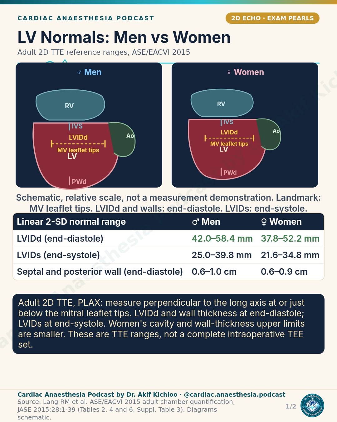

← Back to posts
← Back to posts

High-Risk Pulmonary Embolism: Who Gets Thrombolysis?

Adult education · Sources checked 10 November 2026

View Instagram reel

1. Complete the risk classification

The reel uses ESC 2019 categories. Intermediate-high risk requires haemodynamic stability, PESI III–V or sPESI ≥1, RV dysfunction and positive troponin. Low-score patients with RV dysfunction or elevated biomarkers fall into intermediate-low risk in the ESC framework.

Persistent hypotension must not be due to new arrhythmia, hypovolaemia or sepsis. Obstructive shock includes end-organ hypoperfusion despite adequate filling. ESC 2019, Tables 4 and 8

2. Reperfusion and alteplase

ESC recommends systemic thrombolysis for high-risk PE when appropriate. Alteplase 100 mg over two hours is the standard listed regimen and the U.S. labelled dose for acute massive PE. Check bleeding contraindications and local prescribing information. Activase prescribing information, sections 2.3 and 4.2

The 0.6 mg/kg over 15 minutes regimen (maximum 50 mg) is non-approved accelerated treatment sometimes used in extreme instability, not a routine interchangeable option.

For high-risk PE when thrombolysis is contraindicated or fails, surgical embolectomy is recommended; catheter-directed treatment should be considered where expertise and resources permit. Intermediate-high-risk PE needs monitoring, with rescue reperfusion for haemodynamic deterioration rather than routine primary thrombolysis. ESC 2019, Table 10 and section 7.2

3. Anticoagulation and circulatory support

Avoid a universal UFH-first instruction. Anticoagulant selection, monitoring and peri-thrombolysis timing depend on the clinical setting and local protocol.

A cautious fluid challenge up to 500 mL is considered only with normal–low filling pressure/hypovolaemia; excess fluid can worsen RV failure. Norepinephrine is used for hypotensive cardiogenic shock. ESC 2019, Table 9 and sections 6.1–6.2

4. Newer guideline

The AHA/ACC multisociety 2026 guideline introduces A–E clinical categories and recommends LMWH over UFH for C1–E1 patients needing initial parenteral anticoagulation. It is newer separate guidance, not an ESC replacement. 2026 guideline · Official summary

Key references

  1. Konstantinides SV, Meyer G, Becattini C, et al. 2019 ESC Guidelines for the diagnosis and management of acute pulmonary embolism developed in collaboration with the European Respiratory Society (ERS). Eur Heart J. 2020;41(4):543–603. doi:10.1093/eurheartj/ehz405.
  2. Creager MA, Barnes GD, Giri J, et al. 2026 AHA/ACC/ACCP/ACEP/CHEST/SCAI/SHM/SIR/SVM/SVN Guideline for the Evaluation and Management of Acute Pulmonary Embolism in Adults. Circulation. 2026;153:e977–e1051. doi:10.1161/CIR.0000000000001415.
  3. Genentech. Activase (alteplase) prescribing information. Revised September 2022; checked 10 November 2026. Full prescribing information.

For clinician education and exam study. This reference note is not a standalone bedside protocol; use current local guidance, trained assistance and patient-specific judgement.

← Back to posts
← Back to posts

Unanticipated Difficult Intubation: DAS Plans A to D

Adult education · Sources checked 10 November 2026

View Instagram reel

1. Scope and attempt limits

This reel summarises DAS 2015. Its core Plans A–D sequence and scalpel–bougie–tube equipment are consistent with that guideline; current practice should also use DAS 2025.

Attempt limits are ceilings: up to three intubation attempts, plus one by a more experienced clinician, and up to three second-generation supraglottic airway attempts. Move on earlier if oxygenation or trauma makes further attempts unsafe. DAS 2015

2. Successful rescue ventilation

After successful supraglottic rescue, stop and reassess. Waking is preferred when surgery is not urgent; intubation through the device should be fibreoptically guided. Continuing surgery or choosing front-of-neck access needs a specific senior clinical decision. DAS 2015

If final facemask ventilation succeeds, maintain oxygenation and agree the next step with senior clinicians. If waking is chosen, ensure full reversal and continued ventilation through emergence. Sugammadex can reverse rocuronium but does not reverse anaesthesia or guarantee airway patency. It must not delay emergency front-of-neck access in CICO. DAS 2025

3. Newer guideline

DAS 2025 retains Plans A–D and reinforces continuous oxygen delivery, waveform capnography and videolaryngoscopy whenever possible. Its front-of-neck guidance uses a vertical skin incision as the default when cricothyroid-membrane palpability has not been confidently established. Use the full algorithm and trained team rather than this abbreviated equipment list. Current DAS guideline and algorithms

Key references

  1. Frerk C, Mitchell VS, McNarry AF, et al. Difficult Airway Society 2015 guidelines for management of unanticipated difficult intubation in adults. Br J Anaesth. 2015;115(6):827–848. doi:10.1093/bja/aev371. Open full text.
  2. Ahmad I, El-Boghdadly K, Iliff H, et al. Difficult Airway Society 2025 guidelines for management of unanticipated difficult tracheal intubation in adults. Br J Anaesth. 2026;136(1):283–307. doi:10.1016/j.bja.2025.10.006.

For clinician education and exam study. This reference note is not a standalone bedside protocol; use current local guidance, trained assistance and patient-specific judgement.

← Back to posts
← Back to posts

Reversal Before Urgent Surgery

Adult clinician education · References checked 10 November 2026

View the Instagram reel

Sources and clarifications for this post

The chart brings together reversal options for six commonly encountered anticoagulants. The decision to reverse depends on the urgency and bleeding risk of surgery, the anticoagulant, its last dose, renal function and the likely residual effect. Stop the anticoagulant and involve the surgical, anaesthetic and haematology teams. Where it is clinically safe, allowing time for drug clearance can avoid the thrombotic risks of reversal. ESAIC 2024 reversal guideline, urgent surgery.

Warfarin: use the INR-based PCC dose with vitamin K

The chart’s 4-factor PCC doses match the Kcentra label: 25 IU/kg for INR 2 to <4, 35 IU/kg for INR 4–6, and 50 IU/kg for INR >6, with corresponding maxima of 2500, 3500 and 5000 factor-IX units. These are product-specific dosing instructions, capped at a body weight of 100 kg. Check the locally available product and current INR. Give vitamin K concurrently because PCC provides immediate factor replacement but does not restore endogenous synthesis. The chart’s slow IV vitamin K 5–10 mg regimen is supported for major bleeding requiring urgent reversal. Kcentra prescribing information, §§1–2; ACC 2020 bleeding pathway, VKA reversal.

This dose table should not be read as a requirement to give PCC before every operation in a patient taking warfarin. The available time, INR, bleeding and procedure determine the plan. Check the clinical response and repeat INR after reversal. PCC carries thromboembolic risk; its label does not establish routine repeat dosing. Kcentra prescribing information.

Dabigatran: idarucizumab has an urgent-procedure indication

Idarucizumab 5 g IV, supplied as two 2.5 g vials, is the specific reversal agent for dabigatran when rapid reversal is needed for emergency surgery/urgent procedures or life-threatening or uncontrolled bleeding. The two consecutive 5–10-minute infusions shown are supported by the product information; bolus administration is another labelled option. Dabigatran can reappear after initial reversal, especially with high concentrations or renal impairment, so continue clinical and laboratory assessment. Restart dabigatran no earlier than 24 hours after idarucizumab, and only with clinical stability and adequate haemostasis. Praxbind product information, §§4.1–4.4.

A normal sensitive thrombin time argues against clinically relevant dabigatran effect. It does not quantify the concentration, and a normal aPTT is not equivalent. A calibrated dilute thrombin time or ecarin-based assay is more useful when a level is needed. Haemodialysis can remove dabigatran, but access, time and clinical stability limit its emergency use. Non-specific prohaemostatic treatment when idarucizumab is unavailable is a specialist decision, rather than an interchangeable antidote. EHRA practical guide, laboratory assessment and bleeding management.

Factor Xa inhibitors: PCC is a conditional option, not an automatic next step

For apixaban, rivaroxaban or edoxaban, 4-factor PCC is an off-label option for selected high-risk urgent procedures with clinically relevant residual drug effect. EHRA Figure 9 does support PCC 50 U/kg, with another 25 U/kg if indicated, or aPCC 50 U/kg, maximum 200 U/kg/day. However, that figure concerns life-threatening/critical-site bleeding. It is not an automatic treatment sequence for every nonbleeding surgical patient. Dose and repeat treatment require a local specialist protocol and reassessment of thrombotic risk. EHRA Figure 9 and unplanned-surgery discussion; ESAIC 2024.

Andexanet needs a dated, jurisdiction-specific note. It should not be presented as a generally licensed preoperative reversal agent or as an approved antidote for every factor Xa inhibitor. In the United States, commercial sales of Andexxa ended on 22 December 2025 after FDA concluded that its risks outweighed its benefits. This U.S. regulatory action should not be generalized to other countries. Check the current local licence and availability. FDA safety communication.

The EMA currently lists conditional EU authorisation for adults taking apixaban or rivaroxaban who have life-threatening or uncontrolled bleeding. India’s CDSCO approval list through March 2026 records import/marketing permission dated 19 January 2024 for the same bleeding indication. These records do not verify current hospital stock or exclude subsequent local changes. EMA product record; CDSCO approval record.

The cardiac-surgery warning is important: andexanet can interfere with subsequent heparin anticoagulation. It should be avoided before cardiopulmonary bypass when heparin is required. EACTS 2024 perioperative medication guideline, DOAC reversal.

Unfractionated heparin: account for elapsed time

The 1 mg protamine per 100 units of heparin relationship is supported, but the dose must reflect heparin still active, not simply every unit previously administered. The chart’s recent-infusion example is an estimate; reduce requirements as time elapses after stopping heparin and reassess coagulation. Give protamine slowly. The label specifies administration over 10 minutes in doses not exceeding 50 mg; “max 50 mg” must not be mistaken for a universal total-course limit for every heparin-reversal setting, especially cardiopulmonary bypass. Excess protamine can itself impair coagulation. Protamine prescribing information, dosage and warnings.

Enoxaparin: protamine gives incomplete reversal

The label supports 1 mg protamine per 1 mg enoxaparin within eight hours, and 0.5 mg per 1 mg after eight hours or for a required second dose. At least 12 hours after the last injection, protamine may not be needed, but this is not a guarantee of clearance, particularly with renal impairment. Anti-Xa activity is only partly neutralised, at most about 60%. Lovenox prescribing information, overdosage.

The chart’s repeat-dose wording reflects the label’s prolonged-aPTT instruction at 2–4 hours. However, a normal aPTT does not establish that enoxaparin has cleared or been fully reversed. Clinical haemostasis, renal function and appropriately calibrated anti-Xa testing, when useful and available, are considered together. Lovenox prescribing information, monitoring and overdosage.

Fondaparinux and restarting anticoagulation

Fondaparinux has no established specific antidote. Protamine does not provide effective reversal, consistent with experimental reversal testing. Gatt et al., in-vitro study. Its long, renal-function-dependent elimination makes delay, if safe, and haemostatic support important. Any non-specific rescue treatment needs specialist assessment. Restarting anticoagulation requires both secure haemostasis and a plan matched to the procedure, drug and thrombotic indication; the chart’s closing line is not an instruction to give a full therapeutic dose immediately. Fondaparinux product information; ACC 2020 pathway, restarting anticoagulation.

Newer guidance to read alongside the chart

The 30 ng/mL threshold is a historical consensus consideration for urgent high-bleeding-risk procedures, not a universal reversal trigger. Newer guidance uses different thresholds according to the procedure and clinical context. ISTH SSC 2024 discusses ≥50 ng/mL as a level that may warrant reversal in emergency surgery. ESAIC 2024 states that levels above 50 ng/mL may be considered for haemostatic or antidote intervention in urgent high-risk surgery. Use a drug-appropriate calibrated assay and the relevant procedure-specific guidance; routine normal PT/INR or aPTT does not reliably exclude a factor Xa inhibitor. ISTH 2024 guidance; ESAIC 2024, recommendations 3.1–3.5.

Cardiac-specific EACTS 2024 guidance still uses <30 ng/mL as a low residual anti-FXa drug level for major surgery. Therefore, 30 ng/mL has not simply been superseded everywhere by 50 ng/mL. Neither value alone guarantees safety or mandates reversal. EACTS 2024 perioperative medication guideline.

Key references

Sources identified in the post

  1. Tomaselli GF, Mahaffey KW, Cuker A, et al. 2020 ACC Expert Consensus Decision Pathway on Management of Bleeding in Patients on Oral Anticoagulants. JACC. 2020. DOI: 10.1016/j.jacc.2020.04.053. Acute-bleeding and restart guidance.
  2. Steffel J, Collins R, Antz M, et al. 2021 EHRA Practical Guide on NOACs in Atrial Fibrillation. Europace. 2021;23:1612–1676. DOI: 10.1093/europace/euab065.
  3. Levy JH, Ageno W, Chan NC, et al. When and how to use antidotes for the reversal of direct oral anticoagulants: guidance from the SSC of the ISTH. J Thromb Haemost. 2016;14:623–627. DOI: 10.1111/jth.13227. Historical threshold guidance, read with the 2024 update.
  4. CHEST citation correction: the 2022 perioperative guideline is by Douketis JD, Spyropoulos AC, Murad MH, et al., not Garcia. CHEST. 2022;162:e207–e243. DOI: 10.1016/j.chest.2022.07.025. Its primary scope is elective procedures, not a complete emergency-reversal protocol.
  5. Current product information for 4-factor PCC and idarucizumab. The additional protamine, enoxaparin and fondaparinux labels are linked beside their relevant sections above.

Updates and additional reading

For clinician education and exam study. This page is not a patient-specific reversal prescription. Apply current local protocols, product information and specialist assessment.

← Back to posts
← Back to posts

Neuraxial Blocks and Anticoagulants

Adult clinician education · References checked 10 November 2026

View the Instagram reel

How to read the timing chart

The headline intervals are supported

The chart correctly gives at least 72 hours before neuraxial or deep plexus/peripheral blockade for high-dose apixaban, rivaroxaban and edoxaban. Its minimum 12-hour low-dose and 24-hour high-dose LMWH intervals also agree with the cited guidance. Warfarin requires five days of interruption and a normal INR by the local laboratory before needling; the separate catheter-removal threshold is INR <1.5. These conditions belong with the numbers. ASRA fifth edition, drug-specific recommendations.

High-dose LMWH needs a surgical bleeding-risk check

The high-dose LMWH “after” cell gives at least 24 hours after needle/catheter placement and catheter removal at least four hours before the first postoperative dose. There is another requirement: high-dose LMWH is resumed 24 hours after surgery that is not high bleeding risk, or 48–72 hours after high-bleeding-risk surgery. Give the first postoperative dose only after all applicable intervals have elapsed. Meeting the placement-to-dose interval alone is insufficient. ASRA fifth edition, postoperative LMWH.

The low-dose LMWH rows distinguish once-daily from twice-daily treatment correctly. For twice-daily treatment, the first postoperative dose is on the following day and at least 12 hours after placement; remove the catheter before starting LMWH and leave at least four hours after removal. ASRA fifth edition, postoperative LMWH.

Fondaparinux has an important renal limit

The displayed 36–42-hour interval concerns 2.5 mg once daily with normal renal function; the 58-hour interval concerns creatinine clearance 30–50 mL/min. Below 30 mL/min, ASRA advises against neuraxial or deep plexus/peripheral blocks. Extending the 58-hour number is not the recommended solution. Higher fondaparinux doses have a separate pathway. ASRA fifth edition, fondaparinux.

Read “low dose” with the indication

A smaller tablet dose does not automatically mean the low-dose pathway. The chart appropriately places reduced-dose apixaban for atrial fibrillation in its high-dose group. Low-dose apixaban and rivaroxaban have different interruption intervals; ASRA provides no routine low-dose edoxaban pathway. The caption’s grouping of “high and low dose” drugs should therefore not be read as applying both categories to edoxaban. ASRA educational discussion, questions 2–3.

The graphic also correctly separates the first postoperative DOAC dose from needle placement/catheter removal by at least 24 hours for high-dose treatment and six hours for the listed low-dose regimens. These are regional-procedure minima. Surgical haemostasis and the operation’s bleeding risk can require a later restart. Drug-level testing requires an appropriate assay and correct units; a routine normal INR does not establish that a DOAC has cleared. ASRA fifth edition, DOAC recommendations and perioperative discussion.

IV heparin has a separate catheter-removal sequence

The chart’s one-hour interval after needle placement concerns starting IV heparin. For an indwelling neuraxial catheter, the corrected guideline specifies removal 4–6 hours after the last IV-heparin dose, after assessing coagulation, followed by a one-hour interval before reheparinisation. This differs from the low-dose subcutaneous-heparin row, where dosing may follow catheter removal immediately. Corrected ASRA publication, IV and subcutaneous heparin.

The cardiac-surgery warning remains relevant: neuraxial or deep plexus/peripheral catheters should not be maintained during full anticoagulation for cardiac surgery. Neurological monitoring remains essential. ASRA fifth edition, unfractionated heparin.

The compact rows do not replace a catheter plan

The antiplatelet rows describe drug-specific interruption and loading-dose considerations. Aspirin’s absence of a routine timing restriction should not be extended to combinations with anticoagulants. After thrombolysis, the chart’s at-least-48-hour wait before needling also requires documented recovery of clotting tests, including fibrinogen. Unexpected thrombolysis with a catheter already in place has no single established removal interval. ASRA fifth edition, antiplatelets and thrombolytic therapy.

Guideline version

ASRA’s current index still lists the fifth edition. The publisher has corrected its online publication, including the IV-heparin catheter-removal and motor-monitoring recommendations inadvertently omitted earlier. Use the corrected document rather than an early downloaded copy. No superseding ASRA edition was identified in this check. ASRA guideline index; publisher correction notice.

Key references

  1. Source named in the post: Kopp SL, Vandermeulen E, McBane RD, Perlas A, Leffert L, Horlocker T. Regional anesthesia in the patient receiving antithrombotic or thrombolytic therapy: American Society of Regional Anesthesia and Pain Medicine Evidence-Based Guidelines, fifth edition. Published online 29 January 2025; corrected online version. DOI: 10.1136/rapm-2024-105766. Relevant sections: UFH, LMWH, warfarin, DOACs, antiplatelets, fondaparinux and thrombolytics.
  2. Additional explanation: Pressler M, Clark HJY, Srikalyani S. ASRA Pain Medicine Anticoagulation PBLD: Regional Anesthesia and Pain Procedures in Anticoagulated Patients. 1 September 2025. Official ASRA educational discussion, especially questions 2–3. The guideline remains the primary authority for individual drug pathways.

For clinician education and exam study. This is a non-exhaustive explanation, not a patient-specific stopping or restarting prescription. Apply the complete current guideline, local protocols and specialist assessment.

← Back to posts
← Back to posts

DKA: Diagnosis and the First Hours

Adult education · Sources checked 9 November 2026

View the Instagram reel

1. All three diagnostic components are required

The video's D–K–A thresholds agree with ADA 2026: glucose ≥11.1 mmol/L (200 mg/dL) or established diabetes; β-hydroxybutyrate ≥3.0 mmol/L or urine ketones ≥2+; and pH <7.3 and/or bicarbonate <18 mmol/L. Known diabetes permits diagnosis below the glucose threshold. A glucose result alone cannot exclude euglycaemic DKA. ADA 2026, Table 16.1

2. The fluid rate needs a patient-specific qualification

The video's 500–1000 mL/hour for 2–4 hours applies to adults without cardiac or renal compromise. Saline or balanced crystalloid is acceptable. Older adults and people with heart failure or dialysis-dependent kidney failure may need smaller boluses, such as 250 mL, with frequent reassessment. The displayed rate is not a universal prescription. 2024 consensus, Fluid Therapy

3. Continue insulin, but reassess its rate

The initial IV rate of 0.1 units/kg/hour is supported. Below glucose 13.9 mmol/L, the video correctly adds 5–10% dextrose; it should also specify reducing IV insulin to 0.05 units/kg/hour, with subsequent adjustment under the treatment protocol. Glucose improvement alone is not the endpoint. ADA 2026, Figure 16.1

Keeping established basal insulin going is supported, with review of the prescribed regimen. This does not mean automatically giving an additional basal dose when one is already active. JBDS 2023, Insulin Therapy and Conversion to Subcutaneous Insulin

4. Potassium decisions need repeat measurements

For potassium <3.5 mmol/L, replace potassium and delay insulin until potassium is >3.5 mmol/L. The video's intermediate and high-potassium rows summarize replacement versus withholding. The 2024 consensus targets potassium 4–5 mmol/L and checks it two hours after starting insulin, then four-hourly until resolution. 2024 consensus, Potassium

Assess kidney function and urine output before routine potassium supplementation; oliguria, anuria or dialysis requires individualized specialist management. The video's two-hourly recheck when potassium is high matches the ADA figure. Instability or abnormal potassium can require closer monitoring. ADA 2026, Figure 16.1; JBDS 2023, Monitoring and Kidney Failure

5. A low pH makes bicarbonate a consideration

The pH <7.0 threshold means consider bicarbonate, not administer it automatically. Routine bicarbonate is unsupported and may worsen hypokalaemia. 2024 consensus, Bicarbonate

6. Read the resolution criteria together

Resolution requires blood β-hydroxybutyrate <0.6 mmol/L and either venous pH ≥7.3 or bicarbonate ≥18 mmol/L. The video includes both acid-base alternatives; the caption abbreviates them. Glucose should ideally also be <11.1 mmol/L. Neither anion-gap closure nor urine-ketone disappearance substitutes for these endpoints. 2024 consensus, Criteria for Resolution

7. Insulin overlap requires a defined transition plan

The video's 1–2-hour overlap matches its cited consensus. 2024 consensus, Transition to Maintenance Insulin

ADA 2026 contains an apparent text–figure difference: its DKA prose specifies basal insulin 2–4 hours before stopping IV insulin, while Figure 16.1 shows 1–2 hours after subcutaneous insulin. Avoid treating either interval as universal. Confirm basal coverage and follow the validated local transition protocol. ADA 2026, DKA section and Figure 16.1

8. The short reel is not the complete care pathway

Repeated bedside glucose, blood-ketone, electrolyte and clinical assessment are essential. Identify and treat the precipitant alongside metabolic treatment. JBDS 2023, Management and Monitoring

Before discharge, address insulin access, sick-day advice and ketone testing. SGLT2-inhibitor exposure, fasting and pregnancy are relevant when glucose is not markedly elevated. ADA 2026, Hyperglycaemic Crises

For clinician education and exam study. This adult reference is not a paediatric, pregnancy-specific or patient-specific treatment protocol. Apply current local guidance and specialist assessment.

Key references

  1. Umpierrez GE, Davis GM, ElSayed NA, et al. Hyperglycemic Crises in Adults With Diabetes: A Consensus Report. Diabetes Care. 2024;47:1257–1275. Full text. DOI: 10.2337/dci24-0032.
  2. American Diabetes Association Professional Practice Committee for Diabetes. 16. Diabetes Care in the Hospital: Standards of Care in Diabetes—2026. Diabetes Care. 2026;49(Suppl 1):S339–S355. Full text. DOI: 10.2337/dc26-S016.
  3. American Diabetes Association Professional Practice Committee for Diabetes. 6. Glycemic Goals, Hypoglycemia, and Hyperglycemic Crises: Standards of Care in Diabetes—2026. Full text. DOI: 10.2337/dc26-S006.
  4. Joint British Diabetes Societies for Inpatient Care. The Management of Diabetic Ketoacidosis in Adults. March 2023. Current official listing. Its protocol differs in some thresholds and transition details; this page does not combine those into the 2024 consensus algorithm.
← Back to posts
← Back to posts

Tension pneumothorax: decompress first

View on Instagram

View the Instagram post · Posted 9 November 2026 · References and guidance checked 9 November 2026

When decompression comes before imaging

“Decompress first” applies when suspected tension pneumothorax is causing haemodynamic instability or severe respiratory compromise. Do not delay emergency treatment for a chest radiograph or CT in that setting. NICE makes this qualification explicit for major trauma. In a stable patient, urgent assessment and appropriate imaging help establish the diagnosis and avoid unnecessary invasive treatment. Point-of-care ultrasound can support assessment when immediately available, but a negative ultrasound examination does not reliably exclude pneumothorax. NICE NG39, recommendations 1.3.1–1.3.4 and 1.4.1–1.4.3.

Recognising the clinical pattern

Respiratory distress, tachycardia, hypotension and reduced or absent breath sounds on one side are important warning signs. Hyper-resonance may be difficult to establish. Distended neck veins and tracheal deviation are inconsistent findings: describing them as “late” must not imply that they will eventually appear or are required for diagnosis. Assessment depends on the whole clinical picture, with repeated examination as the situation changes. Waydhas et al., diagnosis recommendations and discussion.

In a ventilated patient, a sudden rise in airway pressure, worsening oxygenation and circulatory deterioration should prompt urgent assessment. These changes are not specific to pneumothorax; tube position and other airway or ventilation problems also need rapid consideration. Positive-pressure ventilation can accelerate deterioration from a pneumothorax. Waydhas et al., Table 5 and diagnosis discussion; ERC 2025, traumatic cardiac arrest: tension pneumothorax, p. 32.

Needle sites depend on the patient and protocol

The adult lateral approach described in the caption should not become a single rule for every patient or procedure. Current trauma guidance includes different needle sites: the WSES-AAST guideline lists the second intercostal space at the midclavicular line or the fifth at the midaxillary line when urgent needle decompression is needed and a chest tube is unavailable. These landmarks are not interchangeable with every finger-thoracostomy or drain-insertion recommendation. Use the applicable protocol, suitable equipment and trained clinical judgement. WSES-AAST 2025, pleural injuries: treatment.

For children, the second intercostal space at the midclavicular line remains an option, but it is not the only current option. RCUK 2025 also lists the fourth or fifth intercostal space at the anterior axillary line for needle thoracocentesis, followed by chest drainage. In paediatric trauma, it describes finger thoracostomy at the fourth or fifth intercostal space at the anterior axillary line, followed by an emergency drain. These are specialist procedures requiring age-appropriate equipment and expertise. RCUK 2025 paediatric guidance, tension pneumothorax.

A needle is a bridge, not a compulsory first step

Needle decompression can be a rapid temporary intervention, but it can fail or lose effectiveness. The sequence is not invariably needle, then finger thoracostomy, then drain. NICE favours open thoracostomy when the relevant expertise is available, with a chest drain through the opening for spontaneously breathing patients in the prehospital setting. In hospital, it recommends open thoracostomy followed by a chest drain. The appropriate immediate approach depends on the patient's condition, ventilation, setting, skills and equipment. Confirm a clinical response and continue checking for recurrence. NICE NG39, recommendations 1.3.5–1.3.6 and 1.4.2; WSES-AAST 2025, pleural injuries: treatment.

Adult traumatic cardiac arrest is a specific setting. ERC 2025 recommends immediate bilateral thoracostomies when the necessary skills and equipment are available; needle decompression is a fallback when they are not. This should not be generalised to bilateral decompression in every patient with suspected pneumothorax. ERC 2025, traumatic cardiac arrest: tension pneumothorax, p. 32.

Guideline update

The caption names ATLS 10th edition (2018). ACS has since released ATLS 11 (2025). Read the post alongside the current paediatric, resuscitation and thoracic-trauma guidance cited above. These sources provide current clinical context; not every recommendation was newly introduced in 2025. ACS ATLS 11 announcement.

References

  1. American College of Surgeons Committee on Trauma. Advanced Trauma Life Support (ATLS): Student Course Manual. 10th ed. Chicago: American College of Surgeons; 2018. Source named in the post. ACS ATLS programme.
  2. National Institute for Health and Care Excellence. Major trauma: assessment and initial management. NICE guideline NG39. Published 17 February 2016. Recommendations 1.3.1–1.3.6 and 1.4.1–1.4.3. Recommendations.
  3. Waydhas C, Prediger B, Kamp O, et al. Prehospital management of chest injuries in severely injured patients: a systematic review and clinical practice guideline update. European Journal of Trauma and Emergency Surgery. 2024;50:1367–1380. doi: 10.1007/s00068-024-02457-3. Table 5 and diagnosis discussion. Open full text.
  4. Lott C, Karageorgos V, Abelairas-Gomez C, et al. European Resuscitation Council Guidelines 2025 Special Circumstances in Resuscitation. Resuscitation. 2025;215(Suppl 1):110753. doi: 10.1016/j.resuscitation.2025.110753. Traumatic cardiac arrest: tension pneumothorax, p. 32. Official full text.
  5. Resuscitation Council UK. Paediatric Life Support (basic and advanced). 2025. Special circumstances: tension pneumothorax. Official guideline.
  6. Coccolini F, Cremonini C, Moore EE, et al. Thoracic trauma WSES-AAST guidelines. World Journal of Emergency Surgery. 2025;20:78. doi: 10.1186/s13017-025-00651-1. Pleural injuries: pneumothorax and haemothorax, treatment.
  7. American College of Surgeons. Trauma Care Gets Major Upgrade with Launch of ATLS 11. 16 September 2025. Official announcement. Edition announcement.

For education. Apply the cited sources in their clinical context and follow current local guidance; this material is not patient-specific advice.
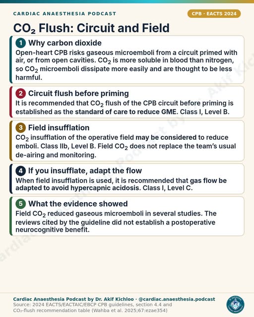
← Back to posts
← Back to posts

Hyponatraemia: Safe Correction Limits

View the Instagram post · References and guidance checked 9 November 2026

Sources supporting the post

Understanding the post

The numerical grade is not the same as clinical severity. The mild 130–135, moderate 125–129 and profound <125 mmol/L categories reproduce the 2014 guideline. Hyponatraemia itself means sodium below 135 mmol/L, so 135 should not be treated as an abnormal result simply because it appears at the edge of the table. Urgency also depends on symptoms and the clinical circumstances. 2014 guideline, §§6.1.1 and 7.1.

The severe-symptom list needs clinical attribution. Vomiting, cardiorespiratory distress, deep somnolence, seizures and coma (GCS ≤8) match the cited classification. These findings can have other causes; their presence alongside a mildly low sodium does not establish hyponatraemic encephalopathy. The 2022 guidance notes that severe symptoms are unlikely above 130 mmol/L. 2014 guideline, Table 5; 2022 emergency guidance, severity assessment.

The early goal is relief of cerebral oedema, not normalisation of sodium. The cited emergency regimen is 150 mL of 3% saline IV over 20 minutes, followed by sodium reassessment and, when needed, up to two further boluses toward an initial rise of about 5 mmol/L within the first hour under the cited protocol. Each repetition requires reassessment. Treatment belongs in a suitable monitored setting with experienced supervision. 2014 guideline, §7.1.1; 2022 guidance, treatment recommendations 1(a–e) and general recommendations.

Stopping after that initial rise depends on the response. The video's instruction to stop 3% saline after a 5 mmol/L rise applies when symptoms improve. Persistent severe symptoms require senior-led reassessment, consideration of another cause, and possible further hypertonic treatment within the correction limits. A sodium number alone should not replace that reassessment. 2014 guideline, §§7.1.2–7.1.3; 2022 guidance, recommendation 1(d) and Figure 2.

Correction ceilings are not targets or guarantees of safety. The post's 10 mmol/L in the first 24 hours and 8 mmol/L in each subsequent 24 hours reflect the European/UK limits. They describe the total rise, including the initial emergency correction. People at high risk of ODS need a stricter ceiling of no more than 8 mmol/L in any 24 hours; expert recommendations aim for only 4–6 mmol/L/day in this group. 2022 guidance; Sterns et al., Practice Guidelines.

The risk factors shown in the video need precise interpretation. Sodium ≤105 mmol/L, hypokalaemia, malnutrition, alcohol use disorder/heavy alcohol use and advanced liver disease identify particular concern. This does not mean that any alcohol exposure or every liver disorder carries the same risk. Potassium replacement can contribute to the rise in sodium and must be included in the correction assessment. Sterns et al., Practice Guidelines; 2014 guideline, practical advice under §7.1.

ODS can present after an initial improvement. The video's 2–6-day interval describes a typical delayed deterioration after correction; it is not an exact countdown, nor does ODS inevitably follow every overshoot. Neurological improvement immediately after treatment does not eliminate the need for continued observation. Sterns et al., Historical Background.

Relowering is an expert-guided rescue decision, not an automatic combined-treatment recipe. Stop active correction and seek experienced advice when correction exceeds the applicable limit. The cited 2014 options include electrolyte-free fluid, such as 5% glucose, 10 mL/kg over one hour, and consideration of 2 micrograms IV desmopressin. The video correctly includes an interval of at least eight hours before another desmopressin dose. Whether fluid, desmopressin or both are appropriate depends on sodium trajectory, chronicity, urine losses and ODS risk, with close fluid-balance and biochemical monitoring. 2014 guideline, §7.5.1; 2022 guidance, Managing over-correction.

Four-hourly sodium checks are not sufficient for every phase. The video's four-hour interval corresponds to ongoing hypertonic infusion in the 2014 pathway. Initial boluses require earlier reassessment. Sudden water diuresis can accelerate correction; the 2014 guideline advises two-hourly sodium checks when urine output suddenly exceeds 100 mL/hour. Monitoring becomes more frequent when the trajectory is unsafe or rescue treatment is needed. 2014 guideline, §§7.1.1, 7.1.3.4 and §7.4.4 practical advice; 2022 guidance, treatment and overcorrection sections.

Newer guidance and publication updates

  • 2022 Society for Endocrinology guidance: Retains the initial 5 mmol/L goal and 10/8 mmol/L limits, favours intermittent boluses and strengthens the emphasis on senior supervision and monitoring. Its emergency pathway also includes moderately severe symptoms. That pathway should not be silently combined with the different moderately symptomatic pathway in the 2014 document. Official guidance.
  • 2024 treatment-standard review: Spasovski's Hyponatraemia—treatment standard 2024 is a newer review, not a replacement edition of the multisociety guideline. Its separate 2024/2025 correction changes a urea dose unit from mg to g, and its July 2026 correction revises Figure 2 and a textual error. Those corrections do not change this post's emergency saline or desmopressin doses; the 2026 notice must be consulted alongside the original article.
  • 2026 European revision: An official joint guideline draft for review is available. A final adopted publication was not verified on 9 November 2026. Its proposed changes should not be presented as established replacement guidance or mixed into the post's 2014 algorithm.

This page explains an educational post about adult emergency management. Clinical care requires assessment of the individual patient and the current local emergency protocol.

For education. Apply the cited sources in their clinical context and follow current local guidance; this material is not patient-specific advice.
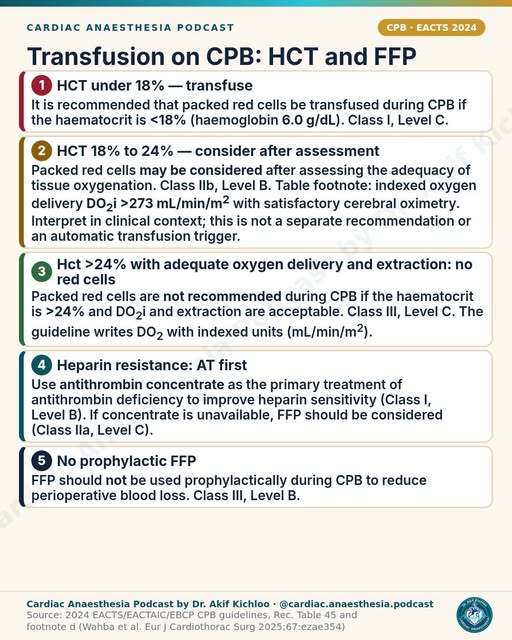
← Back to posts
← Back to posts

Status Epilepticus: The Timed Treatment Ladder

View the Instagram post · References and guidance checked 9 November 2026

Sources supporting the post

Understanding the post

This ladder concerns convulsive status epilepticus. Treat a tonic-clonic seizure lasting at least five minutes, and also recognize recurrent seizures without recovery of consciousness between them. The five-minute threshold is the point to act, not a reason to postpone airway support or other immediate care. Other forms of status epilepticus have different operational time points. ILAE definition; 2025 UK adult guidance, Definitions.

Thirty minutes is an estimate, not the moment injury inevitably begins. ILAE's t2≈30 minutes marks an estimated duration beyond which long-term consequences may occur in tonic-clonic status. Evidence is incomplete and there is substantial variation. It does not guarantee safety before 30 minutes or predict irreversible injury afterwards. ILAE definition.

The stabilization steps in the video belong alongside seizure treatment. Airway, breathing, circulation, oxygenation, monitoring, IV access and bedside glucose are appropriate immediate priorities. Identify and treat hypoglycaemia and other reversible causes while antiseizure treatment proceeds. The AES time bands organize an urgent sequence; they are not instructions to wait for the next labelled minute while seizures continue. AES algorithm, stabilization and treatment phases; NICE NG217, §§7.1.1–7.1.8.

Thiamine should not delay urgent glucose. The video's thiamine-before-glucose instruction reflects older sequencing advice. Give thiamine promptly when alcohol-related deficiency or malnutrition is suspected, ideally before or alongside glucose if readily available. Never withhold or delay treatment of hypoglycaemia while waiting for thiamine. ASAM explicitly permits either order or concurrent administration. ASAM 2020, V.7 and discussion; Schabelman and Kuo, 2012 evidence review.

The repeat instruction differs between the listed benzodiazepines. In the cited AES algorithm:

  • IM midazolam is a single dose: 10 mg if >40 kg, or 5 mg for 13–40 kg.
  • IV lorazepam is 0.1 mg/kg, maximum 4 mg per dose; it may be repeated once.
  • IV diazepam is 0.15–0.2 mg/kg, maximum 10 mg per dose; it may be repeated once.

The trailing repeat-once wording in the post should therefore not be applied indiscriminately to every option. These are alternative first-line regimens, not a list to administer together. AES algorithm, initial-therapy phase.

Use the complete second-line doses shown in the video, including the caps. The studied IV loading regimens are fosphenytoin 20 mg PE/kg (maximum 1500 mg PE), valproate 40 mg/kg (maximum 3000 mg), or levetiracetam 60 mg/kg (maximum 4500 mg). PE means phenytoin equivalents. ESETT infused each allocated drug over ten minutes. These are alternative loading treatments, with selection, preparation, monitoring and contraindications considered for the individual patient. ESETT, Methods.

ESETT found similar observed success, not proof that the drugs are equivalent in every patient. Its primary outcome occurred in 47% with levetiracetam, 45% with fosphenytoin and 46% with valproate. Success meant no clinically apparent seizures and improved responsiveness at 60 minutes, without additional antiseizure medication. The trial found no demonstrably superior drug and stopped for futility; it was not a formal equivalence trial. Participants were aged at least two years with benzodiazepine-refractory convulsive status, and pregnant patients were excluded. These results do not establish interchangeability across all clinical circumstances. ESETT, design, outcome definition and results.

Refractory convulsive status requires critical-care escalation. Failure of an adequate benzodiazepine and a non-benzodiazepine antiseizure medicine warrants urgent specialist involvement, airway/ventilatory support as needed, and continuous EEG. Anaesthetic midazolam, propofol or thiopental/pentobarbital are recognized options. The cited AES third phase also allows another second-line treatment; it does not establish one anaesthetic as universally best. Persistent depressed consciousness after visible convulsions stop can require EEG assessment for continuing seizures. AES algorithm, third phase; 2025 UK guidance, Definitions and General recommendations.

Benzodiazepine underdosing is common, but the superlative is not established. In an ESETT analysis, initial doses were below guideline recommendations in 76% of midazolam and 81% of lorazepam administrations. This selected refractory-status cohort did not compare every possible management error, so it supports describing underdosing as a frequent problem, rather than universally the commonest error. Give an adequate recommended dose promptly with respiratory monitoring and resuscitation capability. Sathe et al., 2021; AES guideline.

Newer guidance and publication updates

  • NICE NG217, chapter 7: The chapter 7 recommendations reviewed here emphasize an individual emergency plan when available, another benzodiazepine dose after 5–10 minutes if needed, and IV levetiracetam, phenytoin or sodium valproate after two benzodiazepine doses. Levetiracetam may be quicker to administer and have fewer adverse effects than the alternatives. A different second-line agent may be tried under expert guidance before phenobarbital or general anaesthesia. Valproate requires attention to the applicable safety restrictions. These are regional practical recommendations, not evidence that the post's AES options are incorrect. NICE, §§7.1.1–7.1.11.
  • 2025 UK multidisciplinary adult guidance: Adds a detailed ICU framework for refractory status, investigation and continuous EEG. It applies to people over 16 and excludes obstetric patients. The publisher's May 2025 correction adds an endorsement footnote, not revised drug doses. Corrected guidance.
  • AES update status: The official AES page still lists the 2016 convulsive-status guideline as published and a new status-epilepticus review/guideline as in development. No completed replacement was verified on 9 November 2026.
  • 2026 neuroprognostication guideline: Addresses counseling about outcomes in critically ill adults with status epilepticus. It does not replace the emergency dosing ladder and reinforces caution about deterministic predictions from individual findings. Guidelines for Neuroprognostication in Critically Ill Adults with Status Epilepticus, 2026.

This page explains an educational post. It is not a substitute for the current local emergency protocol, individualized drug selection or specialist care.

For education. Apply the cited sources in their clinical context and follow current local guidance; this material is not patient-specific advice.
← Back to posts
← Back to posts

Vasoactives by Shock State

View on Instagram

Sources and clarifications for this post

The chart summarizes common adult haemodynamic patterns to help explain drug choice. These patterns can overlap: cardiac output may fall in sepsis, systemic resistance may fall in cardiogenic shock, and RV failure does not always involve raised pulmonary vascular resistance. Filling pressures alone do not establish circulating volume or fluid responsiveness. The pattern becomes useful alongside clinical assessment, echocardiography and repeated assessment of perfusion. ESC heart-failure guideline, acute HF and cardiogenic shock; Harjola et al., acute RV failure.

Understanding the treatment choices

1. Septic shock: thresholds guide treatment rather than trigger it automatically.

Norepinephrine, an initial MAP around 65 mmHg and low-dose vasopressin are supported choices. The norepinephrine 0.25–0.5 µg/kg/min threshold reflects a 2021 panel practice remark: it is a guide to adding vasopressin when pressure remains inadequate or norepinephrine requirements escalate, rather than a compulsory cutoff. Vasopressin 0.03 U/min is a common low-dose sepsis regimen; doses differ in other shock settings. The dobutamine entry applies to cardiac dysfunction with persistent hypoperfusion despite adequate volume and arterial pressure, rather than an abnormal echocardiogram alone. SSC 2021, vasoactive agents and inotropes.

The hydrocortisone entry in the “pitfall” row refers to an adjunctive treatment option, not a drug to avoid. SSC 2021 suggests IV corticosteroids for adults with septic shock and an ongoing vasopressor requirement, typically hydrocortisone 200 mg/day. Its rationale defines that ongoing requirement as noradrenaline or adrenaline at ≥0.25 µg/kg/min for at least four hours after initiation to maintain the target MAP. The newer SSC 2026 guideline retains a conditional suggestion for IV corticosteroids in adults with septic shock. SSC 2021, corticosteroid remark; SSC 2021, full text and rationale; SSC 2026.

2. Cardiogenic shock: pressure, perfusion and the underlying cause all matter.

The ESC 2021 class IIb recommendation supports considering a vasopressor, preferably norepinephrine, and selected inotropic support. Dobutamine is therefore an option for an appropriate low-output state, not a requirement for every patient. A MAP around 65 mmHg is an initial practical target; reaching it does not by itself confirm restored organ perfusion. Congestion influences fluid decisions, while urgent cause-specific treatment and early shock-team involvement remain important when support is escalating. Mechanical circulatory support depends on the shock phenotype, reversibility and risk. ESC 2021, §§11.2–11.3; ACC cardiogenic-shock guidance 2025.

The dopamine warning is justified, but keep this trial distinction in mind: SOAP II found more arrhythmias with dopamine (24.1% versus 12.4%); overall 28-day mortality was not significantly different. The adverse cardiogenic-shock mortality finding was a subgroup result. De Backer et al., SOAP II.

3. Post-CPB vasoplegia: what VANCS and the rescue-drug options tell us.

The 32% versus 49% VANCS result refers to death or severe complications within 30 days, comparing vasopressin with norepinephrine. These are composite-event rates, not mortality rates alone. The trial compared alternative initial vasopressors, rather than specifically testing early vasopressin added to norepinephrine. A cardiac-surgery consensus supports norepinephrine and/or vasopressin, adding the other early if one is inadequate, and considering vasopressin first in pulmonary hypertension or right-heart dysfunction. Hajjar et al., VANCS; Guarracino et al., cardiac-surgery consensus.

Methylene blue 1.5–2 mg/kg and hydroxocobalamin 5 g are reported rescue regimens rather than complete or universally approved vasoplegia prescriptions. Route, infusion time, contraindications and the local specialist protocol are part of the treatment decision. These agents and angiotensin II have different evidence and risk profiles. The newer CPB guideline supports α1 agonists and/or vasopressin during bypass, with selected rescue drugs for refractory vasoplegia; its intra-CPB recommendations need not be considered as a universal postoperative sequence. Their setting is vasoplegia during bypass, while postoperative choices depend on the patient's haemodynamics and response. EACTS/EACTAIC/EBCP 2024 guideline, §9.4 and Recommendation Table 40; Busse et al., treatment options and dose table.

The methylene-blue warning about serotonin syndrome extends beyond SSRIs to other serotonergic medicines and some opioids. G6PD deficiency carries a risk of haemolysis, and pulse oximetry may underestimate oxygen saturation during treatment. Methylene blue prescribing information, §§4–5.

4. RV failure: treatment depends on pressure, output and the cause.

Norepinephrine is reasonable when systemic pressure is inadequate; an inotrope may be needed when output is inadequate. Neither is automatically required for every patient with RV dysfunction. Avoiding overfilling, correcting hypoxaemia/acidosis and treating the cause are also central to RV support. The 2024 acute-cor-pulmonale consensus notes limited evidence for vasopressin in acute RV failure. Its stronger perioperative rationale does not establish the same benefit across every cause of RV failure. Arrigo et al., 2024 consensus; Guarracino et al..

Dobutamine and milrinone can worsen systemic hypotension, so inodilator treatment may need concurrent pressure support. Harjola et al..

“Systemic pressure above PA pressure” is best understood as an emphasis on supporting systemic pressure and RV coronary perfusion, rather than a single numeric target. More precisely, the aim is to maintain systemic pressure and an adequate right-coronary perfusion gradient, individualized to RV pressure, filling pressure and organ perfusion. Mean and systolic pressures are different measurements, so an unspecified comparison between them can be misleading. Harjola et al..

Inhaled nitric oxide or epoprostenol can reduce RV afterload in selected settings. The 5–40 ppm range shown for iNO describes reported treatment doses, not a standard starting dose or an indication to treat all RV failure. Delivery, response, methemoglobin/NO₂ monitoring and gradual withdrawal require a dedicated protocol. INSPIRE-FLO supports comparable RV outcomes for iEPO and iNO after heart transplantation/LVAD surgery; its findings do not establish equivalence across all shock causes. Ghadimi et al., INSPIRE-FLO; 2024 cardiac-surgery iNO review.

5. Anaphylaxis: the IM and IV options belong to different treatment settings.

For the general adult emergency pathway, IM adrenaline 500 µg, repeated after five minutes if needed, remains supported by RCUK 2025. In the perioperative setting, the RCUK algorithm for anaesthetists/intensivists uses an initial 50 µg IV bolus, with careful dilution and monitoring, then an infusion for poor response. The IV option applies to monitored specialist care, not simply to any patient “under GA.” IM and IV doses and preparations are not interchangeable. RCUK 2025, anaphylaxis; RCUK perioperative algorithm.

Crystalloid bolus sizes vary between protocols. The perioperative algorithm starts adults and children over 12 with 500–1000 mL IV crystalloid, reassessing and repeating as needed; its 20 mL/kg instruction is for children under 12. The broader ERC 2025 guidance allows 10–20 mL/kg boluses with reassessment. Thus, 20 mL/kg can be an adult bolus within the applicable protocol, rather than necessarily a paediatric dose. ERC 2025. If shock remains refractory to an adrenaline infusion, specialists may add norepinephrine/vasopressin or consider glucagon 1 mg IV in adults on beta-blockers. Antihistamines do not treat shock, and steroids must not delay adrenaline and fluids. RCUK perioperative algorithm; RCUK 2021, second-line treatment.

6. Bleeding: the TXA regimen and lower pressure targets are trauma-specific.

In the cited European trauma guideline, the 1 g TXA dose is the loading dose of a two-part regimen: 1 g IV over 10 minutes, followed by 1 g IV over eight hours. Treatment is recommended as soon as possible and within three hours of injury, for trauma patients bleeding or at risk of significant bleeding. The loading dose alone is not the full regimen, and this recommendation does not apply automatically to every cause of hypovolaemia or bleeding.

The SBP 80–90 mmHg target applies to bleeding trauma without brain injury, until haemorrhage control. Permissive hypotension is inappropriate with traumatic brain/spinal injury; the guideline recommends MAP ≥80 mmHg in severe TBI. Norepinephrine can temporarily supplement restricted volume replacement when target pressure cannot be achieved, especially with life-threatening hypotension. It does not replace haemorrhage control and blood-volume replacement. Rossaint et al., 2023, Recommendations 13, 14 and 23.

7. Reassessment continues between scheduled reviews.

“Every few hours” is not a waiting interval after deterioration or a drug change. Unstable shock calls for continuous haemodynamic monitoring where indicated and reassessment of perfusion after interventions. Serial lactate, urine output, capillary refill and repeat echo are interpreted according to the clinical course. A low filling pressure alone does not prove fluid responsiveness. ACC 2025 cardiogenic-shock guidance; SSC 2026, resuscitation monitoring.

Newer guidance to read alongside the reel

  • SSC 2026: for adults with sepsis-induced hypoperfusion or septic shock, the suggestion of at least 30 mL/kg IV crystalloid in the first three hours remains conditional and requires individual assessment and frequent reassessment. Initial MAP is generally 65 mmHg, but 60–65 mmHg is suggested for adults aged ≥65. Norepinephrine remains the usual first choice, with vasopressin added for escalating requirements and epinephrine if pressure remains inadequate. With concomitant cardiac dysfunction, either norepinephrine or epinephrine may be first-line, influenced by heart rate/rhythm. Persistent hypoperfusion despite adequate volume and pressure can justify dobutamine plus norepinephrine or epinephrine alone. Official recommendations.
  • Cardiogenic shock, 2026 French multisociety recommendations: norepinephrine is the first vasopressor; either dobutamine or milrinone can be a first inotrope, with insufficient evidence to favour one universally. These recommendations update the cardiogenic-shock context of the reel's ESC 2021 citation; they concern a different patient population from the septic-shock recommendations. Aissaoui et al., recommendations R5–R6.
  • The 2024 CPB guideline, 2024 acute-cor-pulmonale consensus, and 2024 perioperative/2025 general RCUK guidance provide the setting-specific context discussed above.

Full references

  1. Evans L, Rhodes A, Alhazzani W, et al. Surviving sepsis campaign: international guidelines for management of sepsis and septic shock 2021. Intensive Care Med. 2021;47:1181–1247. doi:10.1007/s00134-021-06506-y.
  2. Prescott HC, Antonelli M, Alhazzani W, et al. Surviving Sepsis Campaign: International Guidelines for Management of Sepsis and Septic Shock 2026. Crit Care Med. 2026. doi:10.1097/CCM.0000000000007075.
  3. McDonagh TA, Metra M, Adamo M, et al. 2021 ESC Guidelines for the diagnosis and treatment of acute and chronic heart failure. Eur J Heart Fail. 2022;24:4–131. doi:10.1002/ejhf.2333.
  4. De Backer D, Biston P, Devriendt J, et al.; SOAP II Investigators. Comparison of dopamine and norepinephrine in the treatment of shock. N Engl J Med. 2010;362:779–789. doi:10.1056/NEJMoa0907118.
  5. Sinha SS, Morrow DA, Kapur NK, et al. 2025 Concise Clinical Guidance: An ACC Expert Consensus Statement on the Evaluation and Management of Cardiogenic Shock. J Am Coll Cardiol. 2025;85:1618–1641. doi:10.1016/j.jacc.2025.02.018.
  6. Aissaoui N, Delmas C, Merdji H, et al. Experts' recommendations for the management of adult patients with cardiogenic shock. Ann Intensive Care. 2026;16:100038. doi:10.1016/j.aicoj.2026.100038.
  7. Hajjar LA, Vincent JL, Galas FRBG, et al. Vasopressin versus norepinephrine in patients with vasoplegic shock after cardiac surgery: the VANCS randomized controlled trial. Anesthesiology. 2017;126:85–93. doi:10.1097/ALN.0000000000001434.
  8. Guarracino F, Habicher M, Treskatsch S, et al. Vasopressor Therapy in Cardiac Surgery—An Experts' Consensus Statement. J Cardiothorac Vasc Anesth. 2021;35:1018–1029. doi:10.1053/j.jvca.2020.11.032.
  9. Wahba A, Kunst G, De Somer F, et al. 2024 EACTS/EACTAIC/EBCP Guidelines on cardiopulmonary bypass in adult cardiac surgery. Eur J Cardiothorac Surg. 2025;67:ezae354. doi:10.1093/ejcts/ezae354.
  10. Busse LW, Barker N, Petersen C. Vasoplegic syndrome following cardiothoracic surgery: review of pathophysiology and update of treatment options. Crit Care. 2020;24:36. Full text.
  11. Harjola VP, Mebazaa A, Čelutkienė J, et al. Contemporary management of acute right ventricular failure: a statement from the Heart Failure Association and the Working Group on Pulmonary Circulation and Right Ventricular Function of the European Society of Cardiology. Eur J Heart Fail. 2016;18:226–241. doi:10.1002/ejhf.478.
  12. Arrigo M, Price S, Harjola VP, et al. Diagnosis and treatment of right ventricular failure secondary to acutely increased right ventricular afterload (acute cor pulmonale): a clinical consensus statement of the Association for Acute CardioVascular Care of the European Society of Cardiology. Eur Heart J Acute Cardiovasc Care. 2024;13:304–312. doi:10.1093/ehjacc/zuad157.
  13. Ghadimi K, Cappiello JL, Cooter-Wright M, et al. Inhaled epoprostenol compared with nitric oxide for right ventricular support after major cardiac surgery. Circulation. 2023;148:1316–1329. doi:10.1161/CIRCULATIONAHA.122.062464.
  14. Resuscitation Council UK. Emergency treatment of anaphylaxis: Guidelines for healthcare providers. May 2021. Guideline.
  15. Muraro A, Worm M, Alviani C, et al. EAACI guidelines: Anaphylaxis (2021 update). Allergy. 2022;77:357–377. doi:10.1111/all.15032.
  16. Dodd A, Turner PJ, Soar J, et al. Emergency treatment of peri-operative anaphylaxis: Resuscitation Council UK algorithm for anaesthetists. Anaesthesia. 2024;79:535–541. doi:10.1111/anae.16206.
  17. Resuscitation Council UK. Special circumstances guidelines. Published 27 October 2025. Anaphylaxis section.
  18. Rossaint R, Afshari A, Bouillon B, et al. The European guideline on management of major bleeding and coagulopathy following trauma: sixth edition. Crit Care. 2023;27:80. doi:10.1186/s13054-023-04327-7.
  19. Muenster S, Zarragoikoetxea I, Moscatelli A, et al. Inhaled NO at a crossroads in cardiac surgery: current need to improve mechanistic understanding, clinical trial design and scientific evidence. Front Cardiovasc Med. 2024;11:1374635. doi:10.3389/fcvm.2024.1374635.
  20. Amneal Pharmaceuticals. Methylene blue injection, solution: US prescribing information. DailyMed. Contraindications and Warnings and Precautions. Product label.
  21. Lott C, Karageorgos V, Abelairas-Gómez C, et al. European Resuscitation Council Guidelines 2025: Special Circumstances in Resuscitation. Resuscitation. 2025;215(Suppl 1):110753. doi:10.1016/j.resuscitation.2025.110753.

Adult educational reference, checked against the sources above on 9 November 2026. Follow the applicable local emergency, critical-care and drug-preparation protocols for patient care.

For education. Apply the cited sources in their clinical context and follow current local guidance; this material is not patient-specific advice.
← Back to posts
← Back to posts

12 Vasoactives in 60 Seconds

View on Instagram

Sources and clarifications for this post

These are adult teaching summaries, not complete prescribing instructions. Receptor stars and repeated arrows are schematic, not validated numerical potency comparisons. BP, HR, SVR and CO responses depend on dose, loading conditions, cardiac function and reflex responses. The indication, route, preparation, monitoring and local protocol must accompany a drug dose. Overgaard and Džavík. Circulation 2008;118:1047–1056, physiology and comparison table.

Understanding the twelve vasoactives

1. Noradrenaline: “α1 ≫ β1”, 0.05–0.5 µg/kg/min, first-line sepsis/post-CPB.

Predominant vasoconstriction with some cardiac stimulation is a useful summary. BP and SVR usually rise, but the CO ↑ arrow is not assured: output can be unchanged or decrease, and reflex slowing of HR can occur. The dose strip is an illustrative infusion range, not a universal starting dose or maximum. Norepinephrine label, §§2.2 and 12; Overgaard and Džavík 2008, table p.1049.

Norepinephrine is first-line in septic shock. After cardiac surgery, the consensus supports norepinephrine and/or vasopressin, with vasopressin a possible first choice in pulmonary hypertension or right-heart dysfunction. “First-line” therefore does not describe one sequence for every post-bypass patient. SSC 2021, vasoactive agents; Guarracino et al. 2021, cardiac-surgery consensus.

2. Adrenaline: “arrest & anaphylaxis”, 0.01–0.5 µg/kg/min and IM 500 µg.

The α1/β1/β2 summary is sound, but SVR need not rise at low doses because β2 vasodilation can predominate. The infusion strip is not a dose for every indication: the cited adult septic-shock label uses 0.05–2 µg/kg/min, titrated to response. Epinephrine label, §§2.2 and 12.2.

The emergency doses belong to different routes and treatment settings. IM 500 µg is the RCUK adult anaphylaxis dose; paediatric doses differ. IV treatment of anaphylaxis is for experienced specialists in a monitored setting. Standard adult cardiac-arrest ALS uses 1 mg IV/IO with rhythm-specific timing and repeat dosing, not this infusion strip. RCUK anaphylaxis guidance, pp.29–34; RCUK adult ALS 2025.

A symbol is missing in the lactate line: the reel displays a box before “2”. The relevant mechanism is β2-receptor stimulation in skeletal muscle, which can increase lactate production even when oxygen is available. This can contribute to a lactate rise, but does not exclude poor perfusion; interpretation depends on the whole clinical picture. SSC 2021, vasoactive-agent rationale; epinephrine label, pharmacodynamics.

3. Vasopressin: 0.01–0.03 U/min, “do NOT titrate”, add at noradrenaline 0.25–0.5 µg/kg/min.

V1-mediated vasoconstriction and catecholamine sparing are supported, but V1 is not its only receptor action. HR and CO can fall despite the neutral arrows shown in the reel. The narrow dose strip describes a low-dose approach. The phrase “Do not titrate” reflects a fixed low-dose approach in sepsis, rather than a universal rule: the product label allows adjustment and distinguishes septic from post-cardiotomy shock. The norepinephrine threshold comes from a practice remark in SSC 2021, not a mandatory trigger independent of the pressure target. Vasopressin label, §§2.2, 5.1 and 12; SSC 2021, vasopressin recommendation and remark.

4. Phenylephrine: pure α1, CO →, bolus 50–100 µg, “ideal in HOCM / dynamic LVOTO”.

The α1 pressor mechanism, SVR/BP rise and possible reflex bradycardia are supported. CO is not reliably unchanged: it can fall, and heart failure may worsen. The 50–100 µg IV boluses are within the cited adult perioperative label and are its most commonly reported initial boluses; they are not the whole permitted range. Phenylephrine label, §§2.2 and 5.

The HCM recommendation concerns acute hypotension with obstruction, when fluid administration is insufficient, not routine treatment of all HCM. “Avoid if LV is poor” is a warning about afterload and output, rather than a stand-alone rule that replaces assessment of obstruction and perfusion. AHA 2024 HCM clinical-update slides, slide 22.

5. Dopamine: D1 → β1 → α1, 2–20 µg/kg/min, more arrhythmias than noradrenaline.

The dose-dependent teaching sequence is useful, but dopamine also acts at D2 receptors and the single SVR ↑ arrow does not describe low-dose vasodilation. The 2–20 µg/kg/min strip is a published comparison range, not a fixed receptor-switch schedule or the full product-label range. Dopamine label, §§2.2 and 12; Overgaard and Džavík, table p.1049.

The arrhythmia warning is supported by SOAP II: events occurred in 24.1% with dopamine versus 12.4% with norepinephrine. Overall 28-day mortality did not differ significantly, so the trial did not establish an overall mortality benefit. Norepinephrine remains preferred in septic shock. De Backer et al. SOAP II, NEJM 2010; SSC 2021.

6. Dobutamine: β1-predominant inotropy, 2–20 µg/kg/min, low CO with adequate BP.

The inodilator summary and expected CO increase/SVR reduction are useful. BP → is not guaranteed: pressure may rise or fall, and HR/arrhythmias can increase. The 2–20 µg/kg/min strip matches usual trial infusion rates in the cited label, which starts lower at 0.5–1 µg/kg/min and then titrates. Adequate BP is a useful caution, not a ban on using dobutamine with a separate vasopressor when both are needed. Dobutamine label, clinical pharmacology, warnings and dosing; SSC 2026, inotropes.

7. Milrinone: PDE-III inhibition, 0.375–0.75 µg/kg/min, “often skip load”, RV support and renal-impairment precautions.

PDE3 inhibition, increased output and reduced vascular resistance are supported; hypotension and arrhythmias remain important risks. The maintenance range matches the cited label. The label also specifies a 50 µg/kg loading dose over 10 minutes. “Often skip load” describes an individual clinical decision rather than the label regimen. Renal impairment prolongs elimination and can require lower infusion rates, including below this strip. Milrinone label, pharmacology, precautions and renal dosing.

Milrinone can support RV function and reduce pulmonary vascular resistance. The phrase “great for RV failure” applies only in the appropriate clinical setting: the cause of RV failure and systemic blood pressure matter, and pulmonary hypertension alone is not an indication for an inotrope. Kaplan’s Cardiac Anesthesia, 8th ed., chapter 15, pp.632–633.

8. Levosimendan: calcium sensitiser/K-ATP, 0.05–0.2 µg/kg/min for 24 h, “active metabolite lasts 7–9 days”.

The mechanism and infusion range are supported for the labelled severe decompensated chronic-heart-failure setting; BP may fall and HR may rise as output increases. The 7–9-day statement refers to the duration of effect: haemodynamic effects can persist for up to 7–9 days after a 24-hour infusion. The metabolites have half-lives of about 75–80 hours; the parent drug’s is about one hour. These are different measures. The label contraindicates use with creatinine clearance below 30 mL/min, severe hypotension/tachycardia and significant mechanical filling/outflow obstruction. Simdax SmPC, §§4.2–4.4, 5.1–5.2.

9. Nitroglycerin: NO donor, “0.5–5 µg/kg/min · sublingual / spray OT”, avoid in AS/RV failure.

Predominant venodilation and preload reduction are supported. Despite the HR → and CO → arrows, HR often rises reflexly and output can rise or fall with the starting circulation. IV infusion and sublingual administration use different dosing regimens. The µg/kg/min strip cannot be used as a spray/tablet dose and is not a universal IV starting regimen; the cited US IV label starts at 5 µg/min with non-absorbing tubing and titrates to response. The linked spray contains 400 µg per metered spray. Nitroglycerin IV label, clinical trials and dosing; GTN spray SmPC, §§2 and 4.2.

The AS warning reflects UK product labels, which explicitly list aortic stenosis among contraindications. The “RV failure” warning concerns the danger of low filling pressure/preload dependence and hypotension, rather than every form of RV failure. The actual formulation’s contraindications remain important. PDE5 inhibitors and riociguat are also important nitrate interaction warnings. GTN infusion SmPC, §§4.3–4.5; GTN spray SmPC, §§4.3–4.5.

10. Nitroprusside: arterial and venous dilation, 0.3–2 µg/kg/min, cyanide risk.

The NO-mediated vasodilator mechanism and reductions in preload, SVR and BP are supported. CO ↑ is conditional, not assured; output is variable. Sodium nitroprusside FDA label, §12.1. The dose strip is a low-dose portion of the labelled range: 0.3 µg/kg/min is the starting rate, while 2 µg/kg/min is not a universal maximum. “Short runs” alone does not capture the safety requirements: continuous BP monitoring and adherence to the product’s dose/duration limits are essential. High-rate exposure can cause cyanide toxicity; prolonged treatment, particularly with renal impairment, also risks thiocyanate accumulation. Sodium nitroprusside label, clinical pharmacology, warnings and dosing.

11. Ephedrine: “Indirect α+β”, 3–9 mg IV, tachyphylaxis.

The “Indirect α+β” summary refers to only part of the mechanism: ephedrine combines direct α/β agonism with indirect norepinephrine release. BP, HR and CO usually rise, but the SVR response is variable. The bolus strip corresponds to the UK hydrochloride label, which gives 3–6 mg slowly IV, up to 9 mg per dose and 30 mg cumulatively for spinal/epidural hypotension. A US sulfate label uses 5–10 mg IV with a 50 mg cumulative limit. The formulation and local regimen determine which instructions apply; the two regimens are not interchangeable. Repeated doses may cause tachyphylaxis, making availability of an alternative pressor important. Ephedrine FDA label 2024, §§2.2, 5.2 and 12; Ephedrine UK SmPC, §§4.1–4.2.

12. Isoprenaline: β1/β2 chronotrope, 0.5–5 µg/min, bridge to pacing.

Nonselective β stimulation with increased HR/output and reduced peripheral resistance is supported. The BP ↓ arrow refers to the usual fall in mean and diastolic pressure, while systolic pressure can stay unchanged or rise. The 0.5–5 µg/min range appears in the cited US isoproterenol label for shock/hypoperfusion; it is not the same as a universal bradycardia starting regimen. RCUK 2025 lists 5 µg/min as a starting dose when atropine is ineffective. The bridge concept is reasonable, but pacing must not be delayed in unstable refractory bradycardia; monitoring for arrhythmia and myocardial ischaemia remains essential. The unit is µg/min, not µg/kg/min. Isoproterenol label, §§2.2, 5.1 and 12; RCUK adult ALS 2025, bradycardia.

Guideline update: SSC 2026 compared with SSC 2021

The 2026 Surviving Sepsis Campaign guideline retains norepinephrine as the usual first vasopressor, suggests adding vasopressin with escalating norepinephrine, and adding epinephrine when pressure remains inadequate. It specifically permits norepinephrine or epinephrine first-line when septic shock includes cardiac dysfunction, with tachyarrhythmia favouring norepinephrine and bradyarrhythmia favouring epinephrine.

For cardiac dysfunction with persistent hypoperfusion despite adequate filling and arterial pressure, it suggests dobutamine added to norepinephrine or epinephrine alone. Evidence is insufficient to choose dobutamine over milrinone. It suggests against levosimendan in that septic-shock setting. These recommendations do not make the twelve drugs interchangeable and are not a universal post-bypass algorithm.

For education. Apply the cited sources in their clinical context and follow current local guidance; this material is not patient-specific advice.
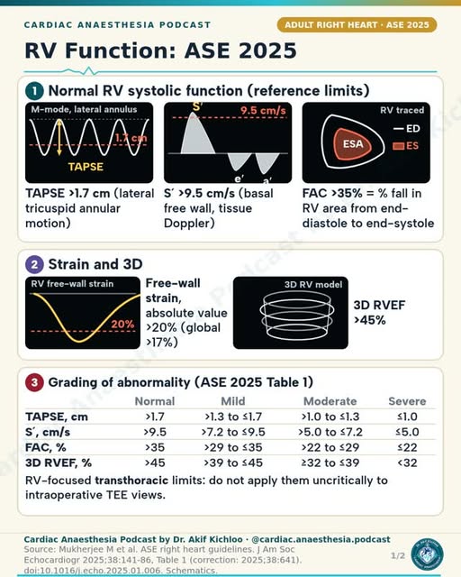
← Back to posts
← Back to posts

Mitral Regurgitation 5/5: Intra-op TEE and Bypass Pearls

View on Instagram

Sources supporting this post

Understanding the clinical context

  • TEE mapping and SAM risk. “ME commissural and long-axis views” are useful parts of a comprehensive multi-view examination, with 3D where helpful. Leaflet lengths, annulus, coaptation, clefts and calcification all matter. The SAM measurements are risk markers, not predictions of certainty: C-sept <2.5 cm, posterior leaflet >1.5 cm, a low anterior/posterior ratio around 1.3, a small LV and basal septal bulge are supported. ASE/SCA/STS 2020, §3.1.2/Table 2; Varghese et al., 2014; Alfieri and Lapenna, 2015, Table 1.
  • Tricuspid size is one part of the concomitant-repair decision. “Tricuspid annulus ≥40 mm (or >21 mm/m²): consider concomitant tricuspid repair (ESC 2021)” uses the correct historical thresholds. That recommendation concerned mild or moderate secondary TR in patients already undergoing left-sided valve surgery, with annular size measured by 2D echo. Size alone does not establish the indication. ESC/EACTS 2021, §8.2/Recommendations H.
  • Persistent, clinically important SAM may require revision. Stopping excess inotropy, appropriate volume, beta-blockade and phenylephrine are supported measures. The phrase “revise the repair if it persists” refers to clinically important SAM with LV outflow obstruction, important MR or haemodynamic compromise despite medical optimization. An isolated residual SAM image does not automatically require revision. Kaplan 8th, printed p. 632; ASE/SCA/STS 2020, §3.1.5, printed p. 702.
  • The gradient is not a pass/fail number. “no more than mild residual MR and mean gradient under 5 mmHg” describes a desirable result. None or trace MR is ideal. Assessment after adequate loading places the gradient alongside heart rate, rhythm, flow, leaflet opening and valve area. A value at or above 5 mmHg alone does not decide reoperation. Hensley 6th, TEE section III.B; Zamper et al., 2022, Iatrogenic mitral stenosis; ASE/SCA/STS 2020, printed p. 702.
  • The post-bypass examination includes the circumflex territory and aortic valve. “check circumflex (lateral RWMA) and the aortic valve” means look for new regional LV dysfunction suggesting circumflex compromise, especially lateral-wall abnormalities, and new or worsened aortic regurgitation. These accompany the full post-bypass examination. Holmes et al., 2017, p. 1; ASE/SCA/STS 2020, Table 3.
  • Afterload mismatch can follow repair too. The phrase “latent LV dysfunction appears; plan inotropic support” describes a possible need for support, rather than an inevitable outcome or routine infusion. Restoring competence changes afterload and preload after repair or replacement. The possible need for support is anticipated, while treatment depends on haemodynamics and TEE findings, including assessment for ischaemia, stunning and SAM. Kaplan 8th, printed p. 633; Quintana et al., 2014.

Guideline update

ESC/EACTS 2025 separates moderate TR, for which concomitant repair should be considered during mitral surgery, from mild TR with annular dilatation. For mild TR plus ≥40 mm or >21 mm/m², repair may be considered after balancing progression risk against pacemaker risk. §11.4.1.2.

For education. Apply the cited sources in their clinical context and follow current local guidance; this material is not patient-specific advice.
← Back to posts
← Back to posts

Mitral Regurgitation 4/5: Anaesthetic Goals and Haemodynamics

View on Instagram

Sources supporting this post

Understanding the clinical context

  • The rate range and mnemonic are teaching guides. “about 80–100” and “Fast, full and forward” have published support. A higher rate can shorten regurgitant systole and limit LV filling/distension, but the target must account for myocardial oxygen demand, ischaemia and perfusion. Preserving sinus rhythm when feasible and avoiding both excessive AF rates and treatment-induced bradycardia remain important. Holmes et al., 2017, p. 8; Paul and Das, 2017, p. 726; Kaplan 8th, printed p. 632.
  • A large LA does not guarantee volume tolerance. The phrase “the dilated LA tolerates volume” refers to the adaptation that can occur in chronic MR. Chronic compensated MR may have greater atrial compliance, but filling should be guided by perfusion, response and congestion. Excess fluid can worsen annular dilatation, MR and RV failure. Kaplan 8th, printed pp. 627 and 632; Hensley 6th, ch. 12, IV.C.5.
  • Forward flow still needs perfusion pressure. “keep afterload low” means avoid excessive resistance while preserving systemic and coronary perfusion. “Avoid myocardial depressants in the dilated LV” means avoid further depression of an impaired ventricle through careful drug selection and titration, not a blanket ban on anaesthetic agents. Dobutamine and milrinone may help when support is needed but can cause hypotension. Kaplan 8th, printed pp. 632–633.
  • Wedge measurements answer different pressure questions. “wedge reads high, use the a wave or pre-v value” applies when estimating LVEDP. With large V waves, mean PAWP may exceed LVEDP. An appropriately timed end-diastolic measurement, approximated by the mean A wave in sinus rhythm or the z/pre-V point in AF, can be more useful for that purpose. Mean PAWP still represents the atrial pressure burden transmitted to the lungs; it is not simply a false reading. Haskell and French, 1988; Tampakakis and Tedford, 2017, waveform-measurement discussion.
  • Phenylephrine is part of reassessment. “test with phenylephrine to awake MAP before grading” describes a studied manoeuvre, not a complete grading rule. Reassessment includes comparison with preoperative echo under appropriate filling and blood-pressure conditions. Matching MAP alone does not restore every awake condition or eliminate misclassification. Mihalatos et al., 2006; Shiran et al., 2007.
  • Shock treatment depends on pressure and the individual patient. The options in “Acute MR with shock: afterload reduction (nitroprusside), inotropes and IABP as a bridge to surgery” depend on blood pressure and patient selection. Vasodilators require adequate pressure. ESC 2021 describes inotropes and IABP as useful in unstable patients, not mandatory for everyone; urgent definitive treatment is central. ESC/EACTS 2021, §§6.1.2–6.1.3; Kaplan 8th, printed pp. 631–632.

Guideline update

ESC/EACTS 2025 makes the acute-MR blood-pressure qualification explicit: nitroprusside bridging is described without hypotension, and IABP is reserved for exceptional acute primary-MR cases. Urgent intervention may be surgical or transcatheter in selected patients. §§9.1.3–9.1.4.

For education. Apply the cited sources in their clinical context and follow current local guidance; this material is not patient-specific advice.
← Back to posts
← Back to posts

Mitral Regurgitation 3/5: Intervention Criteria (ESC 2021 + ACC/AHA 2020)

View on Instagram

Sources supporting this post

  • ESC/EACTS 2021, §6.1.2, recommendation table: supports all six primary-regurgitation rows: lasting repair, symptomatic surgery, ventricular thresholds, atrial fibrillation/lung-pressure triggers, left atrial enlargement and high-risk catheter repair.
  • ESC/EACTS 2021, §6.2.3, recommendation table: supports all six secondary-regurgitation rows: persistent symptoms despite treatment, combined bypass/other cardiac surgery, coronary or aortic-valve catheter treatment followed by reassessment, selected catheter repair, isolated surgery and selected treatment outside usual catheter-response criteria.
  • ACC/AHA 2020, official slides 112–113, recommendations 1–8: supports all eight primary-regurgitation rows, including serial changes on at least three scans, high-risk catheter repair with life expectancy of at least one year, rheumatic repair and the restriction on replacement for limited posterior-leaflet disease. Table 17 on slide 105 defines the stages.
  • ACC/AHA 2020, official slides 120–121, recommendations 1–5: supports all five secondary-regurgitation rows, including catheter-selection thresholds, surgery with bypass, atrial disease, persistent symptomatic ventricular disease and replacement preserving the valve’s supporting chords versus a smaller repair ring.
  • ESC/EACTS 2025: current changes are identified separately below; the original sources remain relevant to what this post quotes.

Clinical clarifications

These recommendations concern severe mitral regurgitation. The on-screen tables give important eligibility conditions shortened in the caption. Class I/1 means recommended; IIa/2a means should be considered or is reasonable; IIb/2b means may be considered. “3: Harm” means the specified action should not be performed in the stated circumstances. The following letters identify the evidence category.

  • The early-repair row uses “LVEF ≥60% and LVESD ≤40 mm”. This faithfully follows ACC/AHA slide 112, recommendation 4. However, its staging table on slide 105 defines C1 using left ventricular ejection fraction >60% and diameter at the end of contraction <40 mm. An ejection fraction of exactly 60% or diameter of exactly 40 mm meets its C2 boundary. C2 uses ejection fraction ≤60% and/or end-contraction diameter ≥40 mm: either abnormality is enough, and the comma in the recommendation row does not require both. Classification follows the staging table, so values exactly at these boundaries do not indicate clearly preserved function. Recommendation 4; Table 17.
  • The ESC row includes “SPAP at rest >50 mmHg”. Its 2021 footnote adds that pulmonary artery pressure should be confirmed by catheter measurement if this is the only reason for surgery. The on-screen row already includes the preserved-function and symptom-free conditions. ESC 2021, primary-regurgitation table, footnote c.
  • “fulfilling criteria suggesting an increased chance of responding to the treatment” refers to patient selection, not guaranteed success. The 2021 footnote points to criteria from the COAPT trial of catheter repair. Being unsuitable for surgery alone is not enough. ESC 2021, §6.2.3, table and footnote e.
  • The atrial-regurgitation row says “despite therapy for HF and associated AF”. The ACC/AHA source also includes treatment of other accompanying illnesses. The treatment context therefore includes those relevant conditions alongside heart failure and atrial fibrillation. ACC/AHA 2020, slide 120, recommendation 3.

Guideline update

The post cites ESC 2021. ESC 2025 provides the following newer recommendations to read alongside it.

  • ESC 2025 upgrades catheter-based edge-to-edge repair to IIa B for symptomatic severe primary mitral regurgitation with suitable anatomy and Heart-Team-assessed high surgical risk. It adds left ventricular diameter at the end of contraction, indexed to body surface area, ≥20 mm/m² as a surgery trigger in symptom-free severe primary disease (I B). For selected low-risk, symptom-free patients with preserved ventricular function and likely lasting repair, surgery is I B when at least three coexist: atrial fibrillation; resting pulmonary artery systolic pressure >50 mmHg; left atrial enlargement (indexed volume ≥60 mL/m² or diameter ≥55 mm); at least moderate secondary tricuspid leakage. ESC/EACTS 2025, §9.1.4; Recommendation Table 6 (advance PDF pp.40–41; journal pp.4674–4675).
  • Catheter repair is now I A for patients with stable circulation, symptoms, persistent severe ventricular secondary mitral regurgitation and left ventricular ejection fraction <50%, despite optimized heart-failure treatment and resynchronization pacing when indicated, who meet Table 7’s selection criteria. Benefits are fewer heart-failure admissions and improved quality of life. Atrial secondary mitral regurgitation has a separate treatment pathway. ESC/EACTS 2025, Table 7 selection criteria; Recommendation Table 7 (advance PDF pp.44–45; journal pp.4678–4679).
  • ESC 2025 also gives atrial secondary mitral regurgitation a separate pathway: surgery should be considered in symptomatic severe disease despite optimal treatment (IIa B); catheter repair may be considered when surgery is unsuitable, after medical and appropriate rhythm treatment (IIb B). ESC/EACTS 2025, Recommendation Table 7; §9.2.5.

For education. Apply the cited sources in their clinical context and follow current local guidance; this material is not patient-specific advice.
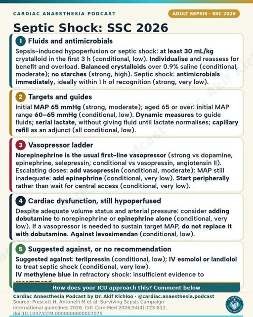
← Back to posts
← Back to posts

Mitral Regurgitation 2/5: Grading Severity on Echo

View on Instagram

Sources supporting this post

  • ASE/SCMR 2017, Table 8, p.332: supports the mild, moderate and severe ranges for jet width, backward-flow volume, backward-flow fraction and effective leaking area. The post correctly combines the two moderate subdivisions into one range. Sections III.C.2.b and III.D.1.a explain the effects of circulatory conditions and the limits of jet area.
  • ESC/EACTS 2021, Table 7: supports the leaflet and Doppler signs, including the mitral-to-aortic velocity-integral ratio above 1.4; the single-view and biplane jet-width thresholds; and the lower secondary-regurgitation thresholds for an elliptical opening or low blood flow. Section 6.2 supplies the treatment context.
  • ESC/EACTS 2025, §9.2.2: current guidance on assessing secondary mitral regurgitation, discussed separately below.

Clinical clarifications

  • The post’s severe cutoffs are not four boxes that must all be ticked. Severity assessment brings together the measurements, cause of leakage and circulatory conditions. Low blood flow can reduce the volume leaking backwards per beat, while an elliptical opening can cause underestimation of the effective leaking area. ASE 2017, Table 8 and §III.J.
  • The panel says “Vena contracta ≥7 mm (≥8 mm biplane) is severe in both primary and secondary MR”. These numbers match ESC 2021 Table 7. ASE 2017 Table 8 instead prints >0.8 cm for the width averaged across two views. The equality sign therefore depends on the source: the panel follows ESC’s threshold. Neither measurement replaces the combined assessment. ESC 2021, Table 7; ASE 2017, Table 8.
  • “Large central jet over 50% of LA, or eccentric wall-impinging jet” is a supporting sign, not a standalone grading rule. Wall-hugging jets may look deceptively small. The early-filling speed and mitral-to-aortic velocity-integral ratio also need the other findings for interpretation. ASE 2017, §III.D.1.a and Table 8; ESC 2021, Table 7.
  • The panel says “Grade under optimal medical therapy and at awake loading: general anaesthesia lowers afterload and underestimates MR”. Anaesthesia can cause underestimation, but this does not happen in every case. Reduced blood pressure, filling or heart-muscle contraction can make leakage look less severe. Comparison with the preoperative scan is most useful under comparable circulatory conditions. ASE 2017, §III.C.2.b, pp.323–324.

Guideline update

ESC 2025 reinforces assessment after optimized treatment, with normal fluid balance and blood pressure. It recognizes lower thresholds for effective leaking area and backward-flow volume in secondary mitral regurgitation. §9.2.2.

For education. Apply the cited sources in their clinical context and follow current local guidance; this material is not patient-specific advice.
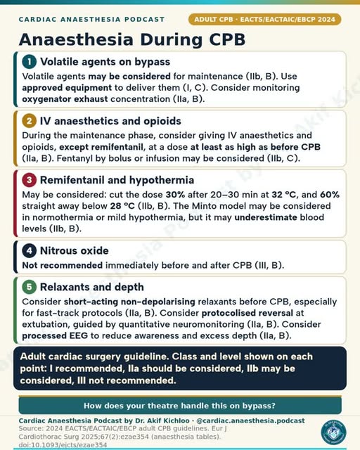
← Back to posts
← Back to posts

Mitral Regurgitation 1/5: Aetiology and Pathophysiology

View on Instagram

Sources supporting this post

Understanding the clinical context

  • Primary does not mean only degenerative. The post’s “Primary (degenerative)” category is more broadly understood as primary (organic) MR. Fibroelastic deficiency and Barlow disease are degenerative causes; rheumatic disease and endocarditis are other primary causes. The disease can involve chordae as well as leaflets. The ventricular and atrial forms of secondary MR are correctly distinguished. Kaplan 8th, ch. 15, printed p. 626.
  • Carpentier describes leaflet motion. Normal motion with annular dilatation, perforation or cleft; excessive motion with prolapse/flail; systolic-and-diastolic restriction with rheumatic, radiation or drug injury; and systolic restriction with ventricular tethering are appropriate examples. This describes leaflet motion, separately from primary versus secondary cause. ACC 2020 checklist, p. 5.
  • The EF threshold applies to severe chronic primary MR. “LVEF ≤60% already means LV dysfunction” applies to severe chronic primary MR. EF includes backward as well as forward ejection and may hide impaired contractility. It is not a general dysfunction threshold for every degree or cause of MR. Kaplan 8th, printed pp. 627–628; ESC/EACTS 2021, Recommendations E.
  • Acute and chronic MR differ in atrial adaptation. “small non-compliant LA, giant v waves, pulmonary oedema and shock” describes abrupt severe MR into an unadapted atrium. Acute-on-chronic MR may already have a large LA, and not every case has every listed finding. V-wave size depends on LA compliance and cannot reliably grade MR. The chronic volume-overload description is supported. Kaplan 8th, printed pp. 627 and 632.
  • Papillary-muscle rupture after myocardial infarction. “Posteromedial papillary muscle ruptures most often: single supply from the PDA” describes the usual pattern after myocardial infarction. Its single posterior descending artery supply helps explain the vulnerability, compared with the anterolateral muscle’s dual supply. The infarct territory also depends on coronary dominance. Labrada et al., 2020, Answer 5.

Guideline update

ESC/EACTS 2025 retains LVEF≤60% as a dysfunction marker in severe primary MR. It also gives a clearer separate definition of atrial secondary MR, including its association with long-standing AF and/or HFpEF. §§9.1.4 and 9.2.1–9.2.3.

For education. Apply the cited sources in their clinical context and follow current local guidance; this material is not patient-specific advice.
← Back to posts
← Back to posts

Tricyclic Overdose: ECG Signs and Sodium Bicarbonate

View on Instagram

Sources supporting this post

How to read the post

  • Bicarbonate dosing and protocol limits. The post’s 8.4% sodium bicarbonate 1–2 mmol/kg IV dose appears in the Ambulance Victoria protocol when blood gases can be measured, with a 100 mmol limit per dose and a 6 mmol/kg total limit. AHA uses a different adult starting-dose table. Follow one complete poison-treatment protocol rather than combining doses and limits. Ambulance Victoria TCA protocol A0723, December 2025; April 2026 edition AHA 2025 Table 4
  • Monitoring during alkalinisation. The post’s arterial pH target of 7.45–7.55 requires monitored treatment, rather than pursuit of a number regardless of other findings. Follow the chosen poison-treatment protocol, checking the ECG, blood pressure, blood gases and electrolytes. The amitriptyline label gives the stated pH range; AHA advises avoiding pH above 7.55 and sodium above 155 mEq/L. FDA amitriptyline label AHA 2025 Part 10
  • The aVR risk marker. A terminal R wave in aVR of 3 mm or more, rather than strictly “over 3 mm,” is associated with a higher risk of seizures or ventricular arrhythmias. It does not mean both will occur. Liebelt et al., 1995
  • Sinus tachycardia and sodium-channel block. The post groups sinus tachycardia and the rightward terminal QRS pattern together, but a fast sinus rhythm alone does not establish sodium-channel block: it can reflect the drug’s anticholinergic effects. QRS widening and the terminal QRS pattern are more relevant ECG clues. Queensland Ambulance Service TCA guideline, January 2020; Liebelt, Francis and Woolf, Ann Emerg Med 1995;26:195–201
  • Lipid emulsion as rescue treatment. The lipid-emulsion entry refers to specialist rescue treatment, with poisons-service advice, for life-threatening poisoning that has not responded to other treatments. It is not routine first-line treatment. AHA 2025 Part 10
  • GABA-A effects. The post lists GABA-A alongside fast sodium-channel, muscarinic, alpha-1 and histamine-receptor effects. Some TCAs inhibit certain GABA-A receptors in laboratory studies, but the effect varies with the drug and receptor type. It is not a uniform effect across all TCAs or all GABA-A receptors. Bampali et al., British Journal of Pharmacology 2022;179:3675–3692

Guideline update: The original 1985 and 1995 studies remain the sources for ECG risk prediction. AHA 2025 Part 10 adds current treatment guidance: bicarbonate for life-threatening TCA heart toxicity, blood-gas/electrolyte monitoring, and avoidance of pH above 7.55 or sodium above 155 mEq/L. It also supports carefully adjusted ventilation in patients already receiving mechanical ventilation. These are treatment additions, not changes to the studies’ results. Each dosing regimen belongs to its named treatment protocol.

For education. Apply the cited sources in their clinical context and follow current local guidance; this material is not patient-specific advice.

← Back to posts
← Back to posts

Sgarbossa Criteria: MI in LBBB or Paced Rhythm

View on Instagram

Sources supporting this post

How to read the post

Concordance and discordance

In the post, the ST segment running opposite to the QRS describes discordance. This is the usual secondary repolarisation pattern in left bundle branch block (LBBB) and right-ventricular pacing. Concordant ST change goes in the same direction as the main QRS deflection; discordant change goes in the opposite direction. The post’s 5-, 3- and 2-point criteria are the original Sgarbossa score. Sgarbossa et al., NEJM 1996;334:481–487

What the original score can detect

The quoted sensitivity of about 20% and specificity of about 98% apply to a score of at least 3. These are pooled results in patients with LBBB, not one universal accuracy estimate for both LBBB and paced rhythms. High specificity makes a positive score concerning; low sensitivity means many infarctions will not meet it. The post correctly warns that a low score does not exclude myocardial infarction. Tabas et al., Ann Emerg Med 2008;52:329–336.e1

The original 5-mm criterion

Discordant ST elevation of at least 5 mm contributes 2 points to the original score. This is the fixed threshold used in the original rule. The modified Sgarbossa rule instead uses at least 1 mm of discordant ST elevation that is at least 25% of the depth of the preceding S wave. Using signed measurements, the ST/S ratio is ≤−0.25. The modified rule is positive if any one criterion is present; it is not a new 2-point item to insert into the old score. Smith et al., Ann Emerg Med 2012;60:766–776

Applying the rule to paced rhythms

Application of the modified rule to right-ventricular pacing has its own evidence base. Dodd and colleagues found greater sensitivity than the original criteria, but performance depended on the comparison group. Neither version replaces symptoms, repeated electrocardiograms, troponin testing and urgent specialist assessment. Dodd et al., Ann Emerg Med 2021;78:517–529

The proportional threshold

The Smith-modified rule uses discordant ST elevation at least 25% of S-wave depth rather than the fixed 5-mm threshold. The modified rule also requires at least 1 mm of ST elevation. “S-wave depth” here means its absolute magnitude; if the S wave is entered as a negative value, the signed ratio is ≤−0.25. Smith et al., Ann Emerg Med 2012;60:766–776

Old ECGs and serial troponin

Comparison with an old ECG and serial troponin testing can inform the assessment. These can help when the diagnosis is uncertain, but they must not delay urgent reperfusion assessment in a patient with ongoing ischaemia or haemodynamic instability. A new LBBB alone is not proof of coronary occlusion; the clinical setting determines urgency. ESC acute coronary syndrome guideline 2023, section 3.2.1

Guideline update: The 1996 score and 2008 accuracy figures remain historical sources; the 2012 proportional rule and 2021 paced-rhythm study are later diagnostic evidence. The ESC acute coronary syndrome guideline 2023, section 3.2.1 does not require a positive Sgarbossa score before urgent management: patients with LBBB, right bundle branch block or a paced rhythm plus symptoms strongly suspicious for ongoing myocardial ischaemia should be managed similarly to patients with clear ST elevation, whether or not the conduction abnormality was previously known.

The Australian ACS guideline 2025, initial ECG assessment, explicitly includes the modified Sgarbossa criteria for LBBB and right-ventricular pacing in its approach to recognising acute coronary occlusion, including patterns beyond conventional ST elevation. This adds a current named clinical pathway; it does not change the populations or accuracy figures of the original studies. A negative score must not delay urgent assessment when symptoms or instability remain concerning.

For education. Apply the cited sources in their clinical context and follow current local guidance; this material is not patient-specific advice.
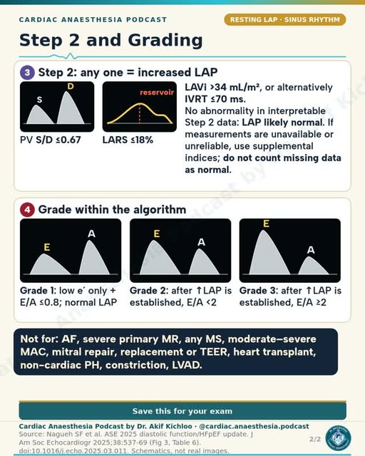
← Back to posts
← Back to posts

Anaphylaxis: Adrenaline Doses That Matter

View on Instagram

Sources supporting this post

How to read the post

  • The Harper citation. The Harper paper cited in the post was published in Anaesthesia in 2009, rather than 2021. Harper et al., Anaesthesia 2009;64:199–211
  • Escalation after two IM doses. Persistent breathing or circulation problems after two appropriate injections into muscle call for urgent expert help for an adrenaline infusion with continuous monitoring. This is not an instruction for untrained staff to give adrenaline into a vein. RCUK Emergency treatment of anaphylaxis, May 2021
  • IV adrenaline during anaesthesia. The 50-microgram IV adult dose belongs to specialist perioperative treatment: the RCUK algorithm starts with this dose, then adjusts treatment to response. Use the correct preparation: the IV dose uses 0.1 mg/mL; the usual IM preparation is 1 mg/mL. RCUK peri-operative anaphylaxis algorithm
  • Infusions, fluids and glucagon. The post’s “after 3 boluses” wording is not a reason to wait for a fixed third bolus if circulation remains unstable. The RCUK perioperative plan starts an infusion after a poor response to boluses; the glucagon 1 mg IV option is for adults on beta-blockers whose reaction remains resistant to the infusion. Its initial fluid bolus is 500–1000 mL in adults and 20 mL/kg in children under 12. The Association’s 2023 handbook uses 20 mL/kg; the fluid regimen therefore depends on the protocol being followed. RCUK peri-operative anaphylaxis algorithm
  • Positioning. Patients with circulation problems need to remain flat. A supported, partly upright position may help breathing when circulation is stable; do not suddenly sit or stand them up. RCUK Emergency treatment of anaphylaxis, May 2021
  • Baseline tryptase timing. In the post’s “third at 24 h or later” entry, the timing is measured from recovery. For the RCUK 2021 three-sample plan, the baseline tryptase sample is at least 24 hours after complete recovery, not simply 24 hours after onset. Earlier samples must never delay resuscitation. RCUK Emergency treatment of anaphylaxis, May 2021

Guideline update: Harper 2009 provides the historical context, while RCUK 2021 addresses general anaphylaxis care. The RCUK peri-operative anaphylaxis algorithm provides a newer, specific plan for anaesthetists: initial adult IV adrenaline 50 micrograms, early crystalloid, an infusion if boluses work poorly, and further treatment for persistent shock. This is a separate specialist setting, not a replacement of general IM treatment with IV treatment. The RCUK Special circumstances 2025 continues to direct readers to detailed anaphylaxis guidance. For Australia and New Zealand, ASCIA acute anaphylaxis guideline, September/October 2026 (Australia/New Zealand) adds new initial and refractory-anaphylaxis flowcharts and information on available adrenaline devices, including nasal sprays. This is separate regional guidance. The post’s UK injection protocols and device-specific nasal doses belong to different treatment pathways; nasal doses cannot be substituted for injection doses.

For education. Apply the cited sources in their clinical context and follow current local guidance; this material is not patient-specific advice.
← Back to posts
← Back to posts

ARDS: Berlin Definition and Lung-Protective Ventilation

View on Instagram

Sources supporting this post

How to read the post

Timing and lung opacities

The one-week onset and bilateral-opacities entries come from Berlin, which requires onset within one week of a known insult or new/worsening respiratory symptoms. The opacities must not be fully explained by effusions, collapse or nodules; respiratory failure must not be fully explained by cardiac failure or fluid overload. Cardiac disease can coexist with acute respiratory distress syndrome (ARDS). ARDS Definition Task Force, JAMA 2012;307:2526–2533; Matthay et al., Global ARDS Definition, AJRCCM 2024;209:37–47

Exact oxygenation boundaries

The card rounds the categories to mild 201–300, moderate 101–200 and severe 100 or less. These are convenient whole-number labels. The exact arterial-oxygen to inspired-oxygen ratio boundaries are 200 < PaO₂/FiO₂ ≤300, 100 < PaO₂/FiO₂ ≤200, and ≤100 mmHg. Berlin requires positive end-expiratory pressure (PEEP) ≥5 cmH₂O; continuous positive airway pressure (CPAP) is allowed in the mild category. The quoted 27%, 32% and 45% mortality figures describe the original validation cohorts, not an individual patient’s predicted outcome. ARDS Definition Task Force, JAMA 2012;307:2526–2533; Matthay et al., Global ARDS Definition, AJRCCM 2024;209:37–47

Lung-protective ventilation

The post starts at 6 mL/kg predicted body weight with plateau pressure 30 cmH₂O or less. This correctly reflects the trial’s starting strategy. Predicted body weight is derived from height and sex, not actual body weight. Tidal volume is adjusted within a lung-protective protocol, rather than fixed at 6 mL/kg; current ATS guidance retains 4–8 mL/kg predicted body weight with plateau pressure below 30 cmH₂O. ARDS Network, NEJM 2000;342:1301–1308; ATS ARDS treatment guideline 2024;209:24–36

When the prone-positioning evidence applies

The 16-hour sessions and PaO₂/FiO₂ below 150 reflect the PROSEVA approach. PROSEVA used sessions of at least 16 hours in selected, invasively ventilated patients with a ratio below 150, inspired oxygen fraction at least 0.60 and PEEP at least 5 cmH₂O after initial stabilisation. The ratio alone is not the entire eligibility rule. Safe turning requires a trained team and attention to airway, lines and haemodynamics. Guérin et al., PROSEVA, NEJM 2013;368:2159–2168

Interpreting driving pressure

The suggested target below 15 is a guide to interpreting pressure, not an independently proven treatment rule. Driving pressure is plateau pressure minus total PEEP in a passive patient. Lower values are associated with better outcomes, but 15 cmH₂O is not a proven universal treatment threshold or a substitute for tidal-volume and plateau-pressure limits. Changing PEEP to achieve that number requires assessment of recruitment, overdistension and circulation. Amato et al., NEJM 2015;372:747–755; Guérin et al., Critical Care 2016;20:384

Calculating predicted body weight

The displayed equations are: male PBW = 50 + 0.91 × (height in cm − 152.4); female PBW = 45.5 + 0.91 × (height in cm − 152.4). These are the standard adult predicted-body-weight equations, with the result in kilograms and height in centimetres. The coefficient is the rounded metric conversion of the ARDS Network’s 2.3 kg-per-inch formula. NHLBI ARDS Network Mechanical Ventilation Protocol Summary

The global definition’s publication date

The post calls this the “2023 global definition.” This is the proposal published online in 2023 and in the January 2024 journal issue, not a different definition from the Matthay 2024 paper in the caption. The pulse-oximetry ratio must be used only when oxygen saturation is ≤97%, and the non-intubated category includes high-flow nasal oxygen at ≥30 L/min or qualifying non-invasive positive-pressure support. Matthay et al., Global ARDS Definition, AJRCCM 2024;209:37–47

Fluids after shock has resolved

The FACTT entry concerns conservative fluids once shock has resolved. The phrase “once shock has resolved” is important: conservative management is not a reason to withhold resuscitation from a patient in shock. FACTT found better lung function and more ventilator-free days, but no significant reduction in 60-day mortality. The fluid plan is individualised according to perfusion and kidney function. ARDS Network, FACTT, NEJM 2006

Guideline update: The Matthay et al., Global ARDS Definition, AJRCCM 2024;209:37–47 broadens the definition to include non-intubated patients receiving high-flow nasal oxygen at ≥30 L/min, permits a pulse-oximetry-based oxygenation ratio under specified conditions, allows lung ultrasound and adds a resource-limited category. It is a consensus definition, not the ATS treatment guideline. The separate ATS ARDS treatment guideline 2024;209:24–36 retains lung-protective ventilation and prolonged prone positioning while adding conditional recommendations for other treatments; it does not erase the original Berlin or trial criteria.

For education. Apply the cited sources in their clinical context and follow current local guidance; this material is not patient-specific advice.
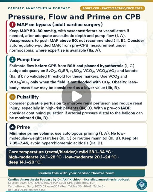
← Back to posts
← Back to posts

Vasopressors and Inotropes at a Glance (infographic)

View on Instagram

Sources supporting this post

Understanding the post

  • The treatment sequence gives context to “2nd agent in sepsis.” In the cited 2021 guideline, add vasopressin when norepinephrine is not achieving the blood-pressure goal; add epinephrine if pressure remains inadequate on both. Poor heart function with continuing poor organ perfusion is a separate treatment decision. SSC 2021, vasoactive agents and inotropes.
  • “not titrated” describes a common fixed-dose sepsis approach, not every use of vasopressin. The product label allows adjustment of the infusion and uses different starting doses for septic shock and shock after cardiac surgery. Vasopressin label, §2.2.
  • “Anaphylaxis, arrest, 2nd agent in sepsis, low output” lists different emergencies beside one infusion range. Do not use that range as the injection dose for anaphylaxis or cardiac arrest. The route, preparation and dose depend on the emergency. RCUK anaphylaxis guidance, pp.29–34; RCUK 2025 adult cardiac-arrest drug guidance; septic-shock infusion label, §2.2.
  • “active metabolite lasts about 7–9 days” is best read as an effect that can persist for days after levosimendan stops. The active metabolites have a half-life of about 75–80 hours; 7–9 days is not their half-life. Simdax SmPC, §4.2 and §5.2.
  • “usual ICU dose” is a quick reference, not one universal regimen. The angiotensin-II starting dose and 80/40 ng/kg/min limits match its label, which also advises clot prevention. Milrinone’s 0.375–0.75 microgram/kg/min range is supported, with reduced infusion rates in renal impairment. GIAPREZA label, §2.2 and §5.1; milrinone label, dosage and renal adjustment.
  • The receptor labels and “pressor” versus “inodilator” distinction are useful summaries. The arrows are typical tendencies, not guaranteed effects or a scale of equivalent potency. Dose, underlying circulation and reflex responses matter. In particular, norepinephrine does not always raise pulmonary resistance at ordinary doses, and low-dose vasopressin can preserve or reduce it rather than having literally no pulmonary effect. Phenylephrine can increase pulmonary resistance, which matters in RV failure. Price et al., Critical Care 2010;14:R169, vasoactive-agent sections.
  • “HOCM / LVOT obstruction, AS” under phenylephrine describes selected hypotensive situations, not treatment of those diagnoses by themselves. “No renal dose benefit” means dopamine should not be given to protect the kidneys; increased urine output alone does not establish kidney protection. “Tolerance after about 72 h” means the dobutamine response may diminish over several days, not that it predictably stops working at 72 hours. Overgaard and Džavík, pp.1047–1049.
  • “Short-term peripheral start is acceptable” requires a monitored infusion protocol and prompt checks for leakage and tissue injury. It does not mean any IV site is safe indefinitely. The 2021 guideline allowed a short peripheral start to avoid delaying pressure support. SSC 2021, monitoring and IV access.
  • “MAP target 65 mmHg” is an initial septic-shock target, then individualised using perfusion. The post’s “Hydrocortisone 200 mg/day if norepi ≥0.25 for ≥4 h” refers to the 2021 guideline’s conditional suggestion for adults with septic shock and an ongoing vasopressor requirement. The full-text rationale describes that requirement as norepinephrine or epinephrine ≥0.25 µg/kg/min for at least four hours after initiation, to maintain the target MAP. This is not an automatic treatment rule based on one dose reading; the guideline also notes uncertainty about the optimal corticosteroid dose, timing and duration. SSC 2021, MAP and corticosteroids; SSC 2021 full guideline, corticosteroid rationale.

Guideline update

The 2026 Surviving Sepsis Campaign guideline retains norepinephrine as the usual first vasopressor and the sequence of adding vasopressin, then epinephrine if pressure remains inadequate. It adds a specific choice of norepinephrine or epinephrine when septic shock includes cardiac dysfunction, with heart rate and rhythm helping guide the choice. For continuing poor perfusion despite adequate filling and blood pressure, it allows dobutamine added to norepinephrine or epinephrine alone; evidence is insufficient to choose dobutamine over milrinone. These are sepsis recommendations, not a universal post-bypass drug sequence.

The 2026 guidance also suggests an initial MAP range of 60–65 mmHg in people aged 65 or older, while retaining 65 mmHg as the usual initial adult target. It still allows peripheral vasopressors to avoid delay, but says evidence is insufficient to specify a universal duration, dose or access-site rule. It continues a conditional recommendation for IV corticosteroids in septic shock. SSC 2026, MAP, vasopressor administration and corticosteroids.

For education. Apply the cited sources in their clinical context and follow current local guidance; this material is not patient-specific advice.
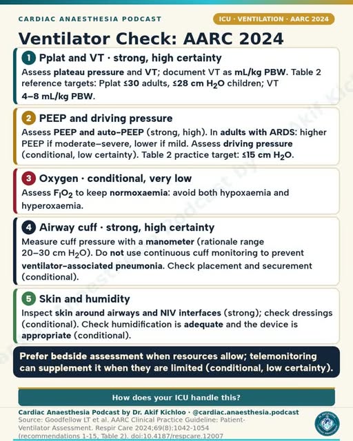
← Back to posts
← Back to posts

CVP Waveforms at a Glance (infographic)

View on Instagram

Understanding the post

The a, c and v waves and the x and y descents describe different stages of right-atrial contraction, filling and emptying. Cannon a waves can occur when the atrium contracts against a closed tricuspid valve; atrial fibrillation removes the organised a wave. Large a waves suggest resistance to right-atrial emptying. A large c-v wave suggests tricuspid regurgitation. A preserved x descent with a reduced y descent is a classic tamponade pattern; brisk descents can occur in constriction. These are useful clues, interpreted with the ECG and echocardiogram. Source: Kaplan’s Cardiac Anesthesia, 8th ed. (2023), chapter 9, printed pp. 291–292.

The statement that “one CVP number won't tell you about fluid responsiveness” is well supported. Marik and Cavallazzi’s 2013 analysis found that a single CVP value was only slightly better than chance at identifying who would increase cardiac output after fluids. The infographic’s figure of about 0.56 is correct. Original meta-analysis.

The catheter, vein and atrial labels explain why a central venous pressure tracing is used to estimate right-atrial pressure. The ECG labels describe the usual sequence: the a wave follows atrial electrical activation, the c wave occurs early in ventricular contraction, and the v wave builds during ventricular systole before atrial emptying. The drawing is schematic, not a timing template for every rhythm. Kaplan, chapter 9, printed pp. 291–292.

How to read the post

  • “Normal CVP: 2–6 mmHg” is a teaching range, not a fluid target. Breathing, ventilation, venous congestion, valve disease and right-heart function all affect CVP. A pressure in or outside that range does not by itself tell you whether to give fluid. Edwards guide, normal haemodynamic parameters; Kaplan, printed pp. 291–292.
  • “At the base of the c wave … ≈ RV end-diastolic pressure” is an approximation. It refers to pressure near the end of filling, not the amount of blood in the RV. Tricuspid obstruction, abnormal waveforms and raised pressure around the heart can make that interpretation misleading. This timed reading differs from the monitor’s mean CVP. Kaplan, chapter 5, printed p. 133, and chapter 9, pp. 291–292.
  • “4th ICS, mid-axillary” is a bedside landmark. The more precise level is the fourth intercostal space at the midpoint between the front and back of the chest. Accurate measurement requires zeroing to atmosphere and re-levelling after position changes. A 10-cm vertical error produces about a 7.4-mmHg pressure error. Edwards guide, sections 2.6–2.8.
  • CVP trends, PLR and PPV provide different kinds of information. CVP trends provide context but do not replace a test of fluid responsiveness. Passive leg raising should be assessed by its effect on stroke volume or cardiac output. Pulse-pressure variation is unreliable in several common settings, including spontaneous breathing and irregular rhythms. Even a positive test does not automatically mean fluids are beneficial. Monnet, Marik and Teboul, 2016.
  • “Kussmaul sign” means the neck-vein pressure rises, rather than falling normally, during inspiration. It supports impaired right-heart filling and can accompany constriction or restrictive heart-muscle disease; it is not specific to constriction. Merck Manual, right-heart failure, signs; Kaplan, chapter 11, Table 11.31, printed p. 515.

References

Sources named on the infographic

  1. Miller’s Anesthesia, central venous pressure monitoring.
  2. Kaplan’s Cardiac Anesthesia. Relevant passages in the 8th edition: chapter 9, printed pp. 291–292; chapter 5, printed p. 133; chapter 11, Table 11.31, printed p. 515.
  3. Marik PE, Cavallazzi R. Does the central venous pressure predict fluid responsiveness? Crit Care Med. 2013;41:1774–1781. Article.

Additional supporting references

  1. Monnet X, Marik PE, Teboul JL. Prediction of fluid responsiveness: an update. Ann Intensive Care. 2016;6:111. Full text.
  2. Edwards Clinical Education. Quick Guide to Cardiopulmonary Care, 5th ed. Sections 1.10, 2.6–2.8 and normal haemodynamic parameters. Guide.
  3. Fine NM, Howlett JG. Right Heart Failure and Cor Pulmonale. Merck Manual Professional, October 2025. Signs and interpretation of Kussmaul sign.

Guideline update

The ESICM circulatory-shock guideline, November 2025, adds current support for the post’s fluid-response warning: after initial resuscitation, assess fluid responsiveness before giving more fluid for persistent shock, using applicable dynamic tests rather than a static filling pressure alone. It also recommends measuring CVP when a central venous catheter is present. Poor prediction of fluid response does not make CVP useless; its waveform, trend and congestion context still matter. This guidance concerns critically ill adults with shock, not a new universal operating-room cutoff.

For education. Apply the cited sources in their clinical context and follow current local guidance; this material is not patient-specific advice.
← Back to posts
← Back to posts

Etomidate for Induction in Cardiac Patients

View on Instagram

Sources supporting this post

How to read the post

Relative cardiovascular stability

The post’s 0.2–0.3 mg/kg induction entry describes relative stability of heart rate, SVR and contractility. Etomidate usually causes less cardiovascular depression than many alternative induction agents. Systemic vascular resistance (SVR) and blood pressure can still fall. Hensley describes a typical 10–15% fall in mean arterial pressure and SVR, with contractility relatively preserved in normovolaemic patients. Dose and rate must be individualised; this is an anaesthetic given with airway and resuscitation support. AMIDATE prescribing information, updated May 2026; Hensley, section B.1, eBook PDF pages 315–316.

Choosing an induction agent

Poor LV function, severe AS, tamponade and shock are the settings highlighted in the post. These are situations in which relative haemodynamic stability may be useful, not a guarantee of safety or a universal preferred drug. Left-ventricular dysfunction, severe aortic stenosis and tamponade each require a tailored induction and ventilation plan. The product label specifically notes insufficient evidence to predict the cardiovascular response in severe trauma or hypovolaemia. AMIDATE prescribing information, updated May 2026

Adrenal suppression after one dose

The post describes cortisol suppression lasting 24 hours or more after a single dose. The mechanism, inhibition of adrenal 11β-hydroxylase, is correct. Duration varies: the label describes suppression for about 6–8 hours, while critically ill cohorts have demonstrated inhibition lasting beyond 24 hours. Biochemical adrenal suppression does not by itself establish increased mortality from one induction dose. AMIDATE prescribing information, updated May 2026; Vinclair et al., Intensive Care Med 2008;34:714–719

Why prolonged infusions differ

Historical prolonged ICU sedation was associated with higher mortality; this does not prove that a single induction dose has the same effect. The label warns against prolonged infusion because of adrenal suppression. Watt and Ledingham, Anaesthesia 1984;39:973–981; AMIDATE prescribing information, updated May 2026

Analgesia and induction effects

Etomidate provides no analgesia; the post also highlights myoclonus and injection pain. Analgesia is tailored to the procedure and patient rather than supplied as an automatic fixed opioid dose: combined etomidate and opioid administration can still cause hypotension. Myoclonus is involuntary movement and is not automatically a seizure. AMIDATE prescribing information, updated May 2026; Hensley, eBook PDF pages 315–316.

Duration of anaesthesia and recovery

The quoted 5–15-minute wake-up estimate is approximate. A fixed wake-up time cannot be predicted. The US label describes approximately 3–5 minutes of anaesthesia after a usual 0.3 mg/kg dose; return to full alertness depends on dose, other medicines, illness and the procedure. Brief hypnosis is not the same as recovery of protective airway reflexes or readiness for discharge. AMIDATE prescribing information, updated May 2026; Hypnomidate 2 mg/mL Summary of Product Characteristics

Adrenal insufficiency and acute porphyria

The post flags both adrenal insufficiency and porphyria as reasons for caution. Known adrenal insufficiency requires an individual anaesthetic and steroid-replacement plan; it is not listed alongside hypersensitivity as an absolute contraindication in the UK product label. The porphyria warning concerns acute porphyrias, for which specialist databases list etomidate as unsafe. Existing adrenal insufficiency is a separate reason for indicated steroid treatment from giving steroids routinely after every etomidate dose. Hypnomidate 2 mg/mL Summary of Product Characteristics; American Porphyria Foundation drug-safety database; SCCM rapid-sequence intubation guideline 2023

Frequency of adverse effects

The post’s “over half” myoclonus estimate concerns unpremedicated patients, and it describes opioid or midazolam premedication to reduce movements. It also lists thrombophlebitis, nausea and vomiting. Involuntary movements, injection pain, nausea and vomiting are recognised adverse effects. Their frequency varies by formulation, premedication and population; “over half” is not a universal incidence. Premedication can reduce movements but may add respiratory or circulatory depression. The UK label includes thrombophlebitis among post-marketing reports of unknown frequency, so it cannot be assumed to be as common as injection pain. AMIDATE prescribing information, updated May 2026; Hypnomidate 2 mg/mL Summary of Product Characteristics

Guideline update: The SCCM rapid-sequence intubation guideline 2023 advises against giving corticosteroids solely to counter etomidate-related adrenal suppression. This is more current than the steroid-supplementation suggestion in the 2019 textbook. The Ketamine versus Etomidate for Tracheal Intubation of Critically Ill Adults, NEJM, online December 2025 is later trial evidence, not a new guideline: ketamine did not significantly reduce 28-day mortality compared with etomidate and produced more peri-intubation cardiovascular collapse. These emergency/critical-care results do not establish one best induction drug for every cardiac surgical lesion.

For education. Apply the cited sources in their clinical context and follow current local guidance; this material is not patient-specific advice.
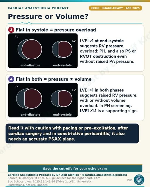
← Back to posts
← Back to posts

Digoxin Toxicity: ECG Clues and Treatment

View on Instagram

Sources supporting this post

How to read the post

  • Fab treatment and the product-label potassium threshold. The post’s potassium cutoff of 5.0 in acute overdose is not a universal threshold for Fab treatment. The DigiFab label specifies potassium above 5.5 mEq/L in adults, or above 6 in children, with rapidly worsening toxicity. Serious heart-rhythm problems can require the antidote regardless of potassium. DigiFab prescribing information, BTG, DailyMed updated 29 September 2026
  • The expert-consensus potassium threshold. A different source, the 2023 expert consensus, lists potassium above 6.5 mmol/L as one reason for immediate antidote treatment. These sources use different thresholds and do not support treating 5.0 as a universal cutoff. Andrews et al., Eur J Emerg Med 2023;30:395–401
  • Electrolytes and urgent antidote treatment. Low potassium or magnesium needs correction when present, but this must not delay the antidote in life-threatening digoxin poisoning. The post’s word “first” does not establish that electrolyte treatment must precede the antidote. The antidote itself can lower potassium, so check it closely during treatment. Andrews et al., Eur J Emerg Med 2023;30:395–401
  • Dialysis. Dialysis does not effectively remove digoxin and is not recommended for that purpose. Dialysis may still be needed for a separate reason. Digoxin prescribing information
  • Digoxin effect versus toxicity. The post’s scooped ‘reverse tick’ ST depression, short QT and flat T waves can occur during treatment without poisoning, but they do not rule poisoning out. Assess symptoms, rhythm, kidney function, electrolytes and the properly timed drug level together. Andrews et al., Eur J Emerg Med 2023;30:395–401; RCEM/NPIS unidentified poisoning guideline, April 2025
  • Timing the digoxin level. Sampling at least 6 hours after a dose helps interpret the concentration after the drug has distributed into tissues. Do not wait for that time or a laboratory result before treating life-threatening toxicity. Andrews et al., Eur J Emerg Med 2023;30:395–401
  • Cardioversion precautions. Electrical shocks can provoke dangerous ventricular rhythms in suspected toxicity, which explains the post’s low-energy precaution. Delay elective cardioversion when possible; if it cannot safely wait, use the lowest effective energy. This is a precaution, not a quantified risk estimate. Digoxin tablets prescribing information, Amneal; revised July 2025

Guideline update: AHA 2025 Part 10: Digoxin and related cardiac glycosides recommends digoxin-specific antibody fragments for life-threatening digoxin or digitoxin poisoning in adults and children. While the antidote is being obtained, specialists may consider lidocaine or phenytoin for ventricular arrhythmias; these are temporary options, not substitutes for the antidote. Dialysis and other blood-filtering treatments are not recommended to remove digoxin. This supports the post’s main treatment advice and adds options while awaiting the antidote; it does not establish a new universal potassium cutoff.

For education. Apply the cited sources in their clinical context and follow current local guidance; this material is not patient-specific advice.
← Back to posts
← Back to posts

Pulsus Paradoxus: Mechanism and Causes

View on Instagram

Understanding the post

The definition “an inspiratory fall in systolic BP of more than 10 mmHg” is a standard description of pulsus paradoxus. In a spontaneously breathing patient with tamponade, increased right-heart filling during inspiration can reduce left-heart filling because the heart is confined within a pressurised pericardial sac. The resulting fall in stroke volume lowers systolic pressure. Schiavone, 2013, pp. 111–114.

It can also occur in severe obstructive lung disease, pulmonary embolism and pneumothorax. Its absence does not exclude tamponade, particularly when ventilation, severe aortic regurgitation, an atrial septal defect or high left-ventricular filling pressure changes the usual physiology. Schiavone, pp. 114–115.

The echo findings support the clinical assessment. “Mitral E velocity varies more than 25%, tricuspid more than 40%” describes marked breathing-related changes in early filling. Current ESC guidance lists mitral variation >25–30% and tricuspid variation >40–60% among possible tamponade findings. These are supporting clues, not a stand-alone diagnosis, and similar variations occur in constriction. 2025 guideline, Table 17 and section 11.9.1.

How to read the post

  • The cause list does not mean every patient with these conditions has pulsus paradoxus. The post’s “hypovolaemia” is better understood as severe volume loss or shock, not ordinary mild dehydration. Very low pressure can also make the sign difficult to detect. Spodick, 2003, pp. 685–686.
  • The usual definition assumes spontaneous breathing. Positive-pressure ventilation can alter or reverse the respiratory pressure pattern. The same bedside rule therefore does not apply unchanged to a ventilated patient. Schiavone, p. 114.
  • “A loculated effusion” is an important exception. After cardiac surgery, a local pocket of fluid or clot can compress one part of the heart without producing all the classic signs. A reassuring pulse examination does not rule it out. 2025 ESC guideline, section 11.8.
  • This is a sign, not a diagnosis or a stand-alone treatment trigger. Its interpretation depends on the clinical condition and echocardiogram. Suspected unstable tamponade needs urgent expert assessment and drainage. 2025 ESC guideline, sections 11.8.2–11.8.3.
  • “The swing reflects volume status” under positive-pressure ventilation is too simple. The swing depends on ventilation settings, rhythm, lung and right-heart function as well as filling. It may help predict a response to fluid only under appropriate conditions; it does not measure circulating volume or prove that fluid is needed. Monnet and colleagues, 2016.
  • “RV infarction” is a possible cause, and “Kussmaul sign … points more to constriction” is a clue, not a separator. A damaged right ventricle can also produce pulsus paradoxus and an inspiratory rise in neck-vein pressure. That sign alone does not identify constriction. Ondrus and colleagues, 2013, p. 27; Kaplan’s Cardiac Anesthesia, 8th ed., chapter 11, Table 11.31, printed p. 515.
  • “Rule out tamponade with TEE” means seek urgent appropriate imaging. After surgery, transoesophageal echo can be particularly useful when transthoracic views are poor or a local clot is suspected. An incomplete or nondiagnostic study must not falsely reassure or delay treatment of an unstable patient. 2025 ESC guideline, section 11.8.

References

  1. Schiavone WA. Cardiac tamponade: 12 pearls in diagnosis and management. Cleve Clin J Med. 2013;80:109–116. Full text.
  2. Spodick DH. Acute cardiac tamponade. N Engl J Med. 2003;349:684–690. Full text.
  3. 2025 ESC Guidelines for the management of myocarditis and pericarditis. Sections 11.8–11.8.3 and Table 17. Guideline.
  4. Ondrus T, et al. Right ventricular myocardial infarction: From pathophysiology to prognosis. Exp Clin Cardiol. 2013;18:27–30. Physical-examination section, p. 27. Full text.
  5. Monnet X, Marik PE, Teboul JL. Prediction of fluid responsiveness: an update. Ann Intensive Care. 2016;6:111. Full text.
  6. Kaplan’s Cardiac Anesthesia, 8th ed. (2023), chapter 9, printed p. 292 (venous waveforms), and chapter 11, Table 11.31, printed p. 515 (constriction and Kussmaul sign). Venous-pressure patterns in constrictive and restrictive disease.

Guideline update

The 2025 ESC guideline provides current clinical context for the older reviews. It defines the inspiratory fall as at least 10 mmHg, whereas the post and Schiavone use more than 10 mmHg. This small wording difference should not turn a borderline measurement into a rule-in or rule-out test. The guideline combines the history, examination and echo findings, explicitly recognises localised postoperative tamponade, and recommends drainage without delay when the patient is unstable. Sections 11.8–11.8.3.

For education. Apply the cited sources in their clinical context and follow current local guidance; this material is not patient-specific advice.
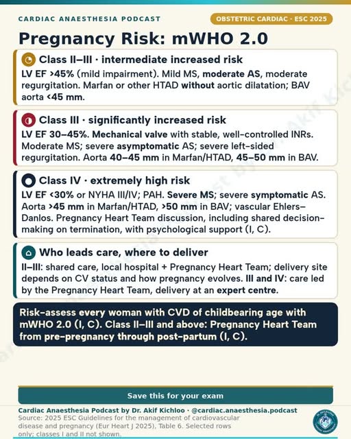
← Back to posts
← Back to posts

CABG Conduits: LIMA, Radial and Vein

View on Instagram

Sources supporting this post

How to read the post

What determines graft durability

Conduit choice is one major determinant of coronary artery bypass graft durability. Target-vessel stenosis, surgical technique, patient factors and secondary prevention also matter; no graft has a fixed expiry date. Goldman et al., JACC 2004;44:2149–2156; ACC/AHA/SCAI coronary revascularization guideline 2021, section 12.2 and Table 12

Interpreting the ten-year patency figures

The comparison in the post is LIMA–LAD patency of about 85% versus saphenous-vein patency of about 61% at ten years. Goldman reported 85% patency for internal mammary artery grafts and 61% for saphenous vein grafts in the study cohort. These are that study’s results, rather than a universal prediction specifically for every left internal mammary artery (LIMA) to left anterior descending artery (LAD) graft. Goldman et al., JACC 2004;44:2149–2156

Radial artery versus vein

The 2018 RADIAL analysis found fewer graft occlusions and fewer combined events of death, myocardial infarction or repeat revascularisation with radial artery grafts at about five years. It did not show a statistically significant reduction in death alone. This supports radial grafting in suitable patients and target vessels, not superiority for every outcome in every patient. Gaudino et al., RADIAL, NEJM 2018;378:2069–2077

Hand perfusion before radial harvest

Assessment before harvest includes objective evaluation of both palmar-arch completeness and whether the ulnar artery can maintain hand perfusion. Prior radial-artery catheterisation, likely future haemodialysis access and the severity of the target coronary stenosis also matter. Competitive flow can impair an arterial graft placed on a less severely narrowed vessel. ACC/AHA/SCAI coronary revascularization guideline 2021, section 12.2 and Table 12

Treating spasm and preventing it later

The post mentions nitrates and calcium-channel blockers for spasm. Treatment around harvesting differs from postoperative prevention. Topical vasodilators are used intraoperatively; current guidance supports oral calcium-channel blockers for the first postoperative year after radial grafting. Long-term nitrate therapy does not have equivalent evidence for graft patency. ACC/AHA/SCAI coronary revascularization guideline 2021, section 12.2 and Table 12; ESC/EACTS conduit-management consensus 2024

Bilateral mammary grafts and wound risk

The bilateral-IMA warning highlights sternal wound infection risk in patients with diabetes or obesity. The level of risk depends on patient selection, glucose control and harvest technique. Skeletonised harvesting, which leaves more surrounding tissue intact, can reduce sternal-wound complications; it does not make the risk disappear. ESC/EACTS conduit-management consensus 2024

How vein graft disease develops

The post outlines early thrombosis, then intimal hyperplasia, then atherosclerosis. This is a useful broad time-course: early clotting, subsequent thickening of the vessel lining and later atherosclerotic disease. These mechanisms overlap and do not occur as an inevitable sequence in every graft. Antiplatelet treatment, lipid lowering, conduit handling and target-vessel factors remain important. ESC/EACTS conduit-management consensus 2024

Assessing flow before closure

Transit-time flowmetry is the graft-flow method highlighted in the post. Intraoperative graft-flow measurement is an important quality-control tool, alongside the ECG and transoesophageal echocardiography. Transit-time flowmetry estimates blood flow through the graft using ultrasound, but readings require interpretation in the context of pressure, target-vessel runoff and graft configuration; one abnormal number does not identify the cause by itself. ESC/EACTS myocardial revascularization guideline 2018, sections 13.1 and 15.1.7

New ischaemic changes after bypass

Graft kinking, spasm or air are possible explanations for new ST changes or wall-motion defects. These are important possibilities, not the full differential or a diagnosis from the ECG alone. Prompt assessment with the surgical team addresses coronary perfusion, mechanical graft problems, inadequate revascularisation and other causes. The advice “no more vasoconstrictor than needed” must not lead to withholding a vasopressor needed to maintain coronary perfusion. Hensley, “Myocardial ischemia,” eBook PDF pages 423–424; ESC/EACTS myocardial revascularization guideline 2018, sections 13.1 and 15.1.7

Guideline update: The 2004 patency study and 2018 RADIAL analysis remain the sources for the quoted comparisons. The ACC/AHA/SCAI coronary revascularization guideline 2021, section 12.2 and Table 12 now recommends a radial artery rather than a vein for the second most important significantly stenosed non-LAD target in suitable isolated-bypass patients. The ESC/EACTS conduit-management consensus 2024 adds practical advice on harvesting and spasm prevention, including consideration of a calcium-channel blocker for one year rather than treating nitrates and calcium-channel blockers as interchangeable long-term regimens.

For education. Apply the cited sources in their clinical context and follow current local guidance; this material is not patient-specific advice.
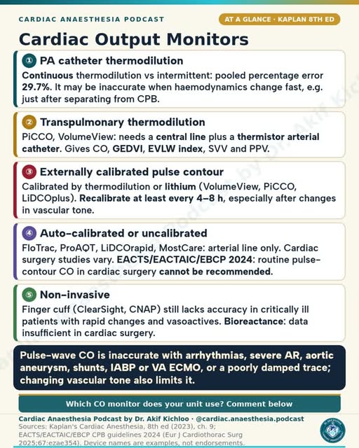
← Back to posts
← Back to posts

Electrolytes on the ECG (infographic)

View on Instagram

Sources supporting this post

How to read the post

Potassium and ECG appearances

Peaked T waves and a sine-wave pattern are among the hyperkalaemic appearances shown. These are classic patterns, not a dependable step-by-step progression. The infographic correctly warns that electrocardiogram (ECG) changes correlate poorly with potassium and a normal ECG does not exclude severe hyperkalaemia. Interpretation combines the measured potassium, clinical condition and ECG. UK Kidney Association hyperkalaemia guideline 2020, retained PDF with 2022 amendment

Low potassium and high calcium

The low-potassium panel shows ST depression, flat T waves and prominent U waves; the high-calcium panel shows a short ST segment and short QT. These are useful typical patterns for low potassium and high calcium, respectively. With low potassium, T–U fusion can produce an apparently long QT that is actually a prolonged QU interval. These appearances are not specific enough to replace electrolyte measurement. ERC 2021: Cardiac arrest in special circumstances, Resuscitation 161:152–219

Low calcium differs from low magnesium

The shared “long flat ST segment, so long QT (T itself normal)” label needs different interpretation for the two electrolytes. The two electrolyte abnormalities have different ECG patterns. Low calcium characteristically lengthens the ST segment and QT. Low magnesium can contribute to QT prolongation and torsades, but its ECG findings are variable and it does not reliably reproduce the same flat-ST, otherwise-normal-T pattern. Associated low potassium often contributes. ERC 2021: Cardiac arrest in special circumstances, Resuscitation 161:152–219

Calcium dose, preparation and infusion time

The adult doses shown are 10 mL of 10% calcium chloride IV or 30 mL of 10% calcium gluconate. These provide approximately equivalent amounts of elemental calcium, but the infusion times and clinical setting matter. Current UK guidance gives calcium chloride over 5 minutes or calcium gluconate over 10 minutes; UK Kidney Association guidance prefers chloride during resuscitation and gluconate for other patients with hyperkalaemic ECG changes. Intravenous calcium protects the heart temporarily; it does not remove potassium. Resuscitation Council UK special-circumstances guideline 2025; UK Kidney Association hyperkalaemia guideline, July 2026 update, section II

Insulin, glucose and salbutamol

The displayed adult regimen is 10 units of soluble insulin plus 25 g glucose IV over 15 minutes, with nebulised salbutamol 10–20 mg. These adult treatments require a monitored protocol. Salbutamol is an adjunct, not sole treatment for severe hyperkalaemia. Glucose monitoring before treatment and repeatedly afterwards is necessary because insulin can cause delayed hypoglycaemia. Current UK Kidney Association guidance monitors glucose through 6 hours and adds a 5-hour glucose infusion when pretreatment glucose is below 7 mmol/L. Potassium also requires repeat measurement for response and rebound, with potassium removal when indicated. UK Kidney Association hyperkalaemia guideline, July 2026 update, section II

Torsades and potassium replacement

The post gives magnesium sulfate 2 g IV over 10 minutes for torsades and a usual IV KCl rate no faster than 20 mmol/hour. Its faster-potassium caveat mentions central access and ECG monitoring. The magnesium wording matches the 2021 adult peri-arrest arrhythmia regimen. Pulseless or unstable rhythms require the appropriate immediate electrical/resuscitation treatment. For potassium chloride, a central line and ECG monitoring alone do not justify a faster rate: the 2021 guidance reserves accelerated replacement for exceptional, immediately life-threatening circumstances under specialist care, with repeated potassium measurements. ERC 2021: Adult advanced life support, Resuscitation 161:115–151; ERC 2021: Cardiac arrest in special circumstances, Resuscitation 161:152–219

Low ionised calcium after bypass or transfusion

The calcium-chloride entry concerns low ionised calcium after CPB or massive transfusion, including citrate-related hypocalcaemia. Cardiopulmonary bypass (CPB) and citrate-containing transfusion can lower ionised calcium, but calcium should be guided by measured ionised calcium and the clinical situation. Routine large calcium boluses for every patient separating from bypass are not advised. Hensley, calcium salts, eBook PDF pages 102–104.

Guideline update: The original 2021 resuscitation and 2020 kidney references provide the infographic’s historical context. Resuscitation Council UK special-circumstances guideline 2025 supplies explicit calcium infusion times and distinguishes treatment during cardiac arrest from treatment with a pulse. The UK Kidney Association hyperkalaemia guideline, July 2026 update, section II retains monitored insulin–glucose treatment and updates the evidence grade for intravenous calcium to reflect the limited evidence; this is not a recommendation to abandon calcium in hyperkalaemic ECG toxicity. Treatment follows a complete current local protocol; arrest and non-arrest doses are not interchangeable.

For education. Apply the cited sources in their clinical context and follow current local guidance; this material is not patient-specific advice.
← Back to posts
← Back to posts

The LV Pressure–Volume Loop (infographic)

View on Instagram

Understanding the post

The four valve events are correctly arranged: A, mitral closure; B, aortic opening; C, aortic closure; D, mitral opening. The intervening phases are contraction without a volume change, ejection, relaxation without a volume change, and filling. Loop width represents stroke volume and the enclosed area represents stroke work. Kaplan’s Cardiac Anesthesia, 8th ed. (2023), chapter 5, printed pp. 127–129, Figures 5.8–5.10.

The example volumes are internally consistent: 120 mL minus 50 mL gives a 70-mL stroke volume, and 70/120 gives an ejection fraction of about 58%. They illustrate an adult loop; they are not normal limits for every body size or patient.

The preload, afterload, contractility and stiff-LV panels show the expected changes when one factor is altered while the others are held reasonably constant. Burkhoff, Mirsky and Suga, 2005, pp. H502–H506; Kaplan, printed pp. 128–129.

How to read the post

  • “Load-independent contractility” is too absolute. The slope of ESPVR, called Ees, is relatively insensitive to loading conditions and is useful for comparing contractile state. It is not completely independent of load, heart rate or the range over which it is measured. Usually it is derived from a family of loops, not one ordinary beat. Burkhoff, pp. H502–H504; Kaplan, printed p. 128.
  • “EDPVR for compliance” means how readily the chamber fills. The curve is not a straight line. At a given volume, a higher pressure suggests a stiffer chamber; its slope represents stiffness, the inverse of compliance. A high filling pressure alone does not prove an intrinsically stiff heart. Burkhoff, diastolic-properties section; Kaplan, printed pp. 128–129.
  • “Ea for afterload” is a useful summary, not the whole arterial system. End-systolic pressure divided by stroke volume gives effective arterial elastance. It combines several arterial-load effects into one number and does not describe every pulsatile pressure effect. Kaplan, chapter 5, printed p. 134.
  • “PVA … tracks myocardial O2 consumption” describes a physiological relationship. Pressure–volume area is stroke work plus the remaining elastic potential energy. Classic experiments found a close relationship with oxygen use when contractile conditions were stable. It is not a direct oxygen-consumption measurement or a universal conversion formula. Suga and colleagues, 1981; Kaplan, Figure 5.8.
  • “Diastolic dysfunction … pulmonary congestion despite normal EF” describes a possibility, not an inevitable outcome. A stiffer chamber can develop a higher filling pressure at the same volume, even with a preserved ejection fraction. Not every patient with impaired relaxation has congestion, and this single schematic does not diagnose heart failure. Burkhoff, pp. H506–H507; Kaplan, printed pp. 128–129.

References

Sources named on the infographic

  1. Burkhoff D, Mirsky I, Suga H. Assessment of systolic and diastolic ventricular properties via pressure-volume analysis: a guide for clinical, translational, and basic researchers. Am J Physiol Heart Circ Physiol. 2005;289:H501–H512. Article.
  2. Hall JE, Hall ME. Guyton and Hall Textbook of Medical Physiology, 14th ed. Cardiac-cycle and ventricular-function teaching.

Additional supporting references

  1. Kaplan’s Cardiac Anesthesia, 8th ed. (2023), chapter 5, printed pp. 127–129 and 133–134.
  2. Suga H, et al. Equal oxygen consumption rates of isovolumic and ejecting contractions with equal systolic pressure-volume areas in canine left ventricle. Circ Res. 1981;49:1082–1091. Article.

For education. Apply the cited sources in their clinical context and follow current local guidance; this material is not patient-specific advice.
← Back to posts
← Back to posts

Pre-op Anaemia Before Cardiac Surgery

View on Instagram

Sources supporting this post

Understanding the post

  • “Pre-op anaemia affects about 1 in 3 cardiac surgical patients and is linked to more transfusion, AKI, stroke and death.” This is supported as a broad summary. The UK audit found 31%, but prevalence varies across populations. The outcome studies show associations; they do not prove that anaemia alone caused each complication or that iron treatment prevents all of them. Klein et al. 2016; Padmanabhan et al. 2019.
  • “Ferritin below 100 µg/L and/or TSAT below 20% means iron deficiency peri-operatively.” is too broad without the surrounding tests. The consensus uses ferritin below 30 for depleted iron stores; the below-100 threshold needs the inflammation/TSAT context. Read ferritin with CRP and transferrin saturation, not as a stand-alone diagnosis. Muñoz et al. 2017, recommendation 3 and Table 2, pp.234 and 236.
  • “Check ferritin, TSAT, CRP, B12, folate and renal function.” These tests help distinguish iron deficiency from other causes. The consensus includes a full blood count, iron tests, inflammation and renal tests in initial assessment, with B12/folate assessment as part of investigating other deficiencies. Muñoz et al. 2017, Figures 2–3 and pp.238–239.
  • “The 2017 international consensus treats Hb below 13 g/dL in both sexes before major surgery.” means investigate the anaemia and treat its cause. The consensus aims for Hb at least 13 g/dL in both sexes; a low Hb does not automatically mean iron deficiency. Muñoz et al. 2017, recommendation 5, p.234; pp.238–239.
  • “Don't transfuse to fix anaemia before elective surgery.” should not be read as “never transfuse.” The cardiac-surgery guideline finds no established benefit from preoperative transfusion simply to correct asymptomatic anaemia. Whether transfusion is needed for a particular patient is a separate clinical decision. STS/SCA/AmSECT/SABM 2021, Table 1 and preoperative anaemia section, PDF pp.3 and 7.
  • “Use oral iron if there are 6–8 weeks or more, otherwise IV iron; Hb rises over 2–4 weeks.” summarises the 2017 approach, but response is not guaranteed. It also recommends IV iron when oral iron is not tolerated or does not work. The measured Hb response guides reassessment; correction within a fixed interval is not guaranteed. Muñoz et al. 2017, recommendations 6 and 8, p.234; treatment discussion, pp.239–240.
  • “Delay elective surgery to treat if you can” refers to non-urgent surgery when postponement is clinically reasonable. The 2017 consensus recommends treating the cause before major non-urgent surgery, but the risks of delay must be weighed against the cardiac condition. Urgent surgery is a different decision; assessment and treatment should still begin when possible. Muñoz et al. 2017, recommendation 4; ICCAMS 2023.

Guideline update

  • ICCAMS 2023 supports Hb below 13 g/dL in both sexes for preoperative anaemia assessment. It recommends screening ideally at least four weeks before surgery and generally prefers IV to oral iron for iron-deficiency anaemia. A short interval before surgery is not a reason to omit assessment or treatment.
  • The 2024 EACTS/EACTAIC cardiac-surgery guideline, §§3.3.2–3.3.4 and Recommendation Table 3, pp.14–15 describes oral iron when enough time is available and IV iron when surgery is within two to four weeks or absorption is impaired. It reinforces avoiding routine transfusion simply to correct preoperative anaemia, while allowing it for emergency or life-threatening anaemia. Its definition uses Hb below 12 g/dL in women and 13 in men; this differs from ICCAMS and from the 2017 treatment target. These Hb thresholds serve different purposes and are not interchangeable.
  • The AHA 2026 scientific statement’s official summary adds emphasis on iron deficiency even without anaemia, including screening people with heart failure regardless of Hb, within coordinated patient blood management. The original 2017 consensus remains the source for the post’s wording.

For education. Apply the cited sources in their clinical context and follow current local guidance; this material is not patient-specific advice.
← Back to posts
← Back to posts

Heart Murmurs and Bedside Manoeuvres

View on Instagram

Understanding the post

The timing patterns are useful starting points: aortic stenosis and obstructive hypertrophic cardiomyopathy usually produce ejection-systolic murmurs; mitral regurgitation and a restrictive ventricular septal defect commonly produce pansystolic murmurs; aortic regurgitation is typically early diastolic; mitral stenosis produces a low-pitched diastolic rumble. MSD Manual, cardiac auscultation.

Inspiration usually makes right-sided murmurs louder. Standing and the strain phase of Valsalva reduce filling and can make the obstructive HCM murmur louder; squatting usually reduces it. Handgrip can increase mitral-regurgitation and ventricular-septal-defect murmurs. These bedside responses are supported by a clinical study, but none is perfectly accurate on its own. Lembo and colleagues, 1988.

The additional PDA row describes the classic continuous “machinery” murmur heard below the left clavicle. With rising pulmonary resistance, its diastolic part can fade and the murmur may disappear. The manoeuvre table also correctly places the MVP click earlier when the LV becomes smaller and later when it fills more. MSD Manual, clicks and continuous murmurs.

How to read the post

  • The locations and radiation are typical patterns, not exclusion rules. “AS … radiating to the carotids” and “MR … radiating to the axilla” are useful descriptions, but the direction and loudness vary with anatomy and blood flow. Obstructive HCM usually lacks the typical carotid radiation of AS. Interpretation depends on the whole examination and echocardiography rather than one listening point. MSD: aortic stenosis, mitral regurgitation and hypertrophic cardiomyopathy.
  • The opening snap in mitral stenosis depends on a mobile valve. The snap can become quiet or disappear when the valve is heavily fibrotic or calcified. Its absence does not exclude mitral stenosis. MSD Manual, opening snap.
  • The slide correctly specifies “Valsalva strain” and includes MVP as an exception. This distinction matters when reading the shorter caption: release increases venous return and can produce different changes. MSD Manual, manoeuvres table.
  • “Handgrip increases MR, AR and VSD” is a usual response, not a severity test. A murmur becoming louder does not mean the disease has suddenly worsened, and a quiet murmur does not exclude severe disease in a low-flow patient. Lembo, 1988; MSD Manual.

References

  1. Gupta JI, Shea MJ. Cardiac Auscultation. MSD Manual Professional Edition, reviewed April 2025 and updated September 2026. Includes timing, listening technique, opening snaps and manoeuvres. Clinical reference.
  2. Lembo NJ, Dell’Italia LJ, Crawford MH, O’Rourke RA. Bedside diagnosis of systolic murmurs. N Engl J Med. 1988;318:1572–1578. Supports the direction and imperfect accuracy of manoeuvre responses, including VSD with handgrip. Article.
  3. Clinical Methods: The History, Physical, and Laboratory Examinations, 3rd ed. Chapter 26, Systolic Murmurs. Classic descriptions of murmur location and radiation. Chapter.
  4. MSD Manual Professional Edition. Symptoms and signs of aortic stenosis, mitral regurgitation and hypertrophic cardiomyopathy. These describe typical sites, radiation and important exceptions.

For education. Apply the cited sources in their clinical context and follow current local guidance; this material is not patient-specific advice.
← Back to posts
← Back to posts

Jones Criteria for Acute Rheumatic Fever (AHA 2015)

View on Instagram

Sources supporting this post

How to read the post

Major manifestations

The listed major manifestations are clinical or echocardiographic carditis, arthritis, chorea, erythema marginatum and subcutaneous nodules. Echocardiographic carditis means pathological valve regurgitation meeting diagnostic Doppler criteria, not any trivial regurgitation. The joint criteria differ by population risk. Gewitz et al., AHA Jones criteria statement, Circulation 2015;131:1806–1818; ICMR Standard Treatment Workflow: Acute Rheumatic Fever, July 2024

Joint findings and population risk

The post highlights monoarthritis and polyarthralgia in moderate/high-risk populations, using India as an example. More precisely, monoarthritis, polyarthritis or polyarthralgia can satisfy a major joint manifestation in moderate/high-risk populations after other causes are excluded. Polyarthralgia is pain in several joints without the objective inflammation of arthritis; it is distinct from arthritis. Risk is population-specific, not determined solely by a country label. Low-risk populations have an annual acute-rheumatic-fever incidence ≤2 per 100,000 school-aged children or an all-age rheumatic-heart-disease prevalence ≤1 per 1,000. ICMR Standard Treatment Workflow: Acute Rheumatic Fever, July 2024

Minor criteria and avoiding double counting

The fever 38.0 °C and ESR 30 entries refer to the lower thresholds in moderate/high-risk populations. The units and population risk are important here. The 2015 moderate/high-risk framework uses fever ≥38.0 °C and erythrocyte sedimentation rate ≥30 mm/hour; C-reactive protein ≥3 mg/dL is another inflammatory-marker threshold. These are minor criteria, not standalone diagnostic tests. The same joint finding cannot count as both major and minor, and a prolonged PR interval cannot count as minor when carditis has already been counted as major. Park, chapter 20; ICMR Standard Treatment Workflow: Acute Rheumatic Fever, July 2024

Initial episodes and recurrence

The initial rule requires two major manifestations or one major plus two minor, with evidence of preceding group A streptococcal infection. Recurrence can also qualify with three minor manifestations under the conditions below. The initial-episode rule is correct. The three-minor option is for a patient with previous acute rheumatic fever or established rheumatic heart disease, still with evidence of preceding group A streptococcal infection and careful exclusion of alternative diagnoses. Park, Box 20.2 and printed page 285.

Chorea as an exception

Sydenham chorea can be an exception because it may appear after other manifestations and evidence of the preceding infection have faded. This is not a rule that any involuntary movement establishes rheumatic fever: other causes must be excluded and cardiac assessment remains important. Park, chapter 20.

Secondary prevention intervals and duration

The post summarises benzathine penicillin every 3–4 weeks for 5 to 10+ years, depending on carditis. The interval and stopping point need an individual secondary-prevention plan. Four-week dosing means every 28 days, not once per calendar month. The AHA-derived schedule reproduced in Park is: no carditis, 5 years or until age 21, whichever is longer; carditis without residual valve disease, 10 years or until age 21, whichever is longer; persistent valve disease, 10 years or until age 40, whichever is longer. Some patients need longer or lifelong prophylaxis. Local protocols may use a 3-week interval or different age cutoffs. Park, Table 20.2; WHO guideline on prevention and diagnosis of rheumatic fever and rheumatic heart disease, 2024

What “whichever is longer” means

The two carditis rows give 10 years or age 21 without residual valve disease, and 10 years or age 40 with residual disease, sometimes lifelong. Both rows mean “whichever is longer,” with duration measured after the last attack. The age limit must not be read as permission to stop earlier than the required number of years. Park, Table 20.2, printed page 287.

Guideline update: The WHO guideline on prevention and diagnosis of rheumatic fever and rheumatic heart disease, 2024 continues to recommend Jones criteria and identifies intramuscular benzathine penicillin as preferred first-line secondary prevention; it does not replace risk-specific diagnosis with a single universal checklist or prescribe one duration for everyone. The ICMR Standard Treatment Workflow: Acute Rheumatic Fever, July 2024 provides local Indian practice: for carditis without residual rheumatic heart disease, it gives 10 years or until age 25, whichever is longer, rather than the age-21 cutoff in Park’s AHA-derived table. The appropriate prophylaxis plan therefore depends on which named protocol applies.

For education. Apply the cited sources in their clinical context and follow current local guidance; this material is not patient-specific advice.
← Back to posts
← Back to posts

Cell Salvage in Cardiac Surgery

View on Instagram

Sources supporting this post

How to read the post

What washed cell salvage returns

Collection with heparin or citrate is followed by centrifugation, washing in 0.9% saline and return of concentrated red cells. Anticoagulant is added during collection, and the returned product is the patient’s concentrated red cells suspended in saline, not whole blood. Collection and washing follow a device-specific protocol. Klein et al., Association of Anaesthetists cell-salvage guideline 2018;73:1141–1150

Understanding the haematocrit range

The post’s approximate haematocrit range is 50–70%. This is an approximate description, not a guaranteed product specification. Haematocrit, the fraction of blood volume occupied by red cells, varies with the device, bowl filling and operating mode. For example, the cited manufacturer’s default-mode bench tests report mean values around 50–56%, while an emergency-mode test is lower. Haemonetics Cell Saver Elite User Manual, P/N 120745-US(AA), pages 14–15 and 215–216

What washing removes

Heparin, free haemoglobin, fat and activated factors are among the components addressed by washing. Washing substantially reduces many unwanted soluble components, but does not guarantee complete removal or sterilise contaminated blood. Fat removal depends on the system and operating mode; the manufacturer’s high fat-washout figures apply to its fat-reduction setting. One device’s test results do not establish performance for every cell saver. Haemonetics Cell Saver Elite User Manual, P/N 120745-US(AA), pages 14–15 and 215–216; Klein et al., Association of Anaesthetists cell-salvage guideline 2018;73:1141–1150

Platelets, clotting factors and replacement

Conventional centrifugation and washing remove platelets and clotting factors as well as unwanted components. Washed red cells do not replace platelets or fibrinogen. Assessment of surgical bleeding and laboratory or viscoelastic clot testing guide replacement; fresh frozen plasma (FFP), platelets or fibrinogen are not automatically required after a predetermined salvaged volume. EACTS/EACTAIC patient blood-management guideline 2024, section 4.2.4

Preventing contamination

Separate suction keeps non-intravenous substances or topical clotting agents out of the salvage system; resuming salvage after contamination requires the local contamination protocol. Washing must not be assumed to make such contamination safe. Klein et al., Association of Anaesthetists cell-salvage guideline 2018;73:1141–1150

Cardiotomy suction and washed salvage

The distinction in the post is between unwashed blood returned by pump suction and blood washed by the cell saver. This is a useful distinction between direct cardiotomy suction back to the cardiopulmonary-bypass circuit and conventional washed salvage. The actual routing matters: shed blood may be returned directly or diverted through a washing system. Unwashed shed blood can carry activated coagulation products and inflammatory mediators. Hensley, cardiotomy suction, eBook PDF pages 379–380.

Individual consent and circuit continuity

The continuous-circuit entry concerns possible acceptance of cell salvage by Jehovah’s Witness patients. Acceptance is an individual decision that must be discussed and documented before surgery. Some patients require continuous connection; others accept different arrangements or decline cell salvage. A continuous circuit is neither universal permission nor a substitute for consent. Klein et al., Association of Anaesthetists cell-salvage guideline 2018;73:1141–1150

Guideline update: The 2018 Association of Anaesthetists guidance remains the technical foundation. The EACTS/EACTAIC patient blood-management guideline 2024, section 4.2.4 adds cardiac-surgery evidence supporting routine consideration of cell salvage within a broader blood-conservation programme, while emphasising coagulation concerns after large-volume reinfusion. Newer filtration systems may preserve platelets or proteins differently; those findings should not be applied to the conventional washed-red-cell process described in this post.

For education. Apply the cited sources in their clinical context and follow current local guidance; this material is not patient-specific advice.

← Back to posts
← Back to posts

AV Block: Every Degree (infographic)

View on Instagram

Sources supporting this post

Understanding the post

  • The ESC recommendation is to observe at least 5 days after surgery, rather than “up to 5 days” as stated in the post. This allows time to see whether the heart’s electrical conduction recovers after surgery. The wait may be shorter if complete block has little or no backup rhythm and recovery is unlikely. 2021 ESC cardiac pacing guideline (Europace co-publication) (recommendations after cardiac surgery, class I C).
  • The post’s warning that atropine often fails below the AV node has an important treatment implication: current UK guidance says not to give atropine for high-degree AV block with a wide QRS, or after a heart transplant. Urgent specialist help and pacing may be needed. RCUK Adult ALS Guidelines 2025 (Bradycardia).
  • “Usually AV node, benign” for first-degree block means generally low risk when there is no structural heart disease. Very long PR intervals can cause symptoms and need assessment. ESC 2021 pacing guideline (§5.2.1.1).
  • A wide QRS can suggest disease below the AV node, but does not prove where the block is. This is the limitation of the post’s “Wide QRS = infranodal conduction or escape” shorthand. In 2:1 block, the level may need further testing; it cannot be labelled Mobitz I or II from that pattern alone. ESC 2021 pacing guideline (§5.2.1.3).
  • Read the “HIGH RISK · PACE” labels as a need for urgent assessment and pacing when indicated. For acquired Mobitz II, high-grade or complete block, permanent pacing recommendations assume there is no reversible cause. ESC 2021 pacing guideline (§5.2.1.3).

Guideline update

The original ESC 2021 pacing guideline and RCUK 2021 algorithm remain listed above. RCUK 2025 retains the UK atropine/interim-drug doses listed in the 2021 algorithm, and explicitly states: do not give atropine after heart transplantation or for high-degree AV block with a wide QRS; arrange timely pacing where indicated. The one-page 2021 algorithm is only part of the 2021 guidance, so the appearance of these warnings in the 2025 guideline does not establish that they were first introduced in 2025. RCUK Adult ALS Guidelines 2025 (Bradycardia). The “at least 5 days” recommendation comes from ESC 2021 itself, rather than a later guideline change.

For education. Apply the cited sources in their clinical context and follow current local guidance; this material is not patient-specific advice.
← Back to posts
← Back to posts

Echo Probability of Pulmonary Hypertension (infographic)

View on Instagram

Echo Probability of Pulmonary Hypertension (infographic)

TR velocity, three echo-sign categories, probability categories, pressure estimation and catheterisation.

Understanding the post

  • The probability algorithm is in Figure 5 of the ESC/ERS guideline. Figure 4 illustrates measurements; Table 10 contains the additional signs. Reference 1.
  • Echo estimates the probability of PH. Right-heart catheterisation is considered when it will provide useful information or change treatment, rather than automatically after every mildly abnormal echo. mPAP >20 mmHg establishes PH haemodynamically; wedge pressure and pulmonary vascular resistance determine its category. Reference 1.
  • Additional echo signs count toward the probability assessment when present in at least two different categories, rather than two findings in one category. A poor TR envelope or severe TR can make the velocity misleading. The estimate 4 × TRV² + RAP represents pulmonary artery systolic pressure only when there is no relevant RV outflow or pulmonary-valve obstruction. Reference 1; Reference 2.

Newer guidance

  • The 2025 ASE right-heart guideline adds a current, comprehensive framework for acquisition quality, RV function and PH assessment. It complements rather than replaces the ESC/ERS probability pathway. Reference 2.

Sources named in the post or series

  1. Humbert M, Kovacs G, Hoeper MM, et al. 2022 ESC/ERS Guidelines for the diagnosis and treatment of pulmonary hypertension. Eur Heart J. 2022;43:3618–3731. DOI: 10.1093/eurheartj/ehac237. Echo probability, additional echo signs, catheterisation and haemodynamic definitions. Figure 5, Table 10, haemodynamic definition table.

Additional supporting sources

  1. Mukherjee M, Rudski LG, et al. Guidelines for the Echocardiographic Assessment of the Right Heart in Adults and Special Considerations in Pulmonary Hypertension. J Am Soc Echocardiogr. 2025;38:141–186. DOI: 10.1016/j.echo.2025.01.006. Current multiparametric RV assessment, Doppler quality, PH assessment and postoperative limitations. Table 1; TAPSE, MPI, dP/dt and TR Doppler sections.

For education. Apply the cited sources in their clinical context and follow current local guidance; this material is not patient-specific advice.
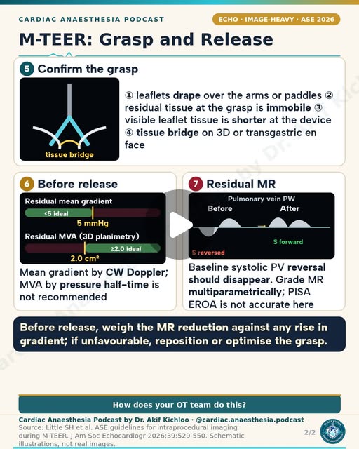
← Back to posts
← Back to posts

Mitral Stenosis 5/5: Intra-op TEE and Bypass Pearls

View on Instagram

Mitral Stenosis 5/5: Intra-op TEE and Bypass Pearls

Pre-bypass thrombus/valve assessment, tricuspid decision-making, prosthetic assessment and post-bypass complications.

Understanding the post

  • An annulus of 40 mm or more, or >21 mm/m², is not a stand-alone reason for tricuspid repair. The decision also depends on TR severity and mechanism, alongside the planned left-sided valve operation. Reference 1; Reference 9.
  • After MVR, assessment includes prosthesis motion, transvalvular and paravalvular regurgitation, gradients, global/regional ventricular function and adjacent valve injury. A gradient is interpreted with heart rate, flow, valve model/size and the postoperative baseline. A single normal-looking cut-off does not exclude dysfunction. Reference 6; Reference 5.
  • Persistent PH or RV dysfunction can occur, especially when present preoperatively; these complications are risks, not inevitable outcomes. Support is tailored to the actual ventricular and pulmonary vascular problem, with avoidance of both systemic hypotension and excessive ventricular loading. Reference 7; Reference 8.

Newer guidance

  • In left-sided valve surgery, ESC/EACTS 2025 recommends concomitant surgery for severe TR (I B), considers repair for moderate TR (IIa B), and allows consideration in selected mild secondary TR with annular dilatation (IIb B). ASE 2024 replaces the 2009 prosthetic-valve document. Reference 9; Reference 5.

Sources named in the post or series

  1. Vahanian A, Beyersdorf F, Praz F, et al. 2021 ESC/EACTS Guidelines for the management of valvular heart disease. Eur Heart J. 2022;43:561–632. DOI: 10.1093/eurheartj/ehab395. Historical valve-disease assessment and intervention recommendations. Sections 4 (AR), 7 (MS), 8 (TR); relevant recommendation tables.
  2. Otto CM, Nishimura RA, Bonow RO, et al. 2020 ACC/AHA Guideline for the Management of Patients With Valvular Heart Disease. J Am Coll Cardiol. 2021;77:e25–e197. DOI: 10.1016/j.jacc.2020.11.018. Valve staging, integrated assessment and US intervention criteria. AR and rheumatic MS sections; official guideline slide set.
  3. Zoghbi WA, Chambers JB, Dumesnil JG, et al. Recommendations for Evaluation of Prosthetic Valves With Echocardiography and Doppler Ultrasound. J Am Soc Echocardiogr. 2009;22:975–1014. DOI: 10.1016/j.echo.2009.07.013. Historical prosthetic-valve source named in the MS series. Historical source, superseded by the 2024 guideline.

Additional supporting sources

  1. Pandian NG, Kim JK, et al. Recommendations for the Use of Echocardiography in the Evaluation of Rheumatic Heart Disease. J Am Soc Echocardiogr. 2023;36:3–28. DOI: 10.1016/j.echo.2022.10.009. Rheumatic valve morphology, mitral valve area methods, associated lesions and procedural assessment. Mitral stenosis and intervention sections; correction DOI: 10.1016/j.echo.2023.01.016.
  2. Zoghbi WA, Jone PN, Chamsi-Pasha MA, et al. Guidelines for the Evaluation of Prosthetic Valve Function With Cardiovascular Imaging. J Am Soc Echocardiogr. 2024;37:2–63. DOI: 10.1016/j.echo.2023.10.004. Integrated assessment of prosthetic valve motion, obstruction, regurgitation and mismatch. Table 11 and mitral prosthesis section.
  3. Nicoara A, Skubas N, Ad N, et al. Guidelines for the Use of Transesophageal Echocardiography to Assist with Surgical Decision-Making in the Operating Room. J Am Soc Echocardiogr. 2020;33:692–734. DOI: 10.1016/j.echo.2020.03.002. Pre- and post-procedure TEE for mitral/aortic surgery, complications, repair geometry and residual lesions. Mitral surgery section and postprocedure table; aortic repair section.
  4. Kaplan’s Cardiac Anesthesia: Perioperative and Critical Care Management. 8th ed. Elsevier; 2023. Chapter 15: Valvular Heart Disease: Repair and Replacement. AR/MS physiology, anaesthetic goals and bypass management. Printed pp. 620–626; Boxes 15.2–15.3.
  5. Gravlee GP, Shaw AD, Bartels K, eds. Hensley’s Practical Approach to Cardiothoracic Anesthesia. 6th ed. Wolters Kluwer; 2019. Chapter 12: Anesthetic Management for Treatment of Mitral and Tricuspid Valve Disease. MS mechanisms, haemodynamics, TEE, perioperative goals and pulmonary vascular/RV complications. Mitral stenosis section.
  6. Praz F, Borger MA, et al. 2025 ESC/EACTS Guidelines for the management of valvular heart disease. DOI: 10.1093/eurheartj/ehaf194. Current European valve intervention recommendations. AR, MS, and TR recommendation tables.

For education. Apply the cited sources in their clinical context and follow current local guidance; this material is not patient-specific advice.
← Back to posts
← Back to posts

Mitral Stenosis 4/5: Anaesthetic Goals and Haemodynamics

View on Instagram

Mitral Stenosis 4/5: Anaesthetic Goals and Haemodynamics

Rate/rhythm, preload, afterload, pulmonary vascular resistance, vasoactive support, PAWP interpretation and neuraxial anaesthesia precautions.

Understanding the post

  • The target is adequate systemic perfusion with afterload in the normal range, not deliberately high SVR. The goals include avoiding tachycardia, preserving sinus rhythm when possible or controlling the ventricular rate, and maintaining adequate preload without pulmonary congestion. Reference 3; Reference 4.
  • A heart rate around 60–70/min is an illustrative target, not a requirement for every patient. Excessive bradycardia can also reduce output. Vasopressor or inotrope choice depends on systemic pressure, RV function and the cause of instability; avoiding unnecessary tachycardia is a goal, not a ban on every drug that can raise heart rate. Reference 3; Reference 4.
  • PAWP reflects pressure upstream of the stenotic valve and may substantially exceed LVEDP. It should not be treated as a direct measure of LV filling pressure or used alone to decide fluid administration. Hypoxaemia, hypercapnia, acidosis and inadequate anaesthesia can worsen pulmonary vascular loading and are important to avoid. Reference 3; Reference 4.
  • A slowly titrated epidural may produce less abrupt haemodynamic change than a single-shot spinal, but this is not a routine recommendation for anticoagulated cardiac surgery. Technique selection must account for the operation, MS severity, haemodynamics and antithrombotic treatment. Reference 3; Reference 6; Reference 7.

Newer guidance

  • ASRA’s 2025 guidance advises against maintaining neuraxial or deep plexus/peripheral catheters during full anticoagulation for cardiac surgery. The bleeding risk and available alternatives need to be considered separately from the haemodynamic advantages of a slowly titrated block. Reference 7.

Sources named in the post or series

  1. Vahanian A, Beyersdorf F, Praz F, et al. 2021 ESC/EACTS Guidelines for the management of valvular heart disease. Eur Heart J. 2022;43:561–632. DOI: 10.1093/eurheartj/ehab395. Historical valve-disease assessment and intervention recommendations. Sections 4 (AR), 7 (MS), 8 (TR); relevant recommendation tables.
  2. Otto CM, Nishimura RA, Bonow RO, et al. 2020 ACC/AHA Guideline for the Management of Patients With Valvular Heart Disease. J Am Coll Cardiol. 2021;77:e25–e197. DOI: 10.1016/j.jacc.2020.11.018. Valve staging, integrated assessment and US intervention criteria. AR and rheumatic MS sections; official guideline slide set.

Additional supporting sources

  1. Kaplan’s Cardiac Anesthesia: Perioperative and Critical Care Management. 8th ed. Elsevier; 2023. Chapter 15: Valvular Heart Disease: Repair and Replacement. AR/MS physiology, anaesthetic goals and bypass management. Printed pp. 620–626; Boxes 15.2–15.3.
  2. Gravlee GP, Shaw AD, Bartels K, eds. Hensley’s Practical Approach to Cardiothoracic Anesthesia. 6th ed. Wolters Kluwer; 2019. Chapter 12: Anesthetic Management for Treatment of Mitral and Tricuspid Valve Disease. MS mechanisms, haemodynamics, TEE, perioperative goals and pulmonary vascular/RV complications. Mitral stenosis section.
  3. Pandian NG, Kim JK, et al. Recommendations for the Use of Echocardiography in the Evaluation of Rheumatic Heart Disease. J Am Soc Echocardiogr. 2023;36:3–28. DOI: 10.1016/j.echo.2022.10.009. Rheumatic valve morphology, mitral valve area methods, associated lesions and procedural assessment. Mitral stenosis and intervention sections; correction DOI: 10.1016/j.echo.2023.01.016.
  4. Kaplan’s Cardiac Anesthesia: Perioperative and Critical Care Management. 8th ed. Elsevier; 2023. Chapter 14: Anesthesia for Myocardial Revascularization. Neuraxial analgesia considerations during cardiac surgery and systemic anticoagulation. Printed pp. 585–586.
  5. Kopp SL, Vandermeulen E, McBane RD, et al. Regional anesthesia in the patient receiving antithrombotic or thrombolytic therapy: ASRA Pain Medicine Evidence-Based Guidelines. 5th edition. Reg Anesth Pain Med. 2025. DOI: 10.1136/rapm-2024-105766. Neuraxial and deep-block precautions with antithrombotic treatment, including full anticoagulation during cardiac surgery. Intravenous heparin recommendations; corrected 2025 online version.

For education. Apply the cited sources in their clinical context and follow current local guidance; this material is not patient-specific advice.
← Back to posts
← Back to posts

Mitral Stenosis 3/5: Intervention Criteria (ESC 2021 + ACC/AHA 2020)

View on Instagram

Mitral Stenosis 3/5: Intervention Criteria (ESC 2021 + ACC/AHA 2020)

Rheumatic MS intervention classes, symptomatic/asymptomatic eligibility, exercise findings and degenerative-MS distinction.

Understanding the post

  • The tables reflect the ESC 2021 and ACC/AHA 2020 recommendations. These are recommendation sets for selected patients, not automatic intervention rules based on valve area alone. Rheumatic commissurotomy requires appropriate anatomy and assessment of MR, LA thrombus, calcification and other disease requiring surgery. Reference 1; Reference 2; Reference 4.
  • The rheumatic commissurotomy pathway does not apply to degenerative stenosis from mitral annular calcification. Commissural fusion and suitability for balloon commissurotomy must be established. Exercise findings and new AF belong to their specific guideline pathways, with the stated eligibility conditions. Reference 5; Reference 2; Reference 1.

Newer guidance

  • ESC/EACTS 2025 retains the main rheumatic-MS commissurotomy/surgery framework. It also allows consideration of transcatheter mitral implantation for symptomatic severe dysfunction with extensive MAC in appropriately selected patients at experienced centres (IIb C). Reference 3.

Sources named in the post or series

  1. Vahanian A, Beyersdorf F, Praz F, et al. 2021 ESC/EACTS Guidelines for the management of valvular heart disease. Eur Heart J. 2022;43:561–632. DOI: 10.1093/eurheartj/ehab395. Historical valve-disease assessment and intervention recommendations. Sections 4 (AR), 7 (MS), 8 (TR); relevant recommendation tables.
  2. Otto CM, Nishimura RA, Bonow RO, et al. 2020 ACC/AHA Guideline for the Management of Patients With Valvular Heart Disease. J Am Coll Cardiol. 2021;77:e25–e197. DOI: 10.1016/j.jacc.2020.11.018. Valve staging, integrated assessment and US intervention criteria. AR and rheumatic MS sections; official guideline slide set.

Additional supporting sources

  1. Praz F, Borger MA, et al. 2025 ESC/EACTS Guidelines for the management of valvular heart disease. DOI: 10.1093/eurheartj/ehaf194. Current European valve intervention recommendations. AR, MS, and TR recommendation tables.
  2. Pandian NG, Kim JK, et al. Recommendations for the Use of Echocardiography in the Evaluation of Rheumatic Heart Disease. J Am Soc Echocardiogr. 2023;36:3–28. DOI: 10.1016/j.echo.2022.10.009. Rheumatic valve morphology, mitral valve area methods, associated lesions and procedural assessment. Mitral stenosis and intervention sections; correction DOI: 10.1016/j.echo.2023.01.016.
  3. Kaplan’s Cardiac Anesthesia: Perioperative and Critical Care Management. 8th ed. Elsevier; 2023. Chapter 15: Valvular Heart Disease: Repair and Replacement. AR/MS physiology, anaesthetic goals and bypass management. Printed pp. 620–626; Boxes 15.2–15.3.

For education. Apply the cited sources in their clinical context and follow current local guidance; this material is not patient-specific advice.
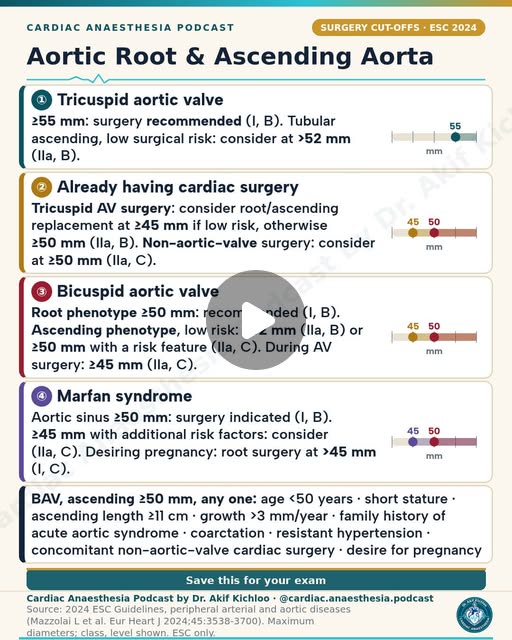
← Back to posts
← Back to posts

Mitral Stenosis 2/5: Grading Severity on Echo

View on Instagram

Mitral Stenosis 2/5: Grading Severity on Echo

Historical versus current severity terminology, planimetry, gradients, PHT and continuity equation.

Understanding the post

  • The <1.0 cm² severe band is the historical 2009 echo table. Current clinical staging uses MVA ≤1.5 cm² for severe rheumatic MS. The apparent difference reflects the framework and year of the recommendations. Mean gradient and pulmonary pressure describe haemodynamic consequences and vary with rate and flow. Reference 1; Reference 3; Reference 4.
  • Pressure half-time does not simply fail whenever any AR, ASD or LV stiffness is present. It becomes unreliable when chamber compliance and pressure decay distort the relation to valve area, particularly just after commissurotomy, in important AR, or in calcific MS. Suitable planimetry and an integrated assessment are therefore more informative than an isolated 220/PHT result. Reference 4; Reference 5.
  • Continuity estimates require comparable forward stroke volumes and become unreliable with important associated regurgitation or shunting. For rheumatic disease, planimetry must be at the smallest leaflet-tip orifice; 3D alignment can help. Reference 4; Reference 7.

Newer guidance

  • The 2023 ASE rheumatic-heart-disease recommendations and the 2025 ESC/EACTS clinical framework provide newer context for the historical 2009 document. The 2009 and 2023 echo documents also have published corrections that form part of their guidance. Reference 4; Reference 1; Reference 6.

Sources named in the post or series

  1. Baumgartner H, Hung J, Bermejo J, et al. Echocardiographic Assessment of Valve Stenosis: EAE/ASE Recommendations for Clinical Practice. J Am Soc Echocardiogr. 2009;22:1–23. DOI: 10.1016/j.echo.2008.11.029. Historical mitral stenosis measurement methods and severity table. Read with published corrections and newer disease-specific guidance. MS section and ASE update notice.
  2. Vahanian A, Beyersdorf F, Praz F, et al. 2021 ESC/EACTS Guidelines for the management of valvular heart disease. Eur Heart J. 2022;43:561–632. DOI: 10.1093/eurheartj/ehab395. Historical valve-disease assessment and intervention recommendations. Sections 4 (AR), 7 (MS), 8 (TR); relevant recommendation tables.
  3. Otto CM, Nishimura RA, Bonow RO, et al. 2020 ACC/AHA Guideline for the Management of Patients With Valvular Heart Disease. J Am Coll Cardiol. 2021;77:e25–e197. DOI: 10.1016/j.jacc.2020.11.018. Valve staging, integrated assessment and US intervention criteria. AR and rheumatic MS sections; official guideline slide set.

Additional supporting sources

  1. Pandian NG, Kim JK, et al. Recommendations for the Use of Echocardiography in the Evaluation of Rheumatic Heart Disease. J Am Soc Echocardiogr. 2023;36:3–28. DOI: 10.1016/j.echo.2022.10.009. Rheumatic valve morphology, mitral valve area methods, associated lesions and procedural assessment. Mitral stenosis and intervention sections; correction DOI: 10.1016/j.echo.2023.01.016.
  2. Kaplan’s Cardiac Anesthesia: Perioperative and Critical Care Management. 8th ed. Elsevier; 2023. Chapter 15: Valvular Heart Disease: Repair and Replacement. AR/MS physiology, anaesthetic goals and bypass management. Printed pp. 620–626; Boxes 15.2–15.3.
  3. Praz F, Borger MA, et al. 2025 ESC/EACTS Guidelines for the management of valvular heart disease. DOI: 10.1093/eurheartj/ehaf194. Current European valve intervention recommendations. AR, MS, and TR recommendation tables.
  4. Gravlee GP, Shaw AD, Bartels K, eds. Hensley’s Practical Approach to Cardiothoracic Anesthesia. 6th ed. Wolters Kluwer; 2019. Chapter 12: Anesthetic Management for Treatment of Mitral and Tricuspid Valve Disease. MS mechanisms, haemodynamics, TEE, perioperative goals and pulmonary vascular/RV complications. Mitral stenosis section.

For education. Apply the cited sources in their clinical context and follow current local guidance; this material is not patient-specific advice.
← Back to posts
← Back to posts

Mitral Stenosis 1/5: Aetiology and Pathophysiology

View on Instagram

Mitral Stenosis 1/5: Aetiology and Pathophysiology

Aetiology, normal and reduced valve area, LA/PH/RV consequences, LV physiology and high-flow/tachycardia effects.

Understanding the post

  • The sequence from raised LA pressure to AF, thrombus, PH and RV failure describes possible consequences, not an inevitable progression in every patient. Reference 5; Reference 6.
  • The LV is often relatively underfilled in isolated MS, but normal LV function cannot be assumed. Coexisting myocardial disease, reduced compliance or systolic dysfunction may be present. Rheumatic commissural fusion and degenerative MAC are different mechanisms and need different assessment and intervention approaches. Reference 5; Reference 6; Reference 4.
  • Tachycardia shortens diastolic filling and can sharply increase the transmitral gradient. High-flow states also raise the gradient, so a gradient should be reported with heart rate and interpreted with valve area and flow. Reference 5; Reference 6.

Newer guidance

  • ESC/EACTS 2025 keeps rheumatic and degenerative MS pathways distinct. This is an important addition to the introductory disease model. Reference 7.

Sources named in the post or series

  1. Baumgartner H, Hung J, Bermejo J, et al. Echocardiographic Assessment of Valve Stenosis: EAE/ASE Recommendations for Clinical Practice. J Am Soc Echocardiogr. 2009;22:1–23. DOI: 10.1016/j.echo.2008.11.029. Historical mitral stenosis measurement methods and severity table. Read with published corrections and newer disease-specific guidance. MS section and ASE update notice.
  2. Vahanian A, Beyersdorf F, Praz F, et al. 2021 ESC/EACTS Guidelines for the management of valvular heart disease. Eur Heart J. 2022;43:561–632. DOI: 10.1093/eurheartj/ehab395. Historical valve-disease assessment and intervention recommendations. Sections 4 (AR), 7 (MS), 8 (TR); relevant recommendation tables.
  3. Otto CM, Nishimura RA, Bonow RO, et al. 2020 ACC/AHA Guideline for the Management of Patients With Valvular Heart Disease. J Am Coll Cardiol. 2021;77:e25–e197. DOI: 10.1016/j.jacc.2020.11.018. Valve staging, integrated assessment and US intervention criteria. AR and rheumatic MS sections; official guideline slide set.

Additional supporting sources

  1. Pandian NG, Kim JK, et al. Recommendations for the Use of Echocardiography in the Evaluation of Rheumatic Heart Disease. J Am Soc Echocardiogr. 2023;36:3–28. DOI: 10.1016/j.echo.2022.10.009. Rheumatic valve morphology, mitral valve area methods, associated lesions and procedural assessment. Mitral stenosis and intervention sections; correction DOI: 10.1016/j.echo.2023.01.016.
  2. Kaplan’s Cardiac Anesthesia: Perioperative and Critical Care Management. 8th ed. Elsevier; 2023. Chapter 15: Valvular Heart Disease: Repair and Replacement. AR/MS physiology, anaesthetic goals and bypass management. Printed pp. 620–626; Boxes 15.2–15.3.
  3. Gravlee GP, Shaw AD, Bartels K, eds. Hensley’s Practical Approach to Cardiothoracic Anesthesia. 6th ed. Wolters Kluwer; 2019. Chapter 12: Anesthetic Management for Treatment of Mitral and Tricuspid Valve Disease. MS mechanisms, haemodynamics, TEE, perioperative goals and pulmonary vascular/RV complications. Mitral stenosis section.
  4. Praz F, Borger MA, et al. 2025 ESC/EACTS Guidelines for the management of valvular heart disease. DOI: 10.1093/eurheartj/ehaf194. Current European valve intervention recommendations. AR, MS, and TR recommendation tables.

For education. Apply the cited sources in their clinical context and follow current local guidance; this material is not patient-specific advice.
← Back to posts
← Back to posts

Perioperative Beta-Blockers: Continue, Don’t Start

View on Instagram

Perioperative Beta-Blockers: Continue, Don’t Start

Continuation versus initiation, POISE interpretation, non-cardiac surgery and postoperative AF prevention.

Understanding the post

  • The “Continue, Don’t Start” message refers to continuing established beta blockers and avoiding routine initiation on the day of surgery. A new, independent indication can justify starting earlier with time to assess tolerance and titrate. Reference 4.
  • POISE tested a particular extended-release metoprolol regimen begun shortly before non-cardiac surgery. Its lower MI rate but greater stroke/mortality risk does not establish that every initiation strategy has the same effects. Hypotension and bradycardia are important concerns, but a single causal explanation is not proved for every event. Reference 2; Reference 4.

Newer guidance

  • AHA/ACC 2024 advises initiation for a new indication sufficiently before elective non-cardiac surgery, optimally >7 days. EACTS 2024 recommends continuing beta blockers through cardiac surgery (I A); low-dose short-term prophylaxis in beta-blocker-naïve patients may be considered (IIb B), with careful titration. Reference 4; Reference 5.

Sources named in the post or series

  1. Halvorsen S, Mehilli J, Cassese S, et al. 2022 ESC Guidelines on cardiovascular assessment and management of patients undergoing non-cardiac surgery. DOI: 10.1093/eurheartj/ehac270. Historical perioperative beta-blocker continuation and initiation recommendations. Beta-blocker section.
  2. POISE Study Group. Effects of extended-release metoprolol succinate in patients undergoing non-cardiac surgery (POISE trial): a randomised controlled trial. Lancet. 2008;371:1839–1847. DOI: 10.1016/S0140-6736(08)60601-7. Benefits and harms of the specific perioperative metoprolol regimen studied.
  3. Hindricks G, Potpara T, Dagres N, et al. 2020 ESC Guidelines for the diagnosis and management of atrial fibrillation developed in collaboration with EACTS. DOI: 10.1093/eurheartj/ehaa612. Historical postoperative AF prevention recommendation.

Additional supporting sources

  1. Thompson A, Fleischmann KE, Smilowitz NR, et al. 2024 AHA/ACC multisociety Guideline for Perioperative Cardiovascular Management for Noncardiac Surgery. DOI: 10.1016/j.jacc.2024.06.013. Continue established beta blockers; new indication and sufficient time for titration; avoid routine day-of-surgery initiation. Perioperative beta-blocker recommendations.
  2. Jeppsson A, Rocca B, et al. 2024 EACTS Guidelines on perioperative medication in adult cardiac surgery. Eur J Cardiothorac Surg. 2025;67:ezae355. DOI: 10.1093/ejcts/ezae355. Cardiac-surgery beta-blocker continuation and selected prophylactic initiation. Recommendation Table 2; beta-blocker discussion.

Guideline update: 2026

The 2024 US perioperative recommendations were reaffirmed in 2026 after evidence review through March 2026. This does not introduce a new beta-blocker starting rule: the existing continuation and carefully timed initiation advice remains applicable. American Heart Association. Guideline Publication Schedule: perioperative cardiovascular management, 2026 reaffirmation..

For education. Apply the cited sources in their clinical context and follow current local guidance; this material is not patient-specific advice.
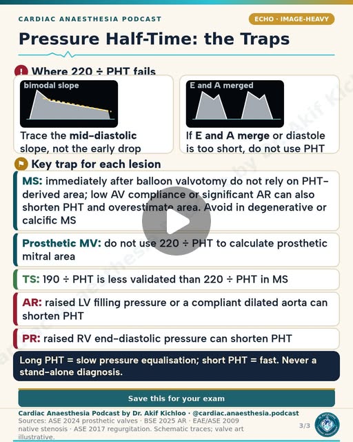
← Back to posts
← Back to posts

Endocarditis Prophylaxis: Who Needs It?

View on Instagram

Endocarditis Prophylaxis: Who Needs It?

Who qualifies, which procedures qualify, adult regimen, allergy caveats and AHA–ESC differences.

Understanding the post

  • European and US recommendations differ. AHA advice does not recommend routine IE prophylaxis for TEE, gastroscopy, colonoscopy or cystoscopy without active infection. ESC 2023 permits consideration for selected high-risk patients having certain invasive non-dental procedures (IIb), so a universal never-needed statement is too broad. Reference 1; Reference 2.
  • Eligible dental procedures include gingival manipulation, manipulation of the periapical region, or perforation of oral mucosa. Adult oral amoxicillin 2 g 30–60 minutes before is a standard regimen when indicated; allergy and inability to take oral medication require the appropriate alternative. Reference 2.
  • High-risk categories are specific to each authority rather than interchangeable parts of a single eligibility list. The AHA wallet card includes transplant recipients with structurally abnormal regurgitant valves and specified congenital defects. The ESC guidance includes its own congenital-repair and device categories. Reference 1; Reference 2.
  • Clindamycin is no longer an AHA dental-prophylaxis alternative. Avoid cephalosporins in patients with previous penicillin/ampicillin anaphylaxis, angioedema or urticaria. Oral hygiene and regular dental care remain central. Reference 2.

Sources named in the post or series

  1. Delgado V, Ajmone Marsan N, de Waha S, et al. 2023 ESC Guidelines for the management of endocarditis. Eur Heart J. 2023;44:3948–4042. DOI: 10.1093/eurheartj/ehad193. High-risk groups, dental prophylaxis and European non-dental recommendations. Prevention sections and recommendation table.

Additional supporting sources

  1. American Heart Association. Prevention of Infective Endocarditis wallet card. 2024. US eligibility, dental procedures, dosing/allergy alternatives and non-dental advice. Both pages.

For education. Apply the cited sources in their clinical context and follow current local guidance; this material is not patient-specific advice.

← Back to posts
← Back to posts

Prostaglandin E1 for Duct-Dependent Lesions

View on Instagram

Sources supporting this post

  • Prostin VR Pediatric (alprostadil) label, revised April 2026. Supports duct-dependent indications, initial 0.05–0.1 microgram/kg/min dosing, reduction to the lowest effective dose and the 10–12% apnoea estimate.
  • Andropoulos et al., Anesthesia for Congenital Heart Disease, 3rd ed. (2015), chapter 24, pp.547–548. Explains why a restrictive atrial opening in TGA may require atrial septostomy despite prostaglandin.
  • Nasr and DiNardo, The Pediatric Cardiac Anesthesia Handbook (2017), chapter 10, pp.80–81. Supports the neonatal coarctation example and shows that lower starting ranges are also used.

Understanding the post

  • “Start 0.05–0.1 mcg/kg/min if the duct is closing, then titrate to the lowest effective dose.” matches the manufacturer’s starting range, followed by dose reduction once circulation improves. This is not a compulsory starting dose for every baby: dose depends on the lesion, duct patency and response. A neonatal coarctation textbook example uses a lower range. Prostin VR Pediatric label, dosage; Nasr and DiNardo 2017, chapter 10, pp.80–81.
  • For “mixing (TGA with intact septum),” keeping the duct open may not provide enough mixing if the opening between the atria is restrictive. Urgent balloon atrial septostomy may be needed. Do not assume prostaglandin alone corrects the low oxygen level. Andropoulos et al., 3rd ed. (2015), chapter 24, pp.547–548.
  • “Apnoea occurs in about 1 in 10 neonates, so be ready to ventilate.” agrees with the label’s 10–12% estimate. It requires continuous breathing monitoring and immediately available ventilatory support, especially early in treatment. Prostin VR Pediatric boxed warning.
  • The listed systemic-flow, pulmonary-flow and mixing lesions are useful examples, but the anatomical diagnosis alone does not establish duct dependence in every baby. The aim is to preserve the particular circulation that depends on the duct. Prostin VR Pediatric, indications; Andropoulos, chapter 24.
  • “Fever, flushing, hypotension, bradycardia, seizures” are recognised reported adverse effects. “Cortical hyperostosis” means extra bone formation reported with prolonged infusions; gastric outlet obstruction can occur through thickening around the stomach outlet. The label advises close observation for the latter during prolonged treatment, especially beyond 120 hours. Prostin VR Pediatric, warnings, precautions and adverse reactions.
  • “Keep PGE1 running until surgery or intervention” means maintain support while the circulation remains duct-dependent, under the treating cardiac team’s plan. Use the lowest effective dose for the shortest necessary time; the endpoint is adequate alternative circulation, not simply that an operation has started. Prostin VR Pediatric, indications, warnings and dosage.

For education. Apply the cited sources in their clinical context and follow current local guidance; this material is not patient-specific advice.
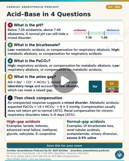
← Back to posts
← Back to posts

CHA2DS2-VASc and HAS-BLED Scores

View on Instagram

CHA2DS2-VASc and HAS-BLED Scores

Stroke and bleeding score components, thresholds, modifiable risks, sex-point change and valvular exceptions.

Understanding the post

  • CHA₂DS₂-VASc estimates stroke risk to inform a treatment decision; it is not an automatic treatment order. The US ≥2-point male/≥3-point female thresholds are examples of an estimated annual thromboembolic risk ≥2%; shared assessment includes contraindications and patient preferences. Reference 2.
  • A high HAS-BLED score identifies issues to address and a need for closer review. It should not be used alone to refuse anticoagulation. Each component has a specific definition, so a renal/liver abnormality or alcohol intake does not automatically earn a point. Reference 2.
  • These scores do not make moderate/severe rheumatic MS or a mechanical valve a low-risk exemption from anticoagulation, and the usual preference for a DOAC does not apply to those groups. Reference 2; Reference 3.

Newer guidance

  • ESC 2024 uses CHA₂DS₂-VA: consider anticoagulation at 1 and recommend it at ≥2, within the full clinical assessment. This is a separate framework from the US CHA₂DS₂-VASc approach. Reference 3.

Sources named in the post or series

  1. Hindricks G, Potpara T, Dagres N, et al. 2020 ESC Guidelines for the diagnosis and management of atrial fibrillation developed in collaboration with EACTS. DOI: 10.1093/eurheartj/ehaa612. Historical CHA₂DS₂-VASc anticoagulation thresholds and bleeding-risk assessment. Stroke prevention and risk-assessment recommendations.
  2. Joglar JA, Chung MK, Armbruster AL, et al. 2023 ACC/AHA/ACCP/HRS Guideline for the Diagnosis and Management of Atrial Fibrillation. DOI: 10.1016/j.jacc.2023.08.017. CHA₂DS₂-VASc/HAS-BLED components, annual stroke-risk assessment, anticoagulation and valve exceptions. Section 6, risk-score tables, antithrombotic recommendations.
  3. Van Gelder IC, Rienstra M, Bunting KV, et al. 2024 ESC Guidelines for the management of atrial fibrillation. DOI: 10.1093/eurheartj/ehae176. CHA₂DS₂-VA and European anticoagulation recommendations. AF-CARE stroke prevention section.

For education. Apply the cited sources in their clinical context and follow current local guidance; this material is not patient-specific advice.
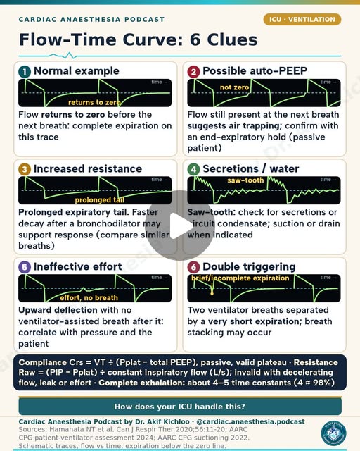
← Back to posts
← Back to posts

The Bypass Circuit: Every Component (infographic)

View on Instagram

The Bypass Circuit: Every Component (infographic)

Circuit blood flow, components, gas and heat exchange, suction/vent/cardioplegia branches, air and temperature safety.

Understanding the post

  • Stopping the pump and clamping the arterial line for air at any step is not a universal rule. Suspected arterial air embolism is an emergency requiring the team’s specific protocol; the response depends on air location, amount and circulation state. A circuit schematic is not an emergency algorithm. Reference 3.
  • Temperature limits depend on measurement sites and conditions: the 2015 guideline specifies arterial-outlet/venous-inflow gradients ≤10°C and, during rewarming at arterial outlet temperature ≥30°C, a gradient ≤4°C and rate ≤0.5°C/min. Its arterial-outlet limit is <37°C. Reference 2.
  • The main blood pathway and accessory circuits have distinct roles. Vent, suction and cardioplegia branches serve different purposes; safety sensors and one-way valves are not interchangeable, and an arterial filter does not make air entry harmless. Reference 4; Reference 3.

Newer guidance

  • The 2024 EACTS/EACTAIC/EBCP guideline, published in 2025, updates the 2019 source. It reinforces team safety systems and context-specific crisis management, and states that cardiotomy suction should stop when protamine administration starts. Reference 3.

Sources named in the post or series

  1. Wahba A, Kunst G, De Somer F, et al. 2019 EACTS/EACTA/EBCP guidelines on cardiopulmonary bypass in adult cardiac surgery. Eur J Cardiothorac Surg. 2020;57:210–251. DOI: 10.1093/ejcts/ezz267. Historical circuit technology, monitoring and safety recommendations.
  2. Engelman R, Baker RA, Likosky DS, et al. STS/SCA/AmSECT Clinical Practice Guidelines for Cardiopulmonary Bypass: Temperature Management During Cardiopulmonary Bypass. Ann Thorac Surg. 2015;100:748–757. DOI: 10.1016/j.athoracsur.2015.03.126. Temperature measurement sites, cooling/rewarming gradients and rewarming rate. Recommendations on pp. 748–749.

Additional supporting sources

  1. Wahba A, Kunst G, De Somer F, et al. 2024 EACTS/EACTAIC/EBCP Guidelines on cardiopulmonary bypass in adult cardiac surgery. Eur J Cardiothorac Surg. 2025;67:ezae354. DOI: 10.1093/ejcts/ezae354. Current CPB safety, components, temperature management, cardioplegia and crisis management. Equipment, cardiotomy suction, temperature and adverse-event sections.
  2. Hessel EA II. Cardiopulmonary Bypass: Equipment, Circuits, and Pathophysiology. In: Hensley’s Practical Approach to Cardiothoracic Anesthesia. 6th ed. Wolters Kluwer; 2019. Chapter 20. Blood pathway, component functions and circuit safety. Figure 20.1 and Part I, Components of the circuit.

For education. Apply the cited sources in their clinical context and follow current local guidance; this material is not patient-specific advice.
← Back to posts
← Back to posts

Cardiac Action Potential: Every Phase (infographic)

View on Instagram

Cardiac Action Potential: Every Phase (infographic)

Ventricular/nodal phases, ionic currents, antiarrhythmic actions, refractoriness and approximate ECG relationships.

Understanding the post

  • Ventricular-myocyte and nodal action potentials have distinct patterns. The phase/current map is a schematic: individual currents overlap in time and vary across tissues, disease states and rate. Class I–IV labels summarise dominant actions; many drugs act on several targets. Reference 2.
  • ECG intervals reflect electrical activity across the whole heart, not one cell’s voltage trace. A single-cell phase does not have a rigid one-to-one equivalence with an ECG segment, and recovery of excitability does not have a universal voltage cut-off. Reference 2.

Newer guidance

  • A 2025 clinical adaptation of the modernised drug classification focuses on established clinical drugs rather than investigational-only subclasses. It is a useful optional update to the classic I–IV teaching framework, not a new prescribing algorithm. Reference 3.

Sources named in the post or series

  1. Hall JE, Hall ME. Guyton and Hall Textbook of Medical Physiology. 14th ed. Elsevier; 2021. Chapters 9–10. Cardiac muscle physiology and rhythmic excitation.
  2. Lei M, Wu L, Terrar DA, Huang CL-H. Modernized Classification of Cardiac Antiarrhythmic Drugs. Circulation. 2018;138:1879–1896. DOI: 10.1161/CIRCULATIONAHA.118.035455. Action-potential ion currents, drug targets and limitations of a one-class/one-target model. Figure 1, Tables 1–2 and mechanism sections.

Additional supporting sources

  1. Lei M, Wu L, Terrar DA, Huang CL-H. The modernized classification of cardiac antiarrhythmic drugs: Its application to clinical practice. Heart Rhythm. 2025;22:e500–e506. DOI: 10.1016/j.hrthm.2025.03.1997. Later clinical adaptation of the 2018 classification.

For education. Apply the cited sources in their clinical context and follow current local guidance; this material is not patient-specific advice.
← Back to posts
← Back to posts

Reversing Anticoagulants Before Urgent Surgery

View on Instagram

Sources supporting this post

How to read the post

  • Andexanet and heparin-based bypass. The post’s andexanet alfa and 4-factor PCC options for apixaban/rivaroxaban are not interchangeable before heart-lung bypass using heparin. Andexanet can stop heparin working properly and allow the bypass circuit to clot, so the guideline advises avoiding it in this setting. 2024 EACTS/EACTAIC PBM guideline with EBCP
  • The setting for 50 U/kg PCC. The 50 U/kg PCC recommendation is conditional: EACTS advises considering it for emergency surgery when a patient is not bleeding but still has a high level of a factor-Xa-inhibitor anticoagulant. This use is outside the product’s approved indication and needs a specialist/local protocol. It is not a blanket dose for every urgent operation. 2024 EACTS/EACTAIC PBM guideline with EBCP
  • US andexanet availability. Availability also matters when interpreting the factor-Xa-inhibitor reversal row: FDA reports that AstraZeneca stopped US commercial sales and manufacture for the US after 22 December 2025. This statement is US-specific. FDA Safety Communication, 18 December 2025
  • Protamine for unfractionated heparin. The ratio of protamine 1 mg per 100 units of heparin is supported, but treatment depends on heparin still active in the body, elapsed time and clotting tests, rather than automatically including every earlier heparin dose. Give protamine slowly; rapid injection can cause serious reactions. Protamine sulfate label, DailyMed
  • Partial reversal of enoxaparin. The approximately 60% anti-Xa reversal in the post is the maximum described for enoxaparin, not a guaranteed percentage for every low-molecular-weight heparin. Reversal remains incomplete. Lovenox prescribing information
  • Vitamin K and PCC timing. Vitamin K is not immediate, but the post’s 6–12 hours is not a fixed onset: the injection label describes INR improvement within 1–8 hours. PCC lowers INR rapidly, but repeat tests and bleeding control still matter. Phytonadione injection prescribing information; Kcentra prescribing information
  • Plasma when PCC is unavailable. The post’s 15–20 mL/kg FFP range is not a fixed dose for every urgent operation. Lifeblood recommends 15 mL/kg when PCC is unavailable for life-threatening warfarin bleeding. Plasma dosing depends on bleeding, clotting tests and the risk of fluid overload; follow the named local reversal protocol. Australian Red Cross Lifeblood: critical bleeding, February 2025; Australian Red Cross Lifeblood: fresh frozen plasma, July 2026

Guideline update: 2024 EACTS/EACTAIC PBM guideline with EBCP adds an important cardiac-surgery qualification to the simple reversal table: avoid andexanet before heparin-based bypass, and use PCC for factor-Xa drugs only in the specified clinical setting. The later FDA Safety Communication, 18 December 2025 reports the end of US Andexxa commercial sales and manufacture after 22 December 2025. This is a US safety and availability change, not a worldwide withdrawal statement.

For education. Apply the cited sources in their clinical context and follow current local guidance; this material is not patient-specific advice.
← Back to posts
← Back to posts

NYHA Class vs ACC/AHA Heart Failure Stage

View on Instagram

NYHA Class vs ACC/AHA Heart Failure Stage

NYHA functional class, HF stages A–D, pre-HF criteria and improvement versus disease history.

Understanding the post

  • Stage B now means pre-HF: no symptoms/signs of HF, with structural disease, evidence of raised filling pressure, or relevant biomarker evidence in an at-risk person after alternative causes are considered. Structural disease alone is not the complete definition. Reference 1.
  • Stage C includes current or previous HF symptoms. Improvement in symptoms or EF does not erase that history or mean the person has never had HF. The phrase stages only move forward should not imply that cardiac function, symptoms or clinical condition cannot improve. Reference 1.
  • NYHA class describes functional limitation at the time of assessment. Class I means no limitation with ordinary activity, not absence of heart disease; class IV includes inability to undertake activity without symptoms and symptoms at rest. Reference 1.

Newer guidance

  • The 2022 guideline’s pre-HF definition and HF with improved EF category are the important additions to this comparison. Improved EF is not, by itself, a reason to stop HFrEF treatment. Reference 1.

Additional supporting sources

  1. Heidenreich PA, Bozkurt B, Aguilar D, et al. 2022 AHA/ACC/HFSA Guideline for the Management of Heart Failure. DOI: 10.1161/CIR.0000000000001063. 2022 HF stages, pre-HF criteria, symptom status and HF with improved EF. Official slide set, Table 3 and HFimpEF recommendations.

Guideline update: 2026

The June 2026 Second Universal Definition of Heart Failure, a joint AHA/ACC/ESC/WHF consensus with HFSA, HFA and JHFS, reaffirms stages A–D and distinguishes improvement, remission and recovery. Symptoms or function can improve without erasing the history of HF. It groups HF as reduced, preserved or improved EF without rigid universal numerical cutoffs. This definition consensus is separate from the August 2026 ESC treatment guideline and its <50%/≥50% categories. The two documents use different frameworks, so their categories are not interchangeable. AHA/ACC/ESC/WHF, with HFSA, HFA and JHFS. Second Universal Definition of Heart Failure (2026)..

For education. Apply the cited sources in their clinical context and follow current local guidance; this material is not patient-specific advice.

← Back to posts
← Back to posts

Humour: Muscle relaxants ranked by how long they stay at the party

View on Instagram

Humour: Muscle relaxants ranked by how long they stay at the party

Five NMBAs, dose/anaesthetic-specific duration, endpoint comparability and safe recovery assessment.

The NIMBEX and PAVULON references below are historical 2010 labels. Their study-specific durations should not replace current prescribing information or quantitative neuromuscular monitoring.

Understanding the post

  • The label-based durations are illustrative rather than a universal shortest-to-longest ranking. The values come from different doses, anaesthetics, populations and endpoints, rather than a head-to-head trial. Reference 1; Reference 2; Reference 3; Reference 4; Reference 5.
  • For atracurium 0.4–0.5 mg/kg under balanced anaesthesia, about 20–35 minutes of clinically required block differs from about 35–45 minutes to 25% control-twitch recovery. This matters when comparing durations: the headline 20–35-minute value is not the same endpoint used for the other nondepolarising drugs. Reference 2.
  • The rocuronium example is 0.6 mg/kg, median 31 minutes (range 15–85) to 25% T1 recovery under opioid/nitrous oxide/oxygen anaesthesia. The historical cisatracurium and pancuronium values apply to their specified doses and anaesthetic conditions. Succinylcholine uses a different recovery-window description. Reference 3; Reference 4; Reference 5; Reference 1.
  • For cisatracurium 0.15 mg/kg, the historical label’s mean 55 minutes to 25% recovery, with a 44–74-minute range, is the propofol-anaesthesia result. The row-specific footnote identifies propofol, so this is not also an opioid/nitrous oxide result. Reference 4.
  • The pancuronium estimates use different starting points as well as different doses: about 22 minutes after 0.05 mg/kg is measured from maximum effect to 25% recovery, whereas about 100 minutes after 0.1 mg/kg is measured from injection to 25% recovery. Both the starting point and the dose therefore matter when comparing these durations. Reference 5.

Newer guidance

  • ASA 2023 recommends quantitative monitoring at the adductor pollicis and confirmation of TOF ratio ≥0.9 before extubation. Elapsed time or 25% recovery of one twitch does not establish readiness for extubation. Reference 6.

Additional supporting sources

  1. ANECTINE (succinylcholine chloride), US prescribing information. DailyMed. Adult single-dose recovery window; patient/dose dependence. Dosage and clinical pharmacology.
  2. Atracurium besylate injection, US prescribing information. DailyMed. Clinical-block duration versus time to 25% twitch recovery. Clinical pharmacology and adult bolus dosing.
  3. Rocuronium bromide injection, US prescribing information. DailyMed. Dose/age/anaesthetic-specific clinical duration and 25% T1 endpoint. Section 12.2, Table 5.
  4. NIMBEX (cisatracurium besylate), FDA prescribing information, historical 2010 version. Historical dose/anaesthetic-specific recovery values. Table 1 and adult dosing.
  5. PAVULON (pancuronium bromide), FDA prescribing information, historical 2010 version. Historical 0.05 and 0.1 mg/kg recovery estimates with different timing origins under balanced anaesthesia. Clinical pharmacology, pp. 1–2.
  6. Thilen SR, Weigel WA, Todd MM, et al. 2023 ASA Practice Guidelines for Monitoring and Antagonism of Neuromuscular Blockade. Anesthesiology. 2023;138:13–41. DOI: 10.1097/ALN.0000000000004379. Quantitative monitoring and confirmation of recovery before extubation. Recommendations for adductor pollicis monitoring and TOF ratio.

For education. Apply the cited sources in their clinical context and follow current local guidance; this material is not patient-specific advice.
← Back to posts
← Back to posts

Shock Types: Haemodynamic Profiles

View on Instagram

Shock Types: Haemodynamic Profiles

Four shock patterns, CO/SVR/filling pressures, tamponade and mixed venous oxygen saturation.

Understanding the post

  • The table describes typical patterns rather than diagnostic rules. Mixed shock, treatment, ventilation and the time of measurement can change output, SVR and filling pressures. Cardiogenic shock from predominant RV failure need not have a high wedge pressure. Reference 1; Reference 2.
  • Tamponade often brings diastolic pressures closer together, but equalisation is not required, particularly with regional postoperative compression. A missing classic pattern should not exclude tamponade. Reference 4.
  • Normal or high SvO₂ does not prove adequate tissue oxygen use. Distributive shock may impair extraction, and associated cardiac dysfunction can lower SvO₂. The value is interpreted with perfusion, haemoglobin, arterial saturation and oxygen demand. Reference 1; Reference 2.

Newer guidance

  • ESICM 2025 recommends echocardiography as first-line imaging to establish shock type, repeated tissue-perfusion assessment, and CO/stroke-volume monitoring when initial therapy is insufficient. Static pressure patterns should not be the sole basis for fluid decisions. Reference 3.

Additional supporting sources

  1. Simmons J, Ventetuolo CE. Cardiopulmonary Monitoring of Shock. Curr Opin Crit Care. 2017;23:223–231. DOI: 10.1097/MCC.0000000000000407. Shock mechanisms, mixed states and interpretation of venous oxygen saturation. The narrative physiology and monitoring sections support this explanation; the distributive-shock CO entry in the source table is inconsistent with the narrative and is not a reliable guide.
  2. Edwards Lifesciences. Hemodynamic Monitoring Clinical Reference Guide. PAC waveform sequence, measurement conditions, oxygen delivery/consumption and illustrative reference values. PAC chapter and venous oximetry chapter.
  3. Monnet X, Messina A, Greco M, et al. ESICM guidelines on circulatory shock and hemodynamic monitoring 2025. Intensive Care Med. 2025;51:1971–2012. DOI: 10.1007/s00134-025-08137-z. Current shock assessment, repeated perfusion assessment, echocardiography and haemodynamic monitoring. Guideline recommendations and official ESICM summary.
  4. Levy JH, Ghadimi K, Bailey JM, Ramsay JG. Postoperative Cardiovascular Management. In: Kaplan’s Essentials of Cardiac Anesthesia. 2nd ed. Elsevier; 2018. Chapter 30, pp. 758–785. Postoperative low-output states and atypical/localised tamponade. Cardiac tamponade section, pp. 777–778.

For education. Apply the cited sources in their clinical context and follow current local guidance; this material is not patient-specific advice.
← Back to posts
← Back to posts

PA Catheter Normal Values

View on Instagram

PA Catheter Normal Values

Pressure and flow reference values, chamber waveform sequence, oxygen saturation and safe interpretation.

Adult reference intervals vary slightly between sources and measurement methods. These values are teaching ranges, not treatment targets.

Understanding the post

  • The displayed numbers are approximate adult resting reference intervals rather than mandatory treatment targets. Published ranges vary. Pressure values use mmHg and cardiac index uses L/min/m²; mixed venous oxygen saturation from the PA and central venous saturation are distinct measurements. Reference 1; Reference 2.
  • The RV-to-PA transition commonly preserves systolic pressure while diastolic pressure rises. Accurate pressures require consistent transducer level/zero and appropriate respiratory timing; PEEP, catheter position and West-zone conditions can distort wedge measurements. Reference 1.
  • PAWP is not always LVEDP and neither pressure is a stand-alone test of fluid responsiveness. This is particularly important in MS and altered ventricular compliance. Balloon inflation/deflation and catheter movement must follow the device instructions and local safety protocol; this post does not provide a catheter-insertion protocol. Reference 4; Reference 1; Reference 3.

Newer guidance

  • ESICM 2025 supports selected haemodynamic monitoring and dynamic fluid-responsiveness assessment rather than routine treatment to fixed normal filling pressures. Reference 3.

Additional supporting sources

  1. Edwards Lifesciences. Hemodynamic Monitoring Clinical Reference Guide. PAC waveform sequence, measurement conditions, oxygen delivery/consumption and illustrative reference values. PAC chapter and venous oximetry chapter.
  2. Baldetti L, Cosenza M, Galdieri C, et al. Invasive Hemodynamic Monitoring in Acute Heart Failure and Cardiogenic Shock. Rev Cardiovasc Med. 2025;26(6):27034. DOI: 10.31083/RCM27034. Independent reference for CI 2.5–4.0 L/min/m² and SvO₂ 65–75%. Table 1; PAC and measurement-method sections.
  3. Monnet X, Messina A, Greco M, et al. ESICM guidelines on circulatory shock and hemodynamic monitoring 2025. Intensive Care Med. 2025;51:1971–2012. DOI: 10.1007/s00134-025-08137-z. Current shock assessment, repeated perfusion assessment, echocardiography and haemodynamic monitoring. Guideline recommendations and official ESICM summary.
  4. Gravlee GP, Shaw AD, Bartels K, eds. Hensley’s Practical Approach to Cardiothoracic Anesthesia. 6th ed. Wolters Kluwer; 2019. Chapter 12: Anesthetic Management for Treatment of Mitral and Tricuspid Valve Disease. MS mechanisms, haemodynamics, TEE, perioperative goals and pulmonary vascular/RV complications. Mitral stenosis section.

For education. Apply the cited sources in their clinical context and follow current local guidance; this material is not patient-specific advice.
← Back to posts
← Back to posts

RV Systolic Function on Echo: Every Method (infographic)

View on Instagram

RV Systolic Function on Echo: Every Method (infographic)

TAPSE, S′, FAC, strain, 3D EF, RIMP, dP/dt, RV dimensions and postoperative caveats.

Understanding the post

  • The numerical table uses the ASE/EACVI 2015 framework. Reference means, abnormal cut-offs and severity categories have different meanings and depend on the source document. For strain, the sign convention matters: a magnitude <20% means a value less negative than about −20%. Reference 1; Reference 3.
  • RV function is assessed using multiple measurements and the clinical setting. TAPSE and S′ reflect longitudinal motion and can fall after cardiac surgery without a proportionate fall in global RV function. RIMP is dimensionless; high RA pressure, rhythm and loading affect it. dP/dt is load-dependent and is not a routine stand-alone RV test. Reference 1; Reference 3.

Newer guidance

  • The 2025 ASE right-heart guideline supersedes the 2010 document. It lists normal pulsed-Doppler RV MPI <0.40 and tissue-Doppler MPI <0.55, rather than the historical 2015 abnormal boundaries >0.43/>0.54. Main TAPSE, S′, FAC and 3D-EF thresholds remain broadly familiar but should be interpreted together. Reference 3.

Sources named in the post or series

  1. Lang RM, Badano LP, Mor-Avi V, et al. Recommendations for Cardiac Chamber Quantification by Echocardiography in Adults. J Am Soc Echocardiogr. 2015;28:1–39. DOI: 10.1016/j.echo.2014.10.003. Adult chamber dimensions, function, LV geometry and reference intervals. Tables 2, 4, 6, 8 and 10; supplementary normal-value tables.
  2. Rudski LG, Lai WW, Afilalo J, et al. Guidelines for the Echocardiographic Assessment of the Right Heart in Adults. J Am Soc Echocardiogr. 2010;23:685–713. DOI: 10.1016/j.echo.2010.05.010. Historical RV dP/dt method; superseded by the 2025 right-heart guideline. RV dP/dt, p. 700.

Additional supporting sources

  1. Mukherjee M, Rudski LG, et al. Guidelines for the Echocardiographic Assessment of the Right Heart in Adults and Special Considerations in Pulmonary Hypertension. J Am Soc Echocardiogr. 2025;38:141–186. DOI: 10.1016/j.echo.2025.01.006. Current multiparametric RV assessment, Doppler quality, PH assessment and postoperative limitations. Table 1; TAPSE, MPI, dP/dt and TR Doppler sections.

For education. Apply the cited sources in their clinical context and follow current local guidance; this material is not patient-specific advice.
← Back to posts
← Back to posts

LV Normals: Men vs Women (infographic)

View on Instagram

LV Normals: Men vs Women (infographic)

LV dimensions, wall thickness, volumes, mass, EF severity bands, LA volume and LV geometry.

These sex-specific reference intervals use the 2015 ASE/EACVI chamber-quantification framework; interpret them with the measurement method, body size and clinical setting.

Understanding the post

  • The 2015 reference intervals are specific to sex and measurement method. Biplane volumes, linear-method mass and relative wall thickness are not interchangeable with measurements from other methods or modalities. Their interpretation depends on the specified indexing of volumes/mass, view and timing. Reference 1.
  • An EF below the reference interval indicates abnormal systolic function by this framework; it does not, by itself, diagnose heart failure. Body size, loading, image quality and the population from which reference values were derived matter. These adult TTE intervals are not automatically paediatric or intraoperative-TEE targets. Reference 1.

Sources named in the post or series

  1. Lang RM, Badano LP, Mor-Avi V, et al. Recommendations for Cardiac Chamber Quantification by Echocardiography in Adults. J Am Soc Echocardiogr. 2015;28:1–39. DOI: 10.1016/j.echo.2014.10.003. Adult chamber dimensions, function, LV geometry and reference intervals. Tables 2, 4, 6, 8 and 10; supplementary normal-value tables.

For education. Apply the cited sources in their clinical context and follow current local guidance; this material is not patient-specific advice.
← Back to posts
← Back to posts

Aortic Regurgitation 5/5: Intra-op TEE and Bypass Pearls

View on Instagram

Aortic Regurgitation 5/5: Intra-op TEE and Bypass Pearls

AR cardioplegia, LV distension, repair geometry and post-repair TEE.

Understanding the post

  • With significant AR, aortic-root cardioplegia can enter the LV, distend it and provide inadequate coronary delivery. The surgical/perfusion team selects direct ostial, retrograde or combined delivery and appropriate venting. Root delivery does not fail identically in every degree of AR. Reference 1; Reference 2.
  • A non-ejecting or fibrillating LV can distend during bypass initiation. Prompt recognition, drainage and myocardial-protection planning are central. Cross-clamp timing belongs within that plan rather than serving as a universal isolated action. Reference 1; Reference 2.
  • Effective height and coaptation length are useful repair-quality markers, not a guarantee of durability. They are interpreted alongside cusp motion, residual AR, transvalvular gradients, coronary perfusion and ventricular function under appropriate loading. The 2020 surgical TEE guidance describes >9 mm effective height and >4 mm coaptation length as favourable measures. Reference 3.

Newer guidance

  • The 2024 CPB guideline provides updated myocardial-protection and bypass safety context. It does not turn a single repair dimension into a complete post-repair acceptance criterion. Reference 4; Reference 3.
  • An October 2026 EACTS expert consensus now addresses aortic valve repair and valve-sparing root replacement specifically, including selection, intraoperative assessment and follow-up. It is an additional repair-focused reference, rather than a reason to treat the two geometric measurements as sufficient on their own. Reference 5.

Additional supporting sources

  1. Kaplan’s Cardiac Anesthesia: Perioperative and Critical Care Management. 8th ed. Elsevier; 2023. Chapter 15: Valvular Heart Disease: Repair and Replacement. AR/MS physiology, anaesthetic goals and bypass management. Printed pp. 620–626; Boxes 15.2–15.3.
  2. Gravlee GP, Shaw AD, Bartels K, eds. Hensley’s Practical Approach to Cardiothoracic Anesthesia. 6th ed. Wolters Kluwer; 2019. Chapter 11: Anesthetic Management for Treatment of Aortic Valve Disease. Acute/chronic AR, coronary perfusion, anaesthesia, cardioplegia and LV distension. Aortic regurgitation section.
  3. Nicoara A, Skubas N, Ad N, et al. Guidelines for the Use of Transesophageal Echocardiography to Assist with Surgical Decision-Making in the Operating Room. J Am Soc Echocardiogr. 2020;33:692–734. DOI: 10.1016/j.echo.2020.03.002. Pre- and post-procedure TEE for mitral/aortic surgery, complications, repair geometry and residual lesions. Mitral surgery section and postprocedure table; aortic repair section.
  4. Wahba A, Kunst G, De Somer F, et al. 2024 EACTS/EACTAIC/EBCP Guidelines on cardiopulmonary bypass in adult cardiac surgery. Eur J Cardiothorac Surg. 2025;67:ezae354. DOI: 10.1093/ejcts/ezae354. Current CPB safety, components, temperature management, cardioplegia and crisis management. Equipment, cardiotomy suction, temperature and adverse-event sections.
  5. De Paulis R, Folino G, Berrebi A, et al. EACTS Expert Consensus Document on aortic valve repair and valve-sparing aortic root replacement procedures. Eur J Cardiothorac Surg. 2026;68(10):ezag182. DOI: 10.1093/ejcts/ezag182. A newer repair-specific consensus addressing mechanisms, patient selection, intraoperative assessment and follow-up. Published 7 October 2026.

For education. Apply the cited sources in their clinical context and follow current local guidance; this material is not patient-specific advice.

← Back to posts
← Back to posts

Aortic Regurgitation 4/5: Anaesthetic Goals and Haemodynamics

View on Instagram

Aortic Regurgitation 4/5: Anaesthetic Goals and Haemodynamics

Rate, preload, afterload and coronary perfusion; integrated AR assessment, IABP, cardioplegia/LV venting and acute severe AR.

Understanding the post

  • Avoiding bradycardia and excessive afterload is a treatment goal, with heart rate and vasoactive support individualised to the patient. A rate of 80–100/min is a teaching target, not a requirement for every patient. Faster heart rate does not always reduce the regurgitant fraction, and excessive tachycardia can increase myocardial oxygen demand. Maintaining contractility is important; inotropic support is used when ventricular dysfunction or low output warrants it. Reference 1; Reference 2.
  • Bradycardia prolongs each diastole and may allow more regurgitant runoff, but it does not by itself establish the coronary driving pressure. Low aortic diastolic pressure together with raised LV diastolic pressure can impair myocardial perfusion. The pressure gradient and the patient’s response determine the interpretation; increasing heart rate does not always improve coronary flow. The goal is to preserve systemic and coronary perfusion while reducing excessive afterload. Reference 1; Reference 2.
  • The preload goal is enough filling to support forward output without causing pulmonary congestion. Underfilling does not have a universal, independent effect on regurgitant fraction: regurgitation also depends on the valve opening, aorta-to-LV pressure gradient and diastolic duration. The instruction to keep the ventricle full should not be read as a reason for indiscriminate fluid loading. Reference 1; Reference 2; Reference 3.
  • Vena contracta >0.6 cm and PHT <200 ms support severe AR, but severity requires an integrated assessment. PHT is affected by LV/aortic pressure and compliance; descending-aortic holodiastolic reversal is not always present in acute severe AR and is not specific to severe AR on its own. These are complementary findings, not three mandatory boxes before induction. Assessment also includes valve/root anatomy, quantitative measures, ventricular response and clinical urgency; acute severe AR may lack chronic LV dilatation. Reference 3; Reference 4.
  • IABP is unsuitable while significant native AR persists because diastolic augmentation can increase regurgitant flow. This does not mean it is permanently prohibited after a competent valve replacement. Root cardioplegia may be inadequate in significant AR, rather than failing across every incompetent valve. Direct ostial and/or retrograde delivery may therefore be appropriate, alongside measures to prevent LV distension during bypass, especially if effective ventricular ejection is lost before cross-clamping. Reference 2; Reference 1; Reference 5.
  • The urgent-surgery message applies to acute severe AR. The 2025 ESC/EACTS guideline states that it usually requires immediate surgery, depending on the cause. Haemodynamic drug support is a bridge, not definitive valve treatment: vasodilatation must be tolerated by the blood pressure, and inotropic support is reserved for inadequate forward output. Suspected dissection or endocarditis requires an urgent cause-specific plan. Reference 6; Reference 1; Reference 2.

Additional supporting sources

  1. Kaplan’s Cardiac Anesthesia: Perioperative and Critical Care Management. 8th ed. Elsevier; 2023. Chapter 15: Valvular Heart Disease: Repair and Replacement. AR loading conditions, limits of heart-rate rules, individualised anaesthesia and myocardial protection. Printed pp. 620–622; Box 15.2.
  2. Gravlee GP, Shaw AD, Bartels K, eds. Hensley’s Practical Approach to Cardiothoracic Anesthesia. 6th ed. Wolters Kluwer; 2019. Chapter 11: Anesthetic Management for Treatment of Aortic Valve Disease. Acute/chronic AR, coronary perfusion, anaesthesia, cardioplegia and LV distension. Aortic regurgitation section: pathophysiology, perioperative goals and postoperative support.
  3. Zoghbi WA, Adams D, Bonow RO, et al. Recommendations for Noninvasive Evaluation of Native Valvular Regurgitation. J Am Soc Echocardiogr. 2017;30:303–371. DOI: 10.1016/j.echo.2017.01.007. Integrated AR grading, quantitative measures and their limitations. AR section, Table 11, pp. 339–340.
  4. Nicoara A, Skubas N, Ad N, et al. Guidelines for the Use of Transesophageal Echocardiography to Assist with Surgical Decision-Making in the Operating Room. J Am Soc Echocardiogr. 2020;33:692–734. DOI: 10.1016/j.echo.2020.03.002. Pre- and post-procedure TEE for mitral/aortic surgery, complications, repair geometry and residual lesions. Mitral surgery section and postprocedure table; aortic repair section.
  5. Wahba A, Kunst G, De Somer F, et al. 2024 EACTS/EACTAIC/EBCP Guidelines on cardiopulmonary bypass in adult cardiac surgery. Eur J Cardiothorac Surg. 2025;67:ezae354. DOI: 10.1093/ejcts/ezae354. Current CPB safety, components, temperature management, cardioplegia and crisis management. Equipment, cardiotomy suction, temperature and adverse-event sections.
  6. Praz F, Borger MA, et al. 2025 ESC/EACTS Guidelines for the management of valvular heart disease. DOI: 10.1093/eurheartj/ehaf194. Current guidance on acute severe AR and intervention. Section 7.4: indications for intervention in AR.

For education. Apply the cited sources in their clinical context and follow current local guidance; this material is not patient-specific advice.
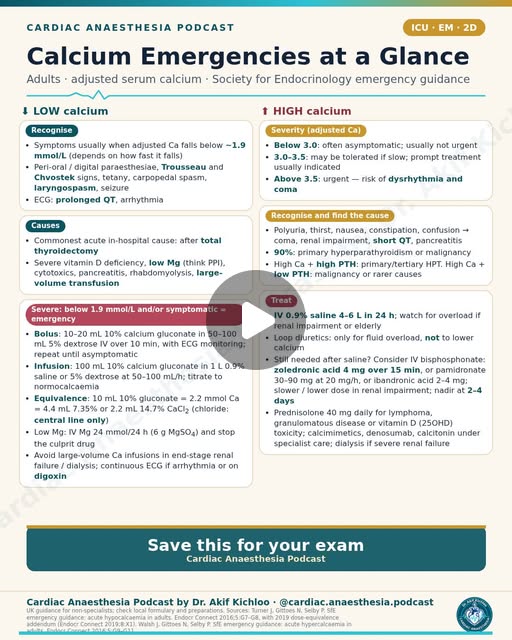
← Back to posts
← Back to posts

Aortic Regurgitation 3/5: Intervention Criteria (ESC 2021 + ACC/AHA 2020)

View on Instagram

Aortic Regurgitation 3/5: Intervention Criteria (ESC 2021 + ACC/AHA 2020)

Historical AR surgical classes, serial ventricular change, aortic thresholds and current European updates.

Understanding the post

  • The historical table reflects ESC 2021/ACC-AHA 2020. The ESC 2021 low-risk early-surgery option is class IIb C, not IIa. Reference 1.
  • The ACC/AHA IIb option is not triggered by one isolated LVEDD >65 mm measurement. In a low-risk asymptomatic patient with severe AR and preserved EF, it concerns progressive serial enlargement into that range or a progressive fall of EF into the low-normal range on serial studies. Reference 2.
  • Aortic thresholds depend on the aortic diagnosis, genetic syndrome, body size, growth and whether valve surgery is already planned. A single 45, 50 or 55 mm threshold is therefore not a universal rule. Reference 1; Reference 3.

Newer guidance

  • ESC/EACTS 2025 retains main severe-AR triggers and broadens the selected low-risk early-surgery discussion to LVESDi >22 mm/m², LVESVi >45 mL/m² or LVEF ≤55% (IIb B). It also supports expert-centre repair in suitable patients and allows selected transcatheter treatment when surgery is unsuitable. These options depend on the guideline’s complete eligibility conditions, alongside the 2024 aortic guidance. Reference 3; Reference 4.

Sources named in the post or series

  1. Vahanian A, Beyersdorf F, Praz F, et al. 2021 ESC/EACTS Guidelines for the management of valvular heart disease. Eur Heart J. 2022;43:561–632. DOI: 10.1093/eurheartj/ehab395. Historical valve-disease assessment and intervention recommendations. Sections 4 (AR), 7 (MS), 8 (TR); relevant recommendation tables.
  2. Otto CM, Nishimura RA, Bonow RO, et al. 2020 ACC/AHA Guideline for the Management of Patients With Valvular Heart Disease. J Am Coll Cardiol. 2021;77:e25–e197. DOI: 10.1016/j.jacc.2020.11.018. Valve staging, integrated assessment and US intervention criteria. AR and rheumatic MS sections; official guideline slide set.

Additional supporting sources

  1. Praz F, Borger MA, et al. 2025 ESC/EACTS Guidelines for the management of valvular heart disease. DOI: 10.1093/eurheartj/ehaf194. Current European valve intervention recommendations. AR, MS, and TR recommendation tables.
  2. Mazzolai L, Teixido-Tura G, Lanzi S, et al. 2024 ESC Guidelines for the management of peripheral arterial and aortic diseases. DOI: 10.1093/eurheartj/ehae179. Disease-specific aortic surveillance and intervention context.

For education. Apply the cited sources in their clinical context and follow current local guidance; this material is not patient-specific advice.
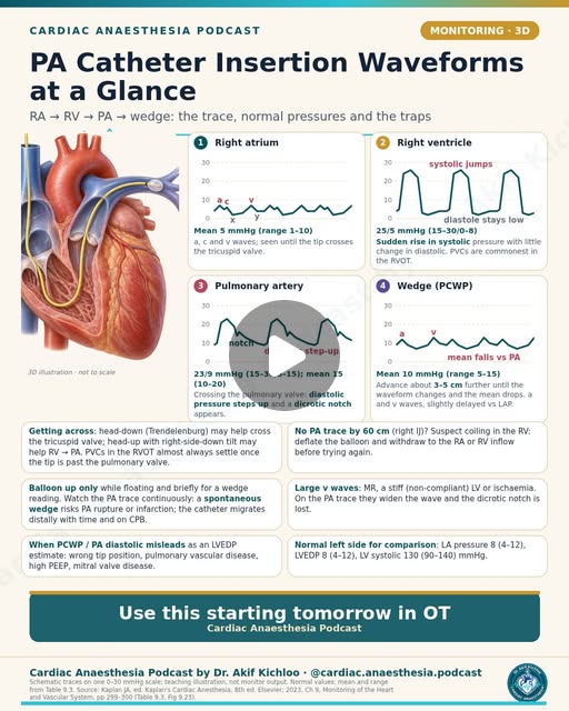
← Back to posts
← Back to posts

Aortic Regurgitation 2/5: Grading Severity on Echo

View on Instagram

Aortic Regurgitation 2/5: Grading Severity on Echo

Vena contracta, jet width, RVol/RF/EROA, PHT, aortic flow reversal and acute-versus-chronic context.

Understanding the post

  • Severe AR is an integrated diagnosis. The quantitative cut-offs are supportive criteria, not a requirement that every value be measurable and simultaneously positive. Jet width/LVOT ratio is chiefly applicable to central jets; eccentric jets need other measures. Reference 1.
  • PHT reflects the rate of aortic-to-LV pressure equalisation, not regurgitant orifice size alone. High LVEDP, altered compliance and vasodilation can shorten it. Vasodilation may do so by lowering aortic diastolic pressure, without necessarily raising LVEDP. Reference 1; Reference 4.
  • A short PHT in acute AR is not automatically an overestimate: acute AR may truly be severe. Likewise, a normal-sized LV cannot exclude severe acute AR. The meaning of aortic flow reversal depends on its sampling site, duration and velocity. Reference 1; Reference 4.

Newer guidance

  • The 2025 ESC/EACTS guideline reinforces integrated imaging and current clinical decision-making. The 2017 ASE grading criteria still require integrated interpretation rather than reliance on one new numeric cut-off. Reference 3; Reference 1.

Additional supporting sources

  1. Zoghbi WA, Adams D, Bonow RO, et al. Recommendations for Noninvasive Evaluation of Native Valvular Regurgitation. J Am Soc Echocardiogr. 2017;30:303–371. DOI: 10.1016/j.echo.2017.01.007. Integrated AR grading, quantitative measures and their limitations. AR section, Table 11, pp. 339–340.
  2. Vahanian A, Beyersdorf F, Praz F, et al. 2021 ESC/EACTS Guidelines for the management of valvular heart disease. Eur Heart J. 2022;43:561–632. DOI: 10.1093/eurheartj/ehab395. Historical valve-disease assessment and intervention recommendations. Sections 4 (AR), 7 (MS), 8 (TR); relevant recommendation tables.
  3. Praz F, Borger MA, et al. 2025 ESC/EACTS Guidelines for the management of valvular heart disease. DOI: 10.1093/eurheartj/ehaf194. Current European valve intervention recommendations. AR, MS, and TR recommendation tables.
  4. Kaplan’s Cardiac Anesthesia: Perioperative and Critical Care Management. 8th ed. Elsevier; 2023. Chapter 15: Valvular Heart Disease: Repair and Replacement. AR/MS physiology, anaesthetic goals and bypass management. Printed pp. 620–626; Boxes 15.2–15.3.

For education. Apply the cited sources in their clinical context and follow current local guidance; this material is not patient-specific advice.
← Back to posts
← Back to posts

Aortic Regurgitation 1/5: Aetiology and Pathophysiology

View on Instagram

Aortic regurgitation: causes and ventricular response

Aortic regurgitation (AR) can result from abnormal valve leaflets, abnormal aortic-root geometry, or both. Bicuspid valve disease, degeneration, rheumatic damage and infective endocarditis belong to the leaflet differential; root enlargement, connective-tissue disease, aortitis and dissection can prevent otherwise mobile cusps from meeting. Hypertension can contribute to root/aortic disease. Dissection, endocarditis and trauma can produce acute, severe AR. [1–3]

Acute and chronic AR behave differently. Abrupt severe regurgitation into a previously normal ventricle can sharply raise LV end-diastolic and pulmonary venous pressures, causing pulmonary oedema or shock before ventricular dilatation develops. A normal-sized LV or apparently normal/hyperdynamic ejection fraction therefore does not exclude severe acute AR. Acute deterioration can also occur in a ventricle already enlarged by chronic disease. The acute-versus-chronic table describes typical patterns: chronic LV filling pressure may be near normal while compensated but rises with decompensation, and a narrow pulse pressure is not mandatory in acute AR. [1–3]

Chronic AR permits gradual chamber enlargement and eccentric remodelling. Increased total stroke volume initially maintains forward output; compensation can eventually fail. The combination of volume overload, increased systolic wall stress and reduced diastolic aortic pressure helps explain the post's discussion of ventricular workload and impaired coronary perfusion. Angina need not imply an obstructive coronary lesion, but coronary disease remains an important alternative. [1–3]

Clinical qualifications

  • A wide pulse pressure, collapsing pulse and other peripheral signs are characteristic teaching findings of established chronic AR. They are neither required nor reliable exclusions, particularly in acute severe disease. [4]
  • The early diastolic murmur and an Austin Flint murmur are clinical clues, not substitutes for imaging. Murmur intensity alone does not grade severity. [4]
  • This post explains mechanism. Decisions about surveillance or intervention require an integrated assessment of symptoms, regurgitation, LV size/function and the aorta using a named current guideline. [2,3]

Original source attribution

The AR series cites the 2021 ESC/EACTS valve guideline, 2020 ACC/AHA valve guideline, and ASE/EACVI guidance. The general society attribution does not identify a particular ASE/EACVI document.

References added for this page

  1. Zoghbi WA et al. Recommendations for Noninvasive Evaluation of Native Valvular Regurgitation. J Am Soc Echocardiogr. 2017;30:303–371. Aortic-regurgitation aetiology, physiology and integrated evaluation. Guideline · DOI
  2. Victor K et al. Echocardiographic assessment of aortic regurgitation: a practical guideline from the British Society of Echocardiography. Echo Res Pract. 2025;12:3. Particularly the acute-AR section and Table 16. Full text
  3. 2025 ESC/EACTS Guidelines for the management of valvular heart disease. The contemporary European comparison for assessment and management, rather than the 2021 edition alone. Guideline
  4. Merck Manual Professional. Aortic Regurgitation. Clinical presentation, murmurs and peripheral signs; updated September 2026. Clinical review

For education. Apply the cited sources in their clinical context and follow current local guidance; this material is not patient-specific advice.
← Back to posts
← Back to posts

Coronary Anatomy: Dominance and Territories

View on Instagram

Coronary dominance and territories

Dominance describes the origin of the posterior descending artery (PDA), with posterolateral supply also relevant to co-dominance. It does not describe which main coronary artery is larger. The approximate 70% right / 10% left / 20% co-dominant split varies by population and definition. [1]

The usual territory map is a useful starting point:

  • LAD: anterior LV, anterior interventricular septum and often the apex, with a variable wrap-around course.
  • LCx: lateral/posterolateral LV, with a larger inferior territory in left dominance.
  • RCA: much of the RV and, in right dominance, inferior LV and posterior septal supply through its distal branches.

Individual branches, collateral flow and revascularisation can alter these typical territories. [1]

Nodal and papillary-muscle qualifications

  • SA and AV nodal arteries commonly arise from the RCA, with LCx variants. The quoted percentages are approximate. [1]
  • Bradycardia and AV block with inferior infarction may reflect nodal ischaemia, increased vagal activity, or both. They do not by themselves prove irreversible nodal infarction. [2]
  • The posteromedial papillary muscle usually depends on the PDA territory, whereas the anterolateral muscle usually has LAD/diagonal and LCx/obtuse-marginal contributions. This contributes to unequal ischaemic vulnerability, but supply varies. [1]

References added for this page

  1. Kesieme et al. Comprehensive Review of Coronary Artery Anatomy Relevant to Cardiac Surgery. Curr Cardiol Rev. 2025; online publication 2024. Anatomical distributions, nodal branches, dominance and papillary-muscle supply. Article · DOI
  2. Conduction Disorders in the Setting of Acute STEMI. Curr Cardiol Rev. 2021. Mechanisms and interpretation of sinus-node and AV conduction disturbances, including vagal effects in inferior infarction. Full text

For education. Apply the cited sources in their clinical context and follow current local guidance; this material is not patient-specific advice.
← Back to posts
← Back to posts

LAST After Chest Wall Blocks

View on Instagram

Sources supporting this post

How to read the post

  • The initial lipid dose. In the Association of Anaesthetists 2023 protocol, the initial 20% lipid 1.5 mL/kg dose is given over 2–3 minutes, rather than the one minute shown in the post. The post’s infusion rate of 15 mL/kg/h and cumulative maximum of 12 mL/kg agree with this protocol. The complete protocol also includes repeat doses and a faster infusion if circulation remains unstable. Association of Anaesthetists QRH June 2023
  • Why the guideline version matters. The difference in initial lipid-bolus duration reflects the guideline version: the Association withdrew its 2010 treatment sheet and directs clinicians to its current handbook, noting slower administration of the first lipid dose. Use a named, current protocol rather than combining instructions from different versions. Association management of severe local anaesthetic toxicity 2023
  • Bupivacaine dose limits. The post’s bupivacaine limit of about 2 mg/kg depends on the product and patient; it is not a guarantee of safety. This product’s information includes a 2 mg/kg limit within four hours and requires adjustment for the injection site and patient; elderly or debilitated patients need lower doses. Bupivacaine 2.5 mg/mL SmPC, emc product 11611
  • Lidocaine product differences. The post’s plain-lidocaine 3 mg/kg figure may be a conservative local limit; it is not a universal product-label maximum. This product specifies 4.5 mg/kg, capped at 300 mg, without a vessel-constricting drug, and 7 mg/kg, capped at 500 mg, with one. Lower doses may be needed. Follow the actual product and local protocol rather than increasing the dose from this comparison. Lidocaine Injection BP 1% SmPC, emc product 15149
  • Warning symptoms. Perioral tingling, tinnitus and agitation need not precede seizures, arrhythmias or cardiovascular collapse. Warning symptoms may be absent during sedation or general anaesthesia, and collapse can occur without an obvious early warning. Bupivacaine 2.5 mg/mL SmPC, emc product 11611
  • Total local-anaesthetic exposure. The reference to bupivacaine 2.5 mg/kg “in some guidance” is not a general allowance for a chest-wall block. Use the actual product’s limit, the injection site and the patient’s condition; this cited product uses 2 mg/kg within four hours. Count all local anaesthetic given, including both sides and other injections. Bupivacaine 2.5 mg/mL SmPC, emc product 11611

Guideline update: The Association explicitly replaced its 2010 treatment sheet with the Association of Anaesthetists QRH June 2023. The first lipid dose is now given over 2–3 minutes, adrenaline doses are smaller, and vasopressin is avoided; its Association management of severe local anaesthetic toxicity 2023 states these changes. RCUK Special circumstances 2025 also supports small adrenaline doses, lipid escalation and possible extracorporeal resuscitation, but does not specify the initial bolus duration. These changes describe the Association’s guidance; the details of other protocols depend on their issuing organisation and version.

For education. Apply the cited sources in their clinical context and follow current local guidance; this material is not patient-specific advice.
← Back to posts
← Back to posts

Oxyhaemoglobin Dissociation Curve Shifts

View on Instagram

Oxygen affinity is not the same as oxygen delivery

A right shift of the oxyhaemoglobin dissociation curve means lower haemoglobin–oxygen affinity at the same oxygen tension. Acidosis, increased carbon dioxide, higher temperature and increased 2,3-DPG/2,3-BPG favour this direction. A left shift means higher affinity; the opposite changes favour it. The Bohr effect concerns the influence of carbon dioxide and hydrogen ions, not every cause of a curve shift. [1]

P50 is the oxygen tension at which haemoglobin is 50% saturated. For normal adult blood under standard conditions it is approximately 26–27 mmHg, or about 3.5 kPa. Temperature and pH must be specified when comparing measurements. The post's 26.6 mmHg and 3.5 kPa are rounded equivalents, not exact interchangeable values. [1]

Important qualifications to the left-shift examples

  • Fetal haemoglobin: reduced interaction with 2,3-BPG contributes to its greater oxygen affinity. [2]
  • Stored red cells: depletion of 2,3-BPG can increase oxygen affinity. The clinical effect after transfusion cannot be inferred from a left-shift diagram alone; circulatory compensation and subsequent metabolic recovery matter. [3]
  • Carbon monoxide: occupied binding sites reduce oxygen-carrying capacity, while remaining sites hold oxygen more tightly. A normal arterial oxygen tension does not exclude dangerous CO poisoning. [1]
  • Methaemoglobinaemia: ferric haem cannot carry oxygen normally, and the remaining ferrous sites have increased oxygen affinity. It is not simply “normal haemoglobin with a left-shifted curve”; both functional oxygen capacity and unloading are impaired. [4]

During hypothermic bypass, lower temperature increases haemoglobin's oxygen affinity, but temperature also changes oxygen demand. Oxygen delivery still depends on blood flow and arterial oxygen content, including haemoglobin concentration. Neither a favourable saturation nor a curve shift alone establishes adequate tissue oxygenation.

References added for this page

  1. Relating oxygen partial pressure, saturation and content: the haemoglobin–oxygen dissociation curve. Breathe. 2015;11:194–201. Curve interpretation, P50, physiological shifts and CO. Article · DOI
  2. Physiology, Fetal Hemoglobin. StatPearls, NCBI Bookshelf. HbF and 2,3-BPG interaction. Chapter
  3. Increased Oxyhemoglobin Affinity After Transfusion of Stored Blood: Evidence for Circulatory Compensation. Primary physiological study illustrating why affinity is not a complete clinical outcome measure. Article
  4. Iolascon A et al. Recommendations for diagnosis and treatment of methemoglobinemia. Am J Hematol. 2021;96:1666–1678. Mechanism and clinical interpretation. DOI

For education. Apply the cited sources in their clinical context and follow current local guidance; this material is not patient-specific advice.
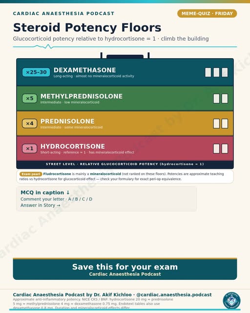
← Back to posts
← Back to posts

Wiggers Diagram: Key Cardiac Cycle Events

View on Instagram

Reading the Wiggers diagram

The diagram aligns the normal cardiac cycle. Pressure gradients open and close valves; ECG deflections do not directly trigger valve motion. [1]

  • Isovolumetric contraction: ventricular pressure rises with both inlet and outlet valves closed. AV-valve closure contributes to S1.
  • Ejection: ventricular pressure exceeds arterial pressure and the semilunar valve opens.
  • Isovolumetric relaxation: semilunar closure precedes a period with both valves closed as ventricular pressure falls.
  • Filling: the AV valve opens when ventricular pressure falls below atrial pressure; rapid filling is followed by slower filling and, in sinus rhythm, atrial contraction. [1]

Understanding valve timing and heart sounds

  • The aortic-pressure dicrotic notch is associated with aortic-valve closure. S2 includes aortic and pulmonary components (A2 and P2); a left-heart Wiggers diagram does not show their complete right-versus-left timing.
  • “All four valves are closed” is a normal-cycle teaching shorthand. Isovolumetric refers to one ventricle with its inlet and outlet valves closed; right- and left-sided valve events are not perfectly simultaneous. Significant regurgitation also changes the pressure–volume relationship. [1]
  • On a right-atrial/CVP waveform, a reflects atrial contraction after atrial depolarisation, c is associated with early ventricular systole and tricuspid-valve motion, and v reflects atrial filling while the tricuspid valve is closed. Their timing should not be transferred uncritically to a left-atrial or wedge-pressure trace. [2]
  • S3 is an early-diastolic sound associated with rapid filling. It can be physiological in younger people and some athletes; in the appropriate older or symptomatic patient it can support ventricular volume overload or dysfunction. It is not synonymous with heart failure. [3]
  • S4 is associated with atrial contraction into a relatively non-compliant ventricle. An organised atrial contraction is required, so a true S4 is not expected in atrial fibrillation. [4]

References added for this page

  1. OpenStax. Anatomy and Physiology 2e, §19.3: Cardiac Cycle. Normal phase sequence, pressures and valve events. Chapter
  2. The Jugular Venous Pressure and Pulse Contour. Clinical Methods, 3rd ed. NCBI Bookshelf. The a, c and v waves and their timing. Chapter
  3. The Third Heart Sound. Clinical Methods, 3rd ed. Physiological and pathological S3. Chapter
  4. The Fourth Heart Sound. Clinical Methods, 3rd ed. Atrial contraction and ventricular compliance. Chapter

For education. Apply the cited sources in their clinical context and follow current local guidance; this material is not patient-specific advice.

← Back to posts
← Back to posts

Stopping DOACs Before Cardiac Surgery

View on Instagram

DOAC interruption before elective cardiac surgery

The stopping interval varies with the direct oral anticoagulant and the patient. Open cardiac surgery has high bleeding risk. The interval depends on the drug, renal clearance, interacting medicines, accumulation risk and the procedure. Urgent operations and neuraxial procedures require their own pathways. [1–3]

The caption's dabigatran sequence of 48 / 72 / 96 hours at progressively lower creatinine clearance resembles the familiar EHRA high-bleeding-risk approach. However, an “EHRA/ESC” label without a year is not enough to distinguish that scheme from later cardiac-surgery-specific guidance. [1]

What newer cardiac guidance changes

The 2024 EACTS perioperative-medication guideline recommends a drug- and renal-function-dependent interval of 48–96 hours before elective open-heart surgery, without routine heparin bridging. Its Table 5 adds renal-specific detail to the caption's minimum “at least 48 hours when CrCl ≥30”: for apixaban, edoxaban and rivaroxaban, the table gives 48 hours for CrCl >50 mL/min, 72 hours for 30–50, and 96 hours for 15–30, with consideration of a drug concentration at the lowest clearance. These intervals apply only where that agent is appropriate for the renal function. [2]

Dabigatran deserves particular care because renal clearance is important and the 2024 cardiac documents do not present all intermediate renal bands identically. The EACTS medication document's dabigatran table and its narrative paragraph also differ. A single institutional protocol based on a specified guideline provides a consistent approach; intervals selected from different tables are not interchangeable. [2,3]

The caption’s “no bridging” refers to avoiding routine bridging. EHRA describes exceptional very-high-thrombotic-risk situations requiring multidisciplinary discussion; this does not justify routine LMWH or UFH substitution for a short DOAC interruption. Renal function is assessed with the measure specified by the protocol, commonly Cockcroft–Gault creatinine clearance for DOAC decisions; laboratory eGFR is not automatically interchangeable with that measure. [1]

Original source attribution

The caption attributes the approach to EHRA/ESC, without identifying a document year.

References added for this page

  1. Steffel J et al. 2021 EHRA Practical Guide on the Use of Non-Vitamin K Antagonist Oral Anticoagulants in Patients with Atrial Fibrillation. Europace. 2021;23:1612–1676. Perioperative interruption, cardiac surgery, renal assessment and exceptional bridging discussions. DOI
  2. 2024 EACTS Guidelines on perioperative medication in adult cardiac surgery. Table 5, §4.4.1 and Recommendation Table 7. DOI
  3. 2024 EACTS/EACTAIC Guidelines on patient blood management in adult cardiac surgery, in collaboration with EBCP. The cardiac-surgery interruption algorithm and renal-function qualifications. Guideline

This is an explanation of the references, not an individual patient's stopping instruction.

For education. Apply the cited sources in their clinical context and follow current local guidance; this material is not patient-specific advice.
← Back to posts
← Back to posts

Oxygen content and delivery on one page

View on Instagram

Sources supporting this post

  • Hensley, 6th ed. (2019), chapter 10, §II.C.1, PDF p.476. Supports the oxygen-content equation using 1.34 for haemoglobin-bound oxygen and 0.003 for dissolved oxygen.
  • Kaplan, 8th ed. (2023), Table 9.5 p.301 and oxygen transport pp.305–306. Supports delivery/consumption calculations and their units; its oxygen-content coefficients differ slightly.
  • 2019 EACTS/EACTA/EBCP CPB guideline, pump-flow recommendations, PDF p.21. The version named in the post. Supports flow adjustment according to arterial oxygen content, but not the exact numbered recommendation as quoted.
  • 2024 EACTS/EACTAIC/EBCP CPB guideline (published 2025), Tables 41 and 43, pp.975–976. Supports the explicit minimum indexed oxygen delivery of 280 and the separate normothermic 280–300 recommendation.
  • Ranucci et al. Ann Thorac Surg 2005;80:2213–2220. Original source for the association between low oxygen delivery during bypass and kidney injury; its identified value was 272 mL/min/m² in 1,048 coronary-operation patients.
  • de Somer et al. Crit Care 2011;15:R192. Original source linking nadir oxygen delivery below 262 mL/min/m² with kidney injury in 359 adults. These observational values help explain the evidence base; they are not universal safe boundaries.

Understanding the post

  • The post’s “DO₂i ≥280 mL/min/m² target on bypass” is an explicit recommendation in the 2024 guideline, published in 2025, to reduce stage-1 acute kidney injury (class I, level A). The 2019 pump-flow recommendation is worded more generally. 2024 CPB guideline, Table 41, p.975 / PDF p.59; 2019 CPB guideline, pump-flow recommendation, PDF p.21.
  • The ×10 factor is correct: convert oxygen content from mL/dL to mL/L, or equivalently flow from L/min to dL/min, so the units cancel. DO₂ = oxygen content × flow × 10. Kaplan, 8th ed., Table 9.5, p.301.
  • “Or transfuse to Hb9” is a worked calculation, not a universal transfusion trigger. Raising haemoglobin would raise calculated oxygen delivery, but the arithmetic alone does not establish that transfusion is the right treatment. Use the patient’s perfusion, bleeding, haemoglobin and safe bypass-flow limits together. 2024 CPB guideline, Tables 41 and 43, pp.975–976.
  • The CaO₂ and DO₂ equations use haemoglobin in g/dL, saturation as a fraction, and PaO₂ in mmHg for the 0.003 coefficient. Hensley supports the graphic’s 1.34/0.003 convention; Kaplan uses 1.39/0.0031. These small coefficient differences reflect different conventions, rather than a calculation error. Hensley, 6th ed., chapter 10, §II.C.1, PDF p.476; Kaplan, 8th ed., Table 9.5, p.301.
  • The worked examples are consistent with the stated formulas after rounding: awake delivery is about 1000 mL/min; the bypass examples are about 272, 294 and 304 mL/min/m² using unrounded oxygen content. Reporting 271 instead of 272 reflects intermediate rounding, not a different physiological result. The dissolved-oxygen values of 0.3 at PaO₂ 100 and 1.5 at 500 are correct with the 0.003 coefficient; 0.0225 is the corresponding coefficient when PaO₂ is in kPa. Hensley, chapter 10, §II.C.1, PDF p.476; direct arithmetic.
  • “VO₂ ≈250”, “O₂ER ≈25%” and “DO₂i ~500–600” are approximate resting-adult examples, not bypass targets for every temperature or clinical state. Lower SvO₂, rising lactate or altered NIRS readings should prompt assessment of the whole circulation; none identifies the cause or mandates transfusion alone. Kaplan, printed pp.305–306.
  • “Raise flow before you transfuse” is a useful blood-conservation aim when extra pump flow can be provided safely, not an absolute order. Check oxygen content, perfusion, venous return, circuit and cannula limits, and bleeding before choosing the intervention. 2024 CPB guideline, Tables 41 and 43.

Guideline update

The 2019 CPB guideline, pump-flow recommendations advises adjusting flow using arterial oxygen content but does not give the quoted 280 target in that recommendation. The 2024 guideline, published in 2025, Tables 41 and 43, pp.975–976 makes this more explicit: a minimum indexed oxygen delivery of 280 mL/min/m² to reduce stage-1 acute kidney injury is class I, level A. A separate recommendation to maintain 280–300 during normothermic bypass is class IIa, level B. It also considers how low oxygen delivery falls and how long it stays low. These are perfusion goals, not automatic transfusion triggers.

For education. Apply the cited sources in their clinical context and follow current local guidance; this material is not patient-specific advice.
← Back to posts
← Back to posts

9 arterial line waveforms to read at a glance

View on Instagram

Arterial waveforms: morphology, measurement and physiology

Waveform interpretation starts with the measurement system. Overdamping tends to lower the displayed systolic pressure and raise diastolic pressure; underdamping tends to exaggerate systolic pressure and lower diastolic pressure. Mean pressure is often less affected by these dynamic errors, but is not guaranteed accurate when levelling, zeroing, calibration or the signal itself is wrong. [1]

The fast-flush response evaluates the combined natural frequency and damping of the catheter–tubing–transducer system. Counting oscillations is a practical clue, not a complete validation test. Air, clot, kinking and compliant connections can impair the response. Tubing length or stiffness alone does not determine damping: tubing length, compliance, resistance, connections and resonance interact. [1]

Interpreting the nine patterns [6]

  • Normal: the dicrotic notch is associated with aortic-valve closure, but its appearance changes with the recording site.
  • Aortic stenosis: a small, delayed arterial upstroke can suggest severe outflow obstruction. Aortic regurgitation: a wide pulse pressure and brisk upstroke can occur in chronic significant disease. Neither contour alone grades a valve lesion.
  • HCM: a spike-and-dome contour can accompany dynamic obstruction; it is not a standalone diagnosis.
  • Pulsus alternans: alternating pulse amplitude at a regular rhythm suggests impaired LV function, after excluding rhythm and recording artefacts.
  • Pulsus paradoxus: an inspiratory systolic fall >10 mmHg describes a finding during spontaneous breathing. Tamponade and severe obstructive respiratory disease are examples, not an exhaustive or exclusive diagnosis.
  • IABP: the teaching target is inflation around aortic-valve closure and deflation before the next systolic ejection. Actual timing must be assessed on the patient's assisted/unassisted traces using the device's instructions. [2]

Pulse-pressure variation is conditional. Michard's 13% threshold came from a selected mechanically ventilated septic population, not all operating-room patients. Interpretation requires appropriate controlled ventilation without spontaneous efforts, a regular rhythm and suitable cardiopulmonary conditions. Low tidal volume, an open chest, RV dysfunction and other factors can make a single threshold misleading. Tidal volume is indexed to predicted, not actual, body weight. A grey zone exists, and fluid responsiveness does not itself mean fluid is needed or safe. [3,4]

Finally, peripheral systolic amplification and a post-bypass radial-to-central gradient can alter comparisons between sites. Transducer levelling to the intended reference point matters: a 10-cm vertical error changes pressure by approximately 7.4–7.5 mmHg, with a transducer too low reading too high. [1,5,7]

Original source attribution

The graphic cites Miller's Anesthesia, Kaplan's Cardiac Anesthesia and Michard et al., AJRCCM 2000;162:134. The textbook editions and pages are not specified. Michard's original study is listed below as [3].

Original study and references added for this page

  1. Added: Saugel B et al. How to measure blood pressure using an arterial catheter: a systematic 5-step approach. Crit Care. 2020;24:172. Dynamic response, zeroing, levelling and measurement errors. Full text
  2. Added: Evaluating the Efficacy of Intra-Aortic Balloon Pump Timing Using the Auto-Timing Mode of Operation With the Datascope CS100. J Extra Corpor Technol. 2007. IABP timing assessment; not a substitute for the current device manual. Article
  3. Original citation: Michard F et al. Relation between respiratory changes in arterial pulse pressure and fluid responsiveness in septic patients with acute circulatory failure. Am J Respir Crit Care Med. 2000;162:134–138. Study abstract · DOI
  4. Added: Prediction of fluid responsiveness: an update. Ann Intensive Care. 2016. PPV validity conditions and alternatives when conditions are not met. Full text
  5. Added: Intraoperative haemodynamic monitoring and management of adults having non-cardiac surgery: guideline of the German Society of Anaesthesiology and Intensive Care Medicine. 2024. Measurement context and contemporary monitoring recommendations. Guideline
  6. Added: The Carotid Pulse. Clinical Methods, 3rd ed. Clinical interpretation of delayed, bounding, bisferiens, alternating and respiratory-variable pulse patterns. Chapter
  7. Added primary study: Radial-to-femoral pressure gradient quantification in cardiac surgery. JTCVS Techniques. 2021;8:446–460. Evidence that radial pressure can differ materially from a more central recording during cardiac surgery. Study

Guideline update: ESICM 2025

ESICM’s 2025 circulatory-shock guideline recommends checking fluid responsiveness before further fluid in persistent shock after initial resuscitation, using applicable dynamic variables rather than static filling-pressure markers. It also supports arterial-catheter monitoring when shock does not respond to initial treatment or requires vasopressors. This is guidance for critically ill adults, not validation of one PPV threshold for every operation. ESICM 2025 guideline.

For education. Apply the cited sources in their clinical context and follow current local guidance; this material is not patient-specific advice.
← Back to posts
← Back to posts

Aortic Dissection: Stanford vs DeBakey

View on Instagram

Classifying aortic dissection without confusing anatomy and management

Stanford A involves the ascending aorta, regardless of the entry-tear location. Stanford B does not involve the ascending aorta. “B = descending only” is an incomplete shorthand: arch-involving dissections that spare the ascending aorta need a more detailed anatomical description, including the contemporary non-A non-B terminology where appropriate. [1–3]

DeBakey I originates in the ascending aorta and extends into the arch and often beyond; it does not necessarily involve the entire aorta. II is confined to the ascending aorta. III originates in the descending aorta; IIIa remains above the diaphragm, while IIIb extends below it. [1,3]

Management qualifications

  • Acute type A dissection generally requires immediate surgical evaluation and emergency repair. Initial anti-impulse treatment is supportive, not an alternative to definitive assessment.
  • Uncomplicated acute type B disease is generally managed initially with medical therapy and close surveillance. Rupture, malperfusion, progression or refractory symptoms/hypertension can require intervention; selected high-risk uncomplicated cases may also be considered for endovascular treatment. “TEVAR only if complicated” is therefore too narrow. [1–3]

Haemodynamic targets differ between guidelines. The caption's HR <60 and SBP 100–120 mmHg are one formulation among several. The 2022 ACC/AHA guideline uses HR 60–80 bpm and SBP <120 mmHg, or the lowest pressure that preserves end-organ perfusion. The 2024 ESC guideline uses HR ≤60 bpm and SBP <120 mmHg; the 2024 EACTS/STS document describes HR 60–80 and SBP 100–120 mmHg. Each set of targets belongs to its respective guideline; averaging them does not produce a guideline-supported target. [1–3]

An IV beta-blocker commonly precedes a vasodilator to avoid reflex tachycardia, but acute severe AR, bradycardia, heart block, shock or malperfusion can change the plan. Adequate organ perfusion takes priority over mechanically reaching a number. [1–3]

References added for this page

  1. 2022 ACC/AHA Guideline for the Diagnosis and Management of Aortic Disease. Acute aortic syndrome classification, initial medical management and type A/B treatment. Full text · DOI
  2. 2024 ESC Guidelines for the management of peripheral arterial and aortic diseases. Contemporary European acute-aortic-syndrome guidance. Guideline
  3. EACTS/STS Guidelines for diagnosing and treating acute and chronic syndromes of the aortic organ. Eur J Cardiothorac Surg. 2024;65:ezad426. Anatomical classification and treatment. Guideline

For education. Apply the cited sources in their clinical context and follow current local guidance; this material is not patient-specific advice.
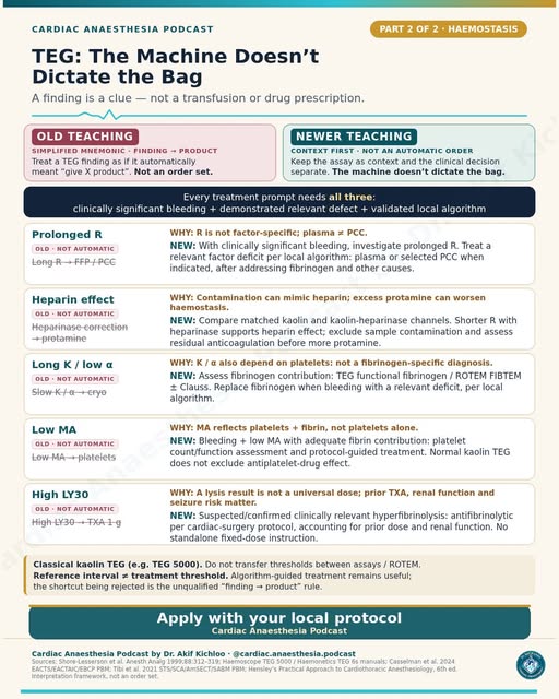
← Back to posts
← Back to posts

Modified Duke Criteria for Endocarditis

View on Instagram

Modified Duke criteria: how the versions differ

The historical modified Duke framework defines definite clinical IE by two major, one major plus three minor, or five minor criteria; possible IE by one major plus one minor, or three minor criteria. Pathological and rejected-diagnosis pathways also matter. [1,2]

Understanding the criteria

  • Fever is >38.0°C, not ≥38.0°C.
  • Positive cultures must meet the specified organism/set requirements.
  • A vegetation and new regurgitation are alternative qualifying imaging findings, not automatically two independent major criteria because they occupy separate rows.
  • New significant regurgitation on imaging is not equivalent to a changed murmur or worsening pre-existing regurgitation. [2]

The 2023 Duke-ISCVID update retains the core counting structure but expands microbiology, cardiac CT and qualifying FDG-PET/CT findings, and adds a conditional surgical-inspection criterion. Coxiella, Bartonella and Tropheryma whipplei molecular testing can qualify in specified circumstances. A positive test alone is not enough: interpretation depends on the complete tables, including definitions, timing and exclusions. [2]

The 2023 ESC guideline uses a separate diagnostic framework. Its individual criteria are not interchangeable with those of Duke-ISCVID. Neither replaces clinical judgement, appropriate microbiology, repeat imaging when indicated or Endocarditis Team assessment. [3]

References added for this page

  1. Li JS et al. Proposed modifications to the Duke criteria for the diagnosis of infective endocarditis. Clin Infect Dis. 2000;30:633–638. Historical source for the modified Duke framework named in the post; the post does not identify a specific publication. PubMed · DOI
  2. Fowler VG Jr et al. The 2023 Duke-International Society for Cardiovascular Infectious Diseases Criteria for Infective Endocarditis: Updating the Modified Duke Criteria. Clin Infect Dis. 2023;77:518–526. Tables 1 and 2 provide the complete definitions and counting rules. Article · Society-hosted full paper · DOI
  3. 2023 ESC Guidelines for the management of endocarditis. Eur Heart J. 2023;44:3948–4042. Guideline

For education. Apply the cited sources in their clinical context and follow current local guidance; this material is not patient-specific advice.
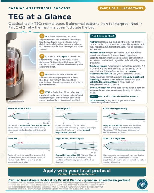
← Back to posts
← Back to posts

Type 5 MI After CABG: Fourth Universal Definition

View on Instagram

CABG-related infarction: the historical definition and the 2026 update

The post teaches the Fourth Universal Definition of Myocardial Infarction (2018). Its “type 5” terminology belongs to that 2018 framework. A troponin rise alone is not sufficient to establish a perioperative myocardial infarction. [1,2]

What the 2018 definition says

Within 48 hours of CABG, the standard type 5 definition requires cardiac troponin >10 times the assay's 99th-percentile upper reference limit (URL), from a normal baseline, plus at least one qualifying finding: new pathological Q waves, angiographically documented new graft/native-coronary occlusion, or imaging evidence of new ischaemic loss of viable myocardium/new regional wall-motion abnormality. [1]

If baseline troponin is already elevated but stable or falling, a >20% increase is required and the absolute postoperative value must still exceed 10 times URL. There is a specific exception for new pathological Q waves with elevated and rising troponin even when the 10-times threshold is not reached. A rising preoperative baseline requires special interpretation rather than automatic application of the stable-baseline rule. [1]

The post also compares PCI-related type 4a MI: under the 2018 definition, >5 times URL likewise requires qualifying new ischaemic ECG, imaging or angiographic evidence. It is not a troponin-only diagnosis, and elevated-baseline qualifications still apply. [1]

Newer definition: published August 2026

The Fifth Universal Definition replaces the numerical categories with primary, secondary and procedure-related MI. Its procedure-related framework addresses suspected coronary complications within 30 days of a cardiac procedure, with acute myocardial injury and relevant clinical/ECG features. Confirmation requires angiographic evidence of a procedural coronary complication and/or an appropriate new ischaemic imaging abnormality; both are required when the complication arises during the procedure or the procedure is performed for an acute MI. [2]

The new document gives examples of continuing postoperative biomarker elevation, including >35 times URL at 24 hours after surgery, within its diagnostic pathway. This is not a replacement rule saying “troponin >35 = MI.” The 2018 48-hour/10-times rule and the new pathway are separate frameworks. The definition used determines how clinical documentation, teaching and study outcomes are interpreted. [2]

Original source and newer reference

  1. Original named source: Thygesen K et al. Fourth Universal Definition of Myocardial Infarction (2018). The CABG/type 5 subsection, including the baseline and Q-wave qualifications. DOI
  2. Added update: Mills NL, Newby LK, Zaman S et al. Fifth Universal Definition of Myocardial Infarction. 2026. DOI · Official ESC document page · Official slide set, comparison and procedure-related diagnostic criteria

For education. Apply the cited sources in their clinical context and follow current local guidance; this material is not patient-specific advice.
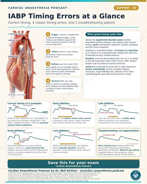
← Back to posts
← Back to posts

Heart Failure by Ejection Fraction

View on Instagram

Heart failure by EF: the guideline year now matters

The post's ≤40% / 41–49% / ≥50% categories reflect the 2021 ESC and 2022 AHA/ACC/HFSA frameworks. They are not the new 2026 ESC classification. [1–4]

In the older framework shown in the post:

  • HFrEF: LVEF ≤40%.
  • HFmrEF: LVEF 41–49%.
  • HFpEF: LVEF ≥50%.
  • HF with improved EF, as defined by the 2022 AHA/ACC/HFSA guideline: previous LVEF ≤40% with a subsequent measurement >40%. [1,2]

EF alone does not diagnose heart failure. The clinical syndrome and supporting evidence matter. In the 2022 American framework, HFmrEF and HFpEF require evidence of spontaneous or provoked increased filling pressures; structural changes, natriuretic peptides and haemodynamic/diastolic assessment help establish the diagnosis in context. A “normal EF” does not exclude HF, and a single EF measurement should not be treated as perfectly precise. [2]

Newer European update: August 2026

The 2026 ESC guideline removes HFmrEF as a separate phenotype, defining HFrEF as LVEF <50% and HFpEF as LVEF ≥50%. These European categories differ from the thresholds in the 2022 American guideline. The guideline and year therefore determine how an EF category is interpreted. [4]

Improvement in EF does not automatically mean that the underlying disease has been cured or that treatment can be stopped. The 2022 American guideline recommends continued HFrEF therapy for HF with improved EF; the 2026 ESC recommendations likewise emphasise continuation of effective therapy after improvement. [2,4]

Treatment evidence now extends beyond reduced EF. The post’s statement that “prognostic drugs are proven mainly in HFrEF” does not reflect the newer recommendations. The 2023 ESC update recommended SGLT2 inhibitors for HFmrEF/HFpEF; the 2026 ESC guideline recommends SGLT2 inhibitors and an appropriate MRA in symptomatic HF irrespective of EF. This does not make every drug class interchangeable across phenotypes or remove renal-function, potassium and other safety requirements. [3,4]

Perioperative qualification: HFpEF is more complex than “a stiff ventricle.” Tachycardia, loss of atrial contribution, congestion and inadequate filling can all matter. “Preload dependent” should not be read as an instruction to give fluid automatically: both hypovolaemia and fluid overload can be harmful. [5]

Original sources and added updates

  1. Original attribution: 2021 ESC Guidelines for the diagnosis and treatment of acute and chronic heart failure. DOI
  2. Original attribution: 2022 AHA/ACC/HFSA Guideline for the Management of Heart Failure. EF definitions, filling-pressure evidence and continued therapy after EF improvement. Official slide set
  3. Added intervening update: 2023 Focused Update of the 2021 ESC Heart Failure Guidelines. Includes SGLT2-inhibitor recommendations for HFmrEF/HFpEF within that earlier classification. DOI
  4. Added current European update: Køber L, Adamo M et al. 2026 ESC Guidelines for the management of heart failure. DOI · ESC explanation of the new EF boundaries · Official slide set
  5. Added perioperative review: Heart failure with preserved ejection fraction: implications for anaesthesia. BJA Education. 2024. Rhythm, filling and perioperative haemodynamic considerations. Article

Guideline update: June 2026 joint definition consensus

The June 2026 Second Universal Definition of Heart Failure, a joint AHA/ACC/ESC/WHF consensus with HFSA, HFA and JHFS, reaffirms stages A–D and distinguishes improvement, remission and recovery. Symptoms or function can improve without erasing the history of HF. It groups HF as reduced, preserved or improved EF without rigid universal numerical cutoffs. This definition consensus is separate from the August 2026 ESC treatment guideline and its <50%/≥50% categories. Each document has its own framework, so the categories depend on which document is being discussed. Official AHA consensus summary.

For education. Apply the cited sources in their clinical context and follow current local guidance; this material is not patient-specific advice.
← Back to posts
← Back to posts

Which ECG leads should be on your screen?

View on Instagram

ECG monitoring: useful leads, with the study context attached

Lead II is often useful for atrial activity and rhythm interpretation, while a left precordial lead such as V5 improves the chance of detecting ischaemic ST change. Neither lead is universally optimal. A five-electrode system provides limb-lead combinations and one chest-lead position; it does not simultaneously record all six standard precordial leads. [3–5]

The quoted percentages describe the study’s ST-change episodes. In London's 1988 study of 105 patients with known or suspected coronary disease undergoing non-cardiac surgery, lead II detected 33% and V5 75% of the study's intraoperative ST-change episodes; II+V5 detected 80%, and II+V4+V5 96%. The analysis assumed the identified ST changes represented true ischaemia. These figures do not mean that the same lead combinations detect 80–96% of clinical myocardial infarctions in every population. [1]

Landesberg's later major-vascular-surgery study adds a different setting: prolonged perioperative/postoperative monitoring, in which V3 was the most sensitive single lead for ischaemic episodes and V4 for infarction-associated changes. It supports attention to the monitoring period and population, rather than treating V5 as an invariant best lead. [2]

Practical qualifications

  • Standard V5 is placed at the anterior axillary line, horizontally level with V4. “Fifth intercostal space” alone is not a sufficiently precise instruction when the chest contour varies.
  • The modified CM5 configuration places the right-arm electrode near the manubrium and left-arm electrode at V5, displaying lead I. It is a bipolar modified lead, not a true unipolar V5. [5]
  • Electrode colours differ between AHA/AAMI and IEC systems. The printed RA/LA/RL/LL/V labels and the actual monitor's instructions identify electrode placement more reliably than colour alone.
  • A horizontal/downsloping ST depression threshold measured 60–80 ms after the J point is a monitoring convention, not a universal MI criterion. Interpretation includes comparison with baseline, assessment for artefact and a diagnostic 12-lead ECG when indicated. [1–4]
  • Conventional monitor filters can distort ST segments, as the post warns. Validated linear-phase digital processing can preserve diagnostic ST information at a higher low-frequency cutoff. The device's validated diagnostic/ST-analysis mode is important for this assessment. [3]

Original source attribution

The graphic names London 1988, Landesberg 2002, and AHA/ACC ECG-monitoring practice standards without an edition for the last item.

Original studies and references added for this page

  1. Original: London MJ et al. Intraoperative myocardial ischemia: localization by continuous 12-lead electrocardiography. Anesthesiology. 1988;69:232–241. Primary study abstract
  2. Original: Landesberg G et al. Perioperative myocardial ischemia and infarction: identification by continuous 12-lead electrocardiogram with online ST-segment monitoring. Anesthesiology. 2002;96:264–270. Article
  3. Added: Kligfield P et al. Recommendations for the standardization and interpretation of the electrocardiogram, Part I: the electrocardiogram and its technology. 2007. Electrode placement and signal-processing qualifications. DOI
  4. Added: Sandau KE et al. Update to Practice Standards for Electrocardiographic Monitoring in Hospital Settings. AHA scientific statement. 2017. DOI
  5. Added: ECG Monitoring in Theatre. e-SAFE/WFSA educational material. Modified leads and monitoring configurations. Chapter

For education. Apply the cited sources in their clinical context and follow current local guidance; this material is not patient-specific advice.
← Back to posts
← Back to posts

A1 to P3 on one page

View on Instagram

A1–P3: anatomy, orientation and leaflet motion

The mitral valve is commonly described in six segments: anterior A1–A3 and posterior P1–P3, with “1” lateral/anterolateral and “3” medial/posteromedial. P2 is the middle posterior scallop. The familiar surgical view looks down from the left atrium, with the aortic valve anteriorly and the left atrial appendage toward the lateral side. Image display and viewing direction must be checked before assigning a segment. [1,3,4]

Anatomical proportions are approximations. The posterior leaflet has the longer annular attachment, while the anterior leaflet is taller. The post's “one-third/two-thirds” descriptions of circumference and leaflet area should not be interpreted as fixed measurements for every heart. The anterior leaflet's relationship to the aortic valve and fibrous continuity is more useful clinically than an exact area fraction. [1,3]

Carpentier classification describes motion, not a single cause:

  • Type I: normal leaflet motion; annular enlargement or leaflet defects may produce MR.
  • Type II: excessive motion, including prolapse or flail.
  • Type IIIa: restriction in systole and diastole, classically rheumatic disease; radiation-related leaflet disease can also restrict motion. The graphic includes carcinoid disease as an example, although carcinoid valve disease predominantly affects the right heart, with left-sided involvement in selected circumstances. [6,7]
  • Type IIIb: predominantly systolic restriction, commonly from ventricular remodelling and leaflet tethering. [2,4]

Barlow disease and fibroelastic deficiency are different degenerative phenotypes, not synonyms for all type II MR. P2 is a common site of degenerative prolapse, not the necessary location of every prolapse lesion. [4]

TEE and surgical qualifications

  • Commissural, long-axis and transgastric short-axis views are complementary. The familiar P3–A2–P1 and A2–P2 teaching patterns depend on probe position and imaging plane. A nominal multiplane angle is not a guarantee that a particular scallop is being imaged; adjacent planes and 3D orientation help establish the segment where appropriate. [1]
  • The LCx, coronary sinus, conduction tissue near the right fibrous trigone/medial commissural region, and adjacent aortic valve are important structures during mitral procedures. Their precise relationship is patient-specific; the diagram is not a procedural safe-distance map. [3]
  • Posteromedial papillary-muscle supply is usually more dependent on a single coronary territory than the anterolateral muscle, but anatomical variants exist. [5]

Original source attribution

  1. Original: Hahn RT et al. Guidelines for Performing a Comprehensive Transesophageal Echocardiographic Examination. ASE/SCA. 2013. Mitral anatomy and standard imaging views. Guideline
  2. Original: Carpentier A. Cardiac valve surgery: the “French correction.” J Thorac Cardiovasc Surg. 1983;86:323–337. Historical source for the functional classification. Article

References added for this page

  1. Pozzoli A et al. Comparative Anatomy of Mitral and Tricuspid Valve: What Can the Interventionlist Learn From the Surgeon. Front Cardiovasc Med. 2018;5:80. Leaflet/annular anatomy and adjacent structures at risk. Full text
  2. Zoghbi WA et al. Recommendations for Noninvasive Evaluation of Native Valvular Regurgitation. ASE. 2017. Mechanisms, leaflet motion and integrated MR assessment. Guideline
  3. Comprehensive Review of Coronary Artery Anatomy Relevant to Cardiac Surgery. 2025. Papillary-muscle blood supply and variation. Article
  4. The assessment of mitral valve disease: a guideline from the British Society of Echocardiography. 2021. Leaflet morphology and mechanisms of restricted motion. DOI
  5. Grozinsky-Glasberg S et al. ENETS 2022 Guidance Paper for Carcinoid Syndrome and Carcinoid Heart Disease. Right- versus left-sided valve involvement. DOI

For education. Apply the cited sources in their clinical context and follow current local guidance; this material is not patient-specific advice.
← Back to posts
← Back to posts

Magnesium in Cardiac Surgery

View on Instagram

Sources supporting this post

Understanding the post

  • The post’s “Torsades de pointes: 2 g IV over 1–2 min, even if Mg is normal” is not a universal dose and administration speed. The UK algorithm gives 8 mmol magnesium over 10 minutes, about 2 g of magnesium sulfate heptahydrate, for this rhythm when associated with a prolonged QT interval. RCUK Adult tachyarrhythmia algorithm, updated March 2026 (V3) (page 1).
  • For “first-line for torsades,” the rhythm’s persistence is crucial: if this abnormal rhythm continues, give an immediate unsynchronised electrical shock. Having a pulse does not make it safe to delay the shock for magnesium. If there is no pulse, start CPR too. Magnesium can help prevent repeated episodes associated with a prolonged QT interval. AHA 2025 Adult Advanced Life Support (§16, polymorphic ventricular tachycardia).
  • The statement “Magnesium sulphate 2 g IV reduces post-op AF and ventricular arrhythmias” does not establish prevention from a single 2-g dose; results depend on the regimen and patient group. A recent trial of a different, continuous-infusion regimen found no reduction in postoperative AF; that does not prove every magnesium regimen is ineffective. Magnesium Sulfate to Prevent Perioperative AF: POMPAE randomized trial (2026) (Results and Discussion).
  • “Reflex loss around 4-5 mmol/L, then respiratory depression and heart block” is not a reliable sequence or a safe threshold to wait for. The label describes weaker reflexes above about 2 mmol/L and potentially absent reflexes with respiratory paralysis around 5 mmol/L. Kidney function, breathing, clinical signs and blood levels all matter. “Treated with IV calcium” also requires stopping magnesium and urgent supportive treatment, not calcium alone. Magnesium sulfate 50% injection DailyMed label (Precautions and Drug interactions).
  • “Even if Mg is normal” is consistent with the historical torsades series: the eight patients with measured pretreatment magnesium all had normal results. This small case series does not establish the general safety of every rapid-injection regimen. Tzivoni et al. Treatment of torsade de pointes with magnesium sulfate (1988) (12-patient case series).
  • Low magnesium can occur after CPB, but cardioplegia does not always lower magnesium. The result depends on the solution and supplementation: magnesium-containing cardioplegia supplies magnesium, yet losses and shifts can still leave levels low. Polderman & Girbes: electrolyte disorders after cardiac surgery (2004) (Methods and Results).

Guideline update

For present-day practice, RCUK’s 2025 guideline algorithm, updated March 2026, gives 8 mmol magnesium over 10 minutes for the prolonged-QT polymorphic-rhythm branch. AHA 2025 emphasizes immediate unsynchronised shock for sustained polymorphic VT, with magnesium considered for recurrent long-QT episodes. These clarify how to use the older magnesium evidence; they do not make the historical series false or establish one rapid-injection rate for every patient. RCUK Adult tachyarrhythmia algorithm, updated March 2026 (V3) (page 1); AHA 2025 Adult Advanced Life Support (§16).

For education. Apply the cited sources in their clinical context and follow current local guidance; this material is not patient-specific advice.
← Back to posts
← Back to posts

Adenosine: Doses and Cautions

View on Instagram

Sources supporting this post

Understanding the post

  • “6 mg ... then 12 mg, then 12 mg again” matches the cited US label for a rapid injection into a peripheral vein, followed by a saline flush. It is not a universal international sequence: the current UK algorithm uses 6, then 12, then 18 mg. Follow one named local protocol rather than mixing them. Adenosine Injection USP, Medical Purchasing Solutions/Mylan label (Adult patients); RCUK Adult tachyarrhythmia algorithm, updated March 2026 (V3) (page 1).
  • The post’s “Start at 3 mg via a central line” and “cut the dose to about a third after heart transplant” are not universal rules. Much smaller doses may work, and treatment requires a monitored specialist protocol. AHA 2025 says 1 mg IV may be enough in either setting; it does not establish one dose for every patient. AHA 2025 Adult Advanced Life Support (§17, supporting text 2).
  • The post combines an avoidance warning with a reduced-dose suggestion for heart-transplant recipients. A transplant is not a blanket prohibition, but sensitivity and safety need special attention. The cited study tested selected healthy children and young adults with central access and pacing backup, not acutely ill patients with a fast rhythm. It does not establish a universal one-third dose. Flyer et al. Prospective Study of Adenosine ... After Heart Transplant (2017) (Methods and Discussion limitations).
  • The warning “Avoid it in asthma and pre-excited AF” remains important. Pre-excited AF means atrial fibrillation with an extra electrical pathway that can carry impulses dangerously quickly. Adenosine can make this rhythm worse. AHA 2025 Adult Advanced Life Support (§17, asthma); 2023 ACC/AHA/ACCP/HRS AF guideline (§10.6, recommendation 4).
  • Read “unmask atrial flutter” as briefly slowing conduction to make flutter waves easier to see, not as reliably converting flutter to a normal rhythm. AHA 2025 ALS (§17).

Guideline update

The US label’s peripheral-vein 6→12→12-mg sequence is a labelled regimen. RCUK’s current algorithm, updated March 2026, uses 6→12→18 mg in its appropriate regular narrow-complex pathway: this is a protocol difference, not proof that the US-label sequence was wrong. AHA 2025 states that 1 mg may suffice through a central line or after transplantation; it does not prescribe a universal replacement dose. RCUK Adult tachyarrhythmia algorithm, updated March 2026 (V3) (page 1); AHA 2025 Adult Advanced Life Support (§17, supporting text 2).

For education. Apply the cited sources in their clinical context and follow current local guidance; this material is not patient-specific advice.
← Back to posts
← Back to posts

Amiodarone in Cardiac Surgery

View on Instagram

Sources supporting this post

Understanding the post

  • The postoperative-AF dose in the post depends on the regimen being followed. ESC 2024 lists 300 mg over 30–60 minutes, followed by 900–1200 mg over 24 hours for AF; it does not support the post’s 20-minute lower limit. Patient suitability, monitoring and product instructions still matter. ESC 2024 AF guideline, tables 12–13.
  • The timing of the post’s 300-mg and 150-mg arrest doses matters: under UK guidance, 300 mg is given after three shocks and 150 mg after five shocks if the shockable rhythm persists or returns. These drugs do not replace CPR or defibrillation. RCUK Adult ALS Guidelines 2025 (Antiarrhythmic drugs).
  • The post highlights thyroid, lung and liver toxicity with long-term treatment, but these problems are not confined to prolonged use. The product information warns that severe liver injury can occur within the first 24 hours of IV treatment, and lung toxicity can also occur with IV use. Hameln amiodarone hydrochloride 50 mg/mL concentrate SmPC (§4.4).
  • Solvent-related vasodilation does not explain both the hypotension and bradycardia listed in the post. Amiodarone itself slows the heart and conduction and has blood-pressure effects; rapid IV administration can worsen circulation. Amiodarone infusion SmPC (§§4.4,5.1).
  • “Corneal deposits” are usually benign and reversible; they are different from the rare, potentially serious optic-nerve injury. New visual symptoms need assessment. Zentiva amiodarone 200-mg tablets SmPC (§4.8).

Guideline update

RCUK 2025 clarifies the arrest sequence for this page: 300 mg after three shocks and another 150 mg after five, including when the shockable rhythm returns between shocks. The original 300/150-mg amounts are correct; the shock timing supplies essential context. Its current tachyarrhythmia algorithm must not be treated as a universal postoperative-AF regimen. RCUK Adult ALS Guidelines 2025 (Antiarrhythmic drugs); RCUK Adult tachyarrhythmia algorithm, updated March 2026 (V3) (page 1).

ESC 2024 AF guidance supplies an AF-specific infusion regimen, separate from arrest doses: 300 mg over 30–60 minutes, then 900–1200 mg over 24 hours. It supports the general dose pattern in the post, while clarifying infusion speed and range; it is not a mandate to give amiodarone to every postoperative AF patient. ESC 2024, tables 12–13.

For education. Apply the cited sources in their clinical context and follow current local guidance; this material is not patient-specific advice.
← Back to posts
← Back to posts

Vasoactive Drugs: Receptor Table

View on Instagram

Vasoactive receptor tables: mechanisms rather than prescribing rules

The table is a useful shorthand for predominant actions, but receptor “plus signs” are not a quantitative comparison of clinical potency, and a zero does not prove no interaction at any concentration. Dose, disease state, endogenous sympathetic tone and concurrent drugs change the response. [1]

  • Noradrenaline/norepinephrine: predominantly vasoconstrictor alpha-adrenergic activity with clinically relevant beta-mediated cardiac effects. The table's beta-2 “0” should not be read as proof of literally no beta-2 activity; published qualitative receptor tables differ. [1,2]
  • Adrenaline/epinephrine: alpha- and beta-adrenergic activity; the balance changes with dose and physiological context. [1]
  • Dobutamine: predominantly beta-mediated inotropic effects, with additional beta-2 and alpha effects. It is not a pure beta-1 agonist, and its net vascular effect is not identical in every patient. [1]
  • Phenylephrine: commonly described clinically as a selective alpha-1 agonist. “Pure alpha” is a therapeutic shorthand rather than a statement about every possible molecular effect. [1]
  • Vasopressin: vascular V1-receptor activation mediates vasoconstriction; the drug also has V2-mediated antidiuretic activity. “V1 only” is incomplete. [3]
  • Milrinone: inhibits phosphodiesterase-3, increasing intracellular cyclic AMP through a non-adrenergic mechanism. It is not a beta-receptor agonist. The chart’s “bypasses receptors” shorthand refers to an action that does not depend on direct beta-receptor stimulation. Its mechanism can remain useful during beta-blockade, but that does not guarantee a safe or adequate response in every patient. [1,4]
  • Dopamine: dopaminergic, beta and alpha effects overlap as exposure rises. The familiar low/middle/high dose bands are approximate, not sharp receptor switches, and a “renal-dose” band should not be interpreted as evidence of renal protection. [1]

A receptor chart alone cannot determine drug selection or dose. Inotropy, vascular resistance, arrhythmia risk, myocardial oxygen demand, blood pressure, organ perfusion and clearance must be considered together.

References added for this page

  1. Overgaard CB, Džavík V. Inotropes and vasopressors: review of physiology and clinical use in cardiovascular disease. Circulation. 2008;118:1047–1056. Mechanistic Table 1 and accompanying discussion; its older therapeutic recommendations should not be mistaken for a current treatment guideline. DOI · Article PDF
  2. Norepinephrine prescribing information. FDA, 2026 label. Mechanism and haemodynamic effects. Label
  3. Vasopressin injection prescribing information, §12.1. V1 and V2 effects. DailyMed
  4. Milrinone lactate injection prescribing information. Mechanism, haemodynamic effects and safety information. DailyMed

For education. Apply the cited sources in their clinical context and follow current local guidance; this material is not patient-specific advice.
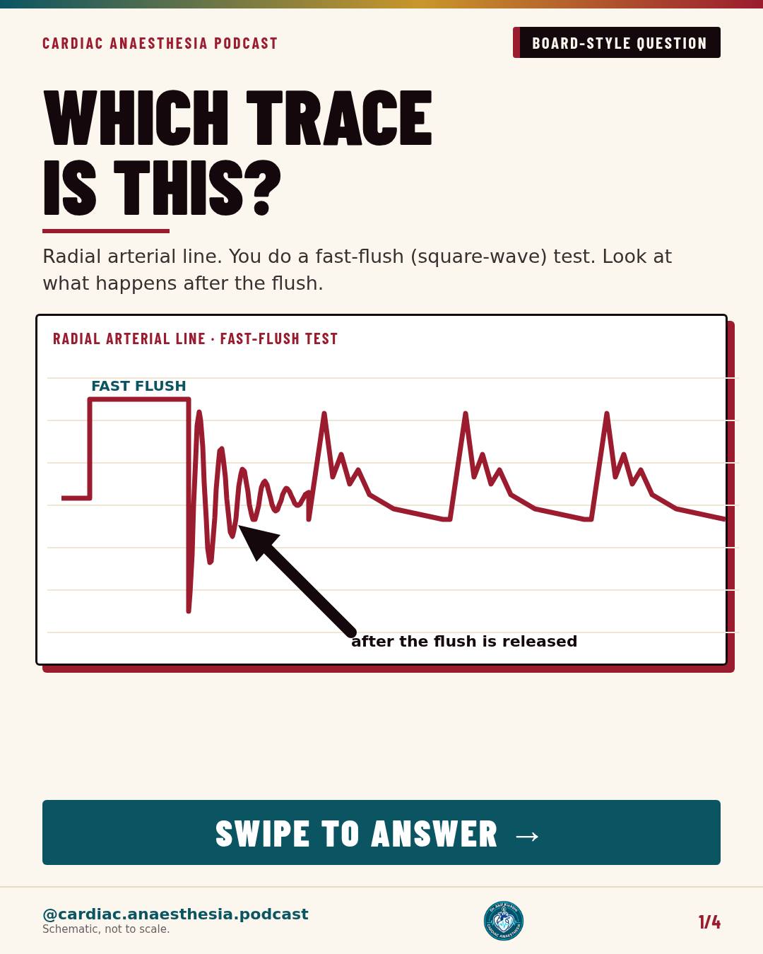
← Back to posts
← Back to posts

Why inhaled NO drops PVR without dropping SVR

View on Instagram

Inhaled nitric oxide: selective pulmonary action, with important limits

Inhaled NO reaches ventilated lung units, activates soluble guanylate cyclase in vascular smooth muscle and increases cyclic GMP, producing pulmonary vasodilatation. Rapid binding/inactivation by haemoglobin limits its systemic vasodilator effect. Preferential action in ventilated regions can improve ventilation–perfusion matching. The post’s “no effect on SVR” describes a usually limited direct systemic effect; the patient's systemic pressure still depends on ventricular function and the clinical situation. [1,2]

Dose and licence are context-specific. A perioperative adult range such as 5–20 ppm is a protocol example, not a universal approved regimen. In the US, INOmax's labelled indication and 20-ppm recommendation concern term/near-term neonates with hypoxic respiratory failure associated with pulmonary hypertension; adult cardiac-surgery use is off-label there. In the EU, INOmax also has an authorised peri-/postoperative cardiac-surgery pulmonary-hypertension indication in adults and children. The relevant product information and local delivery protocol therefore matter. [2,3]

Mechanism and safety qualifications

  • Oxyhaemoglobin reacts predominantly to form methaemoglobin and nitrate; nitrosylhaemoglobin is an intermediate associated with deoxygenated haemoglobin, not a mandatory single sequence for all NO uptake. [2]
  • Safe delivery requires an approved system with validated circuit positioning and NO/NO2 monitoring, with methaemoglobin checked by co-oximetry. The US label's 3-ppm NO2 action threshold is not a universal acceptable concentration. [2,3]
  • Withdrawal must be stepwise and monitored. Tapering reduces, but does not eliminate, rebound risk; increasing FiO2 alone is insufficient. The post’s metHb treatment shorthand does not justify an unplanned abrupt shutdown or an automatic methylene-blue prescription. [2,3,6]
  • The US label contraindicates use in neonates dependent on right-to-left shunting. In LV dysfunction, pulmonary vasodilatation can raise left-sided filling pressure and cause pulmonary oedema; this is not an uncomplicated rescue drug for every form of pulmonary hypertension. [2,3]

Vasoreactivity testing and perioperative RV support have different purposes. Acute vasoreactivity testing for identifying calcium-channel-blocker candidates is recommended in selected idiopathic, heritable or drug-associated PAH, not all forms of pulmonary hypertension. The 2022 ESC/ERS positive-response definition is an mPAP fall ≥10 mmHg to ≤40 mmHg, with unchanged or increased cardiac output. That diagnostic test is different from perioperative RV support. [4]

A 2023 randomised comparison found similar RV-failure outcomes with inhaled epoprostenol and iNO in adults undergoing heart transplantation or LVAD implantation. It does not establish equivalence for every cardiac operation, nor prove routine iNO improves survival after all bypass surgery. [5]

Original source attribution

The graphic cites INOmax US prescribing information, the 2022 ESC/ERS pulmonary-hypertension guideline, and Griffiths & Evans, NEJM 2005.

Original references and added updates

  1. Original: Griffiths MJD, Evans TW. Inhaled Nitric Oxide Therapy in Adults. N Engl J Med. 2005;353:2683–2695. Mechanism and adult clinical context. DOI
  2. Original source family, specified label: INOmax US prescribing information, revised January 2023. Indication, dosing, monitoring and rebound warnings. FDA label
  3. Added jurisdictional comparison: European Medicines Agency. INOmax EPAR and product information. Adult and paediatric cardiac-surgery indication; product information updated May 2025. EMA · Product information
  4. Original: 2022 ESC/ERS Guidelines for the diagnosis and treatment of pulmonary hypertension. Vasoreactivity testing and its restricted indications. DOI
  5. Added newer trial: Ghadimi K et al. Inhaled Epoprostenol Compared With Nitric Oxide for Right Ventricular Support After Major Cardiac Surgery. Circulation. 2023. INSPIRE-FLO trial. Article
  6. Added safety context: Iolascon A et al. Recommendations for diagnosis and treatment of methemoglobinemia. Am J Hematol. 2021. DOI

For education. Apply the cited sources in their clinical context and follow current local guidance; this material is not patient-specific advice.

← Back to posts
← Back to posts

Four CCS classes, exact wording — the pre-op and viva staple

View on Instagram

CCS angina classes: a concise summary, not an exact quotation

The Canadian Cardiovascular Society classification describes the activity threshold for angina, not coronary anatomy, EF, or NYHA breathlessness class. Despite its “exact wording” title, the graphic is an abridged teaching summary rather than a complete verbatim transcription of the original definitions. [1,3]

  • Class I: ordinary activity does not provoke angina; symptoms occur with strenuous, rapid or prolonged exertion.
  • Class II: slight limitation of ordinary activity, such as faster walking or stair climbing, uphill effort, or activity affected by cold, wind, meals, emotional stress or the first hours after waking; the full description also includes walking more than two blocks or climbing more than one flight under normal conditions.
  • Class III: marked limitation; the conventional examples include walking one or two level blocks or climbing one flight of stairs at a normal pace under normal conditions.
  • Class IV: inability to undertake physical activity without discomfort; angina may occur at rest. Rest angina is possible, not mandatory, in the general class description. [1,3]

Preoperative and risk-score qualifications

  • CCS class can make symptom history more reproducible, but it does not independently establish how recent the symptoms are, the severity of coronary stenosis, or whether an acute coronary syndrome is present.
  • Class III or IV is not automatically “recent ischaemia” and does not replace a complete perioperative risk assessment. Functional limitation, symptom trajectory, operation type and other clinical variables matter.
  • EuroSCORE II uses a CCS class IV variable, defined in its source paper as inability to perform activity without angina or angina at rest. It is not a graded I–IV score input. An intravenous-nitrate requirement belongs to a different or older risk-score definition, not this EuroSCORE II variable. [2]
  • New, worsening or ongoing rest symptoms require urgent clinical assessment for ACS and other dangerous causes. However, a stable class label alone is not a diagnosis of unstable angina. The descriptive class and the assessment of clinical stability answer different questions. [3]

Original sources

  1. Campeau L. Grading of angina pectoris. Circulation. 1976;54:522–523. Historical source of the CCS classification. PubMed
  2. Nashef SAM et al. EuroSCORE II. Eur J Cardiothorac Surg. 2012;41:734–744. The model's CCS-IV variable and its definition. Official paper · DOI

Reference added for this page

  1. 2024 ESC Guidelines for the management of chronic coronary syndromes. CCS symptom grading in the context of evaluating chronic coronary syndromes and changing symptoms. Guideline

For education. Apply the cited sources in their clinical context and follow current local guidance; this material is not patient-specific advice.
← Back to posts
← Back to posts

Aortic Stenosis 5/5: Intra-op TEE and Post-bypass Pearls

View on Instagram

Sources supporting this post

How to read the post

Choosing views and interpreting measurements

The post locates planimetry in the ME AV short-axis view, LVOT measurement in ME AV long-axis and gradient measurement in deep transgastric views. These are starting views for the examination. In transoesophageal echocardiography (TEE), the mid-oesophageal aortic-valve views show anatomy; transgastric views align Doppler with blood flow. Calcification can obscure the opening, and poor alignment can underestimate velocity and gradient. Interpretation combines the measurements rather than relying on planimetry alone. ASE/SCA 2013, pp945–946

Myocardial protection in a thickened ventricle

A thickened left ventricle needs carefully planned, adequately distributed myocardial protection. The post’s word “generous” does not mean an unspecified larger dose: solution, delivery route, pressure, temperature and repeat dosing must fit the operation and coronary anatomy. CPB guideline, pp26–27

Dynamic obstruction after valve replacement

A hypertrophied septum can cause dynamic LVOT obstruction after AVR. The listed options include volume, slowing the heart, stopping inotropes and phenylephrine. This describes treatment of confirmed dynamic obstruction of the left-ventricular outflow tract after aortic valve replacement, not all post-bypass hypotension. TEE helps identify the mechanism. Treatment involves optimising filling, reducing aggravating inotropic stimulation and considering carefully titrated rate/contractility reduction and an appropriate vasoconstrictor. An unstable patient may need a different sequence or surgical correction. Makhija et al., Management

Complications after TAVI

Coronary obstruction, annular rupture, heart block and paravalvular leak are the complications highlighted. These are recognized complications of transcatheter aortic valve implantation. Assessment combines imaging with electrocardiography, pressure/flow assessment and clinical findings. A leak around the prosthesis, coronary obstruction, annular injury and conduction block require different responses. ASE 2024, pp11–12; manufacturer safety information

Assessment after replacement includes leaflet motion, seating, leakage, ventricular function and the measured hemodynamic profile. A high gradient can reflect increased flow, a prosthesis too small for the patient, obstruction below the valve, pressure recovery or true prosthetic dysfunction. The thickened ventricle remodels over time; that change is not instantaneous. ASE/SCA/STS 2020, p707; ASE 2024, p23; Kaplan, pp611–612

Assessment after AVR

The post highlights gradients, paravalvular leak, coronary flow and new wall-motion defects. Comparison with the pre-bypass examination also includes prosthetic seating/motion, ventricular function and the aortic root. A new regional wall-motion abnormality can suggest compromised coronary perfusion but is not specific for coronary obstruction. ASE/SCA/STS 2020, §3.2.4, p707; ASE 2024, pp11–12

Gradient relief and ventricular remodelling

The post contrasts prompt gradient reduction with LVH regression over months. Successful relief of obstruction generally reduces the valve gradient promptly; regression of left-ventricular hypertrophy (LVH) and improvement in relaxation take longer and may be incomplete. Appropriate filling remains important, but this does not mean fluid is automatically needed. A residual high gradient needs interpretation for flow, prosthesis size, outflow obstruction and valve function. Kaplan, pp611–612; ASE 2024, p23

Recognising a new post-TAVI problem

Sudden hypotension, ST change and new RWMA are the warning signs listed for coronary obstruction. These are warning clues to coronary obstruction after TAVI, not a diagnostic combination unique to it. RWMA means regional wall-motion abnormality; ST change refers to the electrocardiogram. The table’s new effusion/tamponade warning should prompt urgent assessment for annular or other cardiac injury. Complete heart block requires rhythm confirmation and prompt pacing assessment; bradycardia alone is not diagnostic. A paravalvular jet arises around the valve frame and must be assessed from multiple views. Savvoulidis et al., immediate post-deployment assessment; Evolut safety information

Guideline update: The 2024 ASE guideline replaces the 2009 prosthetic-valve guideline and strengthens integrated assessment using valve type/size, flow-dependent and less flow-dependent measures, morphology and comparison with a baseline study. It complements the 2013 examination views. ESC/EACTS 2025 retains detailed CT-based TAVI planning and Heart Team selection. A falling gradient alone does not establish a successful, normally functioning replacement.

For education. Apply the cited sources in their clinical context and follow current local guidance; this material is not patient-specific advice.
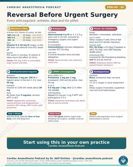
← Back to posts
← Back to posts

Aortic Stenosis 4/5: Anaesthetic Goals and Haemodynamics

View on Instagram

Sources supporting this post

Understanding the post

  • The post’s statements “CPR rarely works in severe AS” and “chest compressions are often ineffective, so prevention is everything” are unsupported. Severe AS alone is not a reason to regard CPR as futile. If cardiac arrest occurs, start immediate high-quality CPR and give electrical shocks when indicated; severe aortic stenosis is not a reason to withhold them. RCUK Adult ALS Guidelines 2025 (Key points and treatment of in-hospital cardiac arrest).
  • “Sinus; cardiovert new AF early” depends on haemodynamic stability. When AF is causing unstable blood pressure or circulation, urgent synchronised electrical cardioversion is indicated to restore the normal rhythm. In a stable patient, choose treatment individually and assess clot and stroke risk before planned cardioversion. 2023 ACC/AHA/ACCP/HRS AF guideline (§8.2.1–8.2.2).
  • “Keep the heart rate low-normal ... keep it full, and keep the SVR up” means avoid very fast or very slow rates, maintain enough filling, and prevent low blood pressure. It does not mean give unlimited fluid or deliberately raise pressure in every patient. Tailor treatment to the patient’s circulation. Hensley’s Practical Approach to Cardiothoracic Anesthesia, 6th ed (2019), chapter 11, section V.E (PDF pages 532–533).
  • The post contrasts single-shot spinal with slowly titrated epidural or CSE, but neither an absolute ban nor a blanket assurance of safety follows from that distinction. These techniques may be considered in selected patients, but monitoring does not remove the risk of a dangerous blood-pressure fall. Valve severity, symptoms, blood-thinning medicines and the planned dose/technique require specialist assessment. Tabrizi et al.: neuraxial anaesthesia in AS (2024); APSF summary.

Guideline update

Alongside the 2019 textbook and 2023 AF guideline, RCUK 2025 reinforces immediate high-quality CPR and indicated defibrillation in cardiac arrest. The statement “CPR rarely works in severe AS” is unsupported; the guidance does not demonstrate a reversal of an older AS-specific resuscitation recommendation. Heart-rate goals and spinal/epidural decisions still need to be individualized. RCUK Adult ALS Guidelines 2025 (Key points; treatment of in-hospital cardiac arrest).

ESC 2024 adds a specific safety limit to “cardiovert new AF early”: in a stable patient with AF lasting more than 24 hours, cardioversion should wait for at least three weeks of therapeutic anticoagulation, or a transoesophageal echo must exclude a heart clot first. This is the ESC protocol, not a reason to delay emergency cardioversion when AF is causing unstable circulation. ESC 2024, §7.2.1 and table 15, p3355.

ASRA’s fifth-edition 2025 guidance adds a separate caution to “a slowly titrated epidural or CSE is acceptable with invasive monitoring”: neuraxial or deep plexus/peripheral catheters should not be maintained during full anticoagulation for cardiac surgery. This addresses bleeding risk, not just the blood-pressure effects of anaesthesia in AS. It is not a blanket ban on every regional technique for every operation. ASRA 2025, unfractionated-heparin recommendations.

For education. Apply the cited sources in their clinical context and follow current local guidance; this material is not patient-specific advice.
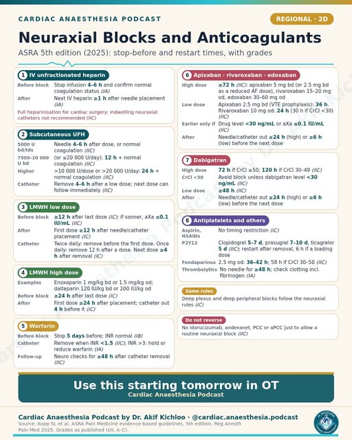
← Back to posts
← Back to posts

Aortic Stenosis 3/5: Intervention Criteria (ESC 2021 + ACC/AHA 2020)

View on Instagram

Sources supporting this post

How to read the post

Symptomatic severe disease in ESC 2021

The original European table gives Class I B to symptomatic severe high-gradient AS and to symptomatic low-flow, low-gradient AS with reduced EF and flow reserve. These are the 2021 European recommendations. AS means aortic stenosis; EF is the proportion of blood ejected by the left ventricle with each beat. In the low-flow group, the original recommendation requires symptoms, confirmed severe stenosis, reduced EF and demonstrable flow reserve. Class I means recommended; B describes the evidence level. The 2025 change is described below. ESC/EACTS 2021, p26

Asymptomatic indications in ESC 2021

The table lists LVEF <50% as Class I B and symptoms on exercise testing as Class I C. These indications apply to severe aortic stenosis; reduced left-ventricular ejection fraction must be without another cause. Exercise-induced symptoms are themselves an indication for intervention in the 2021 guideline. These are not thresholds for patients with any degree of stenosis. ESC/EACTS 2021, p26

Reduced EF and exercise blood pressure

LVEF <55% is Class IIa B and a sustained exercise BP fall >20 mmHg is Class IIa C in the 2021 European table. These 2021 European Class IIa recommendations mean intervention should be considered. The reduced ejection fraction must be without another cause. The exercise threshold is specific to the European guideline: the 2020 US recommendation uses a systolic fall of at least 10 mmHg from baseline to peak exercise, or reduced exercise tolerance, in an apparently asymptomatic patient with severe disease and low surgical risk. ESC 2021, p26; ACC/AHA 2020, slide 61

Additional high-risk features in ESC 2021

For LVEF >55% and low risk, the post lists very severe AS (MG ≥60 mmHg or Vmax >5 m/s), rapid progression with severe calcification or BNP >3× normal as Class IIa B. The 2021 European recommendation also requires a normal exercise test. Mean gradient (MG) and peak jet velocity (Vmax) are Doppler measurements. Rapid progression means a velocity increase of at least 0.3 m/s/year with severe calcification. The B-type natriuretic peptide (BNP) threshold is more than three times the age- and sex-adjusted normal range, repeated and without another explanation. ESC/EACTS 2021, p26

The historical European procedure-choice thresholds

ESC 2021 lists SAVR below age 75 with low risk (STS/EuroSCORE II <4%) as Class I B; TAVI at age ≥75 or high risk >8% is Class I A. This is the historical 2021 European framework. Surgical aortic valve replacement (SAVR) and transcatheter implantation (TAVI) still require assessment of anatomy, vascular access, operative suitability and expected benefit. STS-PROM and EuroSCORE II are surgical-risk estimates; age or a score alone does not select the procedure. ESC/EACTS 2021, pp24–27

Understanding the US disease stages

The post’s US table gives AVR for D1 symptoms Class I A; C2 LVEF <50%, other cardiac surgery, D2 and D3 carry Class I B-NR. These are US disease stages: D1 is symptomatic high-gradient severe stenosis; D2 and D3 are symptomatic low-flow, low-gradient severe stenosis with reduced and preserved ejection fraction, respectively. In D3, stenosis must be the most likely cause of symptoms. C2 is asymptomatic severe stenosis with reduced ejection fraction. The “other cardiac surgery” Class I row refers to severe, not moderate, stenosis. B-NR means nonrandomized evidence. ACC/AHA 2020, slides 59–60

Age and life expectancy in ACC/AHA 2020

The age summary is SAVR below 65, shared decision-making at 65–80 and transfemoral TAVI above 80, all Class I A within the applicable conditions. The 2020 US framework also uses life expectancy: surgery is preferred below 65 or with expected survival beyond 20 years. The 65–80 and over-80 recommendations refer to symptomatic severe disease with anatomy suitable for transfemoral TAVI, meaning access through the femoral artery. A life expectancy below 10 years can favour transfemoral TAVI at a younger age. The separate high/prohibitive-risk recommendation also requires symptomatic severe disease, predicted survival beyond 12 months and acceptable quality of life. ACC/AHA 2020, slides 67–69

The US very-severe threshold is Vmax ≥5 m/s; the European 2021 wording is >5 m/s or mean gradient ≥60 mmHg. These boundaries belong to their respective guidelines and are not interchangeable. US recommendations based on high BNP, rapid progression, abnormal exercise testing or very severe stenosis also require the relevant asymptomatic severe-disease and low-surgical-risk conditions. ACC/AHA 2020, slides 61–62

Low-flow, low-gradient disease with preserved EF

The preserved-EF, low-flow, low-gradient entry concerns consideration of intervention after careful confirmation of severe AS. These rows apply to symptomatic patients. In the 2021 European guideline, both this preserved-ejection-fraction row and the adjacent reduced-ejection-fraction row without flow reserve are Class IIa C. For the latter, CT calcium scoring can support confirmation of severe disease. Neither row means that a small valve area alone establishes severity. ESC/EACTS 2021, p26

When intervention is unlikely to help

The severe-comorbidity entry addresses intervention unlikely to improve quality of life or survival beyond one year. This correctly reflects the 2021 Class III C recommendation about lack of expected benefit. QoL means quality of life. It requires an individualized estimate of benefit, not exclusion merely because someone has a comorbidity. ESC/EACTS 2021, p26

Heart Valve Centres and the Heart Team

The original European Class I C recommendations call for an experienced centre with on-site interventional cardiology and cardiac surgery, and a multidisciplinary recommendation discussed with the patient. The remaining-patient choice of surgery or TAVI is Class I B. Non-transfemoral TAVI was Class IIb C for patients both inoperable and unsuitable for transfemoral access. These conditions accompany the age/risk rows. ESC/EACTS 2021, pp26–27

Valve replacement during other cardiac surgery

The moderate-AS entry concerns CABG, ascending-aorta surgery or other valve surgery, with Heart Team assessment. The post correctly preserves the different original recommendations: European 2021 Class IIa C for moderate disease during these operations, versus US 2020 Class IIb C-EO during other cardiac surgery. CABG is coronary artery bypass grafting; C-EO means expert opinion. European surgery for severe stenosis during these operations is Class I C. The moderate-disease rows do not make replacement an automatic requirement. ESC/EACTS 2021, pp26–27; ACC/AHA 2020, slide 63

Balloon valvotomy as a bridge

The post describes bridging to SAVR/TAVI in instability or before urgent high-risk non-cardiac surgery. This is a selected bridging option, European 2021 Class IIb C, not definitive routine treatment. The urgent-surgery branch requires severe stenosis and feasibility; procedural risk requires Heart Team discussion. ESC/EACTS 2021, pp26–27

A progressive fall in ejection fraction

The C1 entry concerns progressive LVEF decline to <60% on at least three serial imaging studies. The 2020 US Class IIb B-NR row requires asymptomatic severe high-gradient stenosis and a progressive decrease over at least three studies. It is not a one-off EF below 60% trigger. Unlike the preceding Class IIa rows, this particular original recommendation does not explicitly require low surgical risk, although it sits under that shared heading in the post. The rapid-progression Class IIa row also specifies high-gradient severe disease. ACC/AHA 2020, slides 62–63

High or prohibitive surgical risk

The TAVI entry applies at any age when predicted post-TAVI survival exceeds 12 months with acceptable quality of life, within the disease criteria below. “At any age” applies to symptomatic severe aortic stenosis in the original US Class I A recommendation. The predicted survival beyond 12 months and acceptable quality-of-life conditions also apply. The US age-based procedure-choice table is also framed for patients in whom a bioprosthetic replacement is appropriate and an indication for valve replacement is already established. ACC/AHA 2020, slides 67–69

Guideline update: The post’s tables describe the original 2021 European and 2020 US recommendations. In ESC/EACTS 2025, Recommendation Table 4, TAVI is recommended from age 70 for a three-cusp aortic valve when anatomy and transfemoral access are suitable, while surgery is recommended below 70 when surgical risk is low; other cases require Heart Team assessment. Intervention should now be considered for low-procedural-risk, asymptomatic severe high-gradient stenosis with ejection fraction at least 50%, as an alternative to close active surveillance (Class IIa A; normal exercise testing if feasible). In symptomatic low-flow, low-gradient disease with reduced ejection fraction, the Class I recommendation now requires careful confirmation of severe stenosis rather than requiring flow reserve. The preserved-ejection-fraction symptomatic low-flow recommendation changes from IIa C to IIa B, and non-transfemoral TAVI for patients unsuitable for both surgery and transfemoral access changes from IIb C to IIa B.

For education. Apply the cited sources in their clinical context and follow current local guidance; this material is not patient-specific advice.
← Back to posts
← Back to posts

Aortic Stenosis 2/5: Grading Severity on Echo

View on Instagram

Sources supporting this post

How to read the post

The 2017 severe-stenosis thresholds

The post gives Vmax ≥4 m/s, mean gradient ≥40 mmHg, AVA <1.0 cm² (indexed <0.6 cm²/m²) and velocity ratio <0.25. These reproduce the 2017 echocardiographic severity cutoffs. Vmax is peak velocity, AVA is aortic valve area, and the velocity ratio compares outflow-tract with valve velocity. The measurements should agree, but low flow can produce severe stenosis without a high gradient. Severity is assessed from the combined findings, without requiring every threshold to be met or relying on one measurement. EACVI/ASE 2017, Table 3

Why the LVOT diameter matters

The diameter is squared in the area calculation, making the post’s 1-mm example a useful warning about measurement error. LVOT diameter is a major potential source of error; a particular 1 mm error is not established as the commonest measured cause. The guideline describes diameter as the greatest potential error source; Doppler sampling, beam alignment and assuming a circular outflow tract also matter. EACVI/ASE 2017, p381; ASE/SCA 2013, p946

When area and gradient disagree

Assessment starts with rechecking the images and measurements, blood pressure and rhythm. Stroke volume indexed to body surface area then helps classify the flow pattern. A small calculated valve area with a low gradient is not automatically severe stenosis. ESC/EACTS 2025, Figure 6

Low-flow assessment with dobutamine and CT

The post pairs classical low flow (EF <50%) with low-dose dobutamine echo and paradoxical low flow (EF ≥50%, SVi ≤35 mL/m²) with CT calcium scoring (men ≥2000, women ≥1200 AU). This is a useful outline, not a rigid either/or rule. Ejection fraction below 50% alone does not establish classical low-flow, low-gradient stenosis: a small valve area, low gradient and reduced indexed stroke volume must also be present. Dobutamine stress echocardiography and computed tomography (CT) calcium scoring can be complementary. In the 2017 source, the quoted calcium values make severe stenosis likely; they are not stand-alone proof. AU means Agatston units. EACVI/ASE 2017, Table 5; ESC/EACTS 2025, pp29–30

The continuity equation

AVA = LVOT area × LVOT VTI / AV VTI. This correctly expresses conservation of forward stroke volume. VTI is the velocity–time integral, the distance represented by the Doppler trace. Accurate calculation requires an outflow-tract area measurement matched to its pulsed-Doppler sampling level, and continuous-wave Doppler aligned with the highest-velocity valve jet. Misalignment underestimates velocity and the calculated gradient. Kaplan, pp612–613; ASE/SCA 2013, p946

Reading rounded severity ranges

The moderate-velocity row is printed as “3.0–3.9.” The moderate-velocity entry and the “20–39” mean-gradient entry are shorthand bins. For continuous measurements, these correspond to 3.0 to <4.0 m/s and 20 to <40 mmHg, including values such as 3.95 or 39.5. The 2017 source table prints 3.0–4.0 and 20–40, with ≥4.0 and ≥40 in the severe column. The remaining mild/moderate entries match that table: valve area >1.5 / 1.0–1.5 cm²; indexed area >0.85 / 0.60–0.85 cm²/m²; velocity ratio >0.50 / 0.25–0.50. The source table’s mild peak-velocity range of 2.6–2.9 m/s is also rounded shorthand: for continuous measurements, this corresponds to 2.6 to <3.0 m/s, including a value such as 2.95. Mild mean gradient is <20 mmHg. The parameters are interpreted together. EACVI/ASE 2017, Table 3, p383

Normal-flow, low-gradient findings

The post defines normal flow here as SVi >35 mL/m². Rechecking measurements is appropriate for every discordant study, not only this group. Normal-flow low-gradient stenosis is less likely to be severe, but a persistent discrepancy is not resolved by flow alone. Current guidance concerns patients with preserved ejection fraction and considers blood pressure, slow heart rate, symptoms and, when needed, CT calcium scoring. ESC/EACTS 2025, Figure 6 and footnote c

Interpreting the dobutamine response

The listed true-severe response is MG ≥40 mmHg or Vmax ≥4 m/s with AVA ≤1.0 cm²; the listed contractile-reserve threshold is a stroke-volume rise ≥20%. This is a useful response pattern during low-dose dobutamine testing, provided flow has increased sufficiently and the measurements are reliable. Stroke volume (SV) rising by at least 20% defines flow reserve in the current algorithm. If valve area increases above 1.0 cm² as flow improves, pseudo-severe stenosis is suggested. Inadequate flow augmentation can leave the test inconclusive; it does not prove that the valve is non-severe or preclude intervention. EACVI/ASE 2017, pp384–385 and 388; ESC/EACTS 2025, Figure 6

Guideline update: The 2025 ESC/EACTS algorithm, Figure 6 uses valve area ≤1.0 cm² (indexed ≤0.6) and indexed stroke volume ≤35 mL/m². This boundary convention differs from the original 2017 table; each set of thresholds belongs to its named source. It explicitly treats CT and dobutamine testing as complementary when one is inconclusive; flow reserve is a stroke-volume increase of at least 20%, with projected valve area considered for a 10%–20% rise. Calcium scores >2000 in men and >1200 in women support severe disease, but interpretation needs particular caution in bicuspid, predominantly fibrotic and other less-calcific disease.

For education. Apply the cited sources in their clinical context and follow current local guidance; this material is not patient-specific advice.
← Back to posts
← Back to posts

Aortic Stenosis 1/5: Aetiology and Pathophysiology

View on Instagram

Sources supporting this post

How to read the post

Common causes of stenosis

Calcific degeneration, bicuspid valves and rheumatic disease are the three causes highlighted. A bicuspid valve tends to degenerate earlier than a normal three-cusp valve, but “10-20 years earlier” is a teaching approximation, not a predictable onset age for an individual. Their relative frequency also varies by population. EACVI/ASE 2017, p373; Kaplan, pp608–610

Valve area and the onset of symptoms

The post quotes a normal AVA of 3–4 cm² and symptom onset below about 1 cm². These are approximate teaching values. Kaplan and Hensley give normal aortic valve area as 2.6–3.5 cm². A value around 1 cm² is used in severity assessment, but symptoms do not start at a fixed area: flow, ventricular function, activity and other disease matter. Symptoms require direct assessment. Kaplan, p610; Hensley, Chapter 11, §V.A; ESC/EACTS 2025, §8.2

Pressure overload and ventricular filling

The post links pressure overload to concentric LVH and a stiff ventricle, with atrial contraction contributing up to 40% of filling. The mechanism is correct, and Hensley does use the “up to 40%” teaching figure. Left-ventricular hypertrophy (LVH) and impaired relaxation can make coordinated atrial contraction important for filling. The percentage is not a fixed contribution in every patient. Hensley, Chapter 11, key point 3; Kaplan, pp610–611

Why ischaemia can occur with unobstructed coronaries

Raised left-ventricular end-diastolic pressure (LVEDP), increased muscle oxygen demand and impaired coronary reserve can reduce oxygen supply to the inner heart muscle despite unobstructed major coronary arteries. Low diastolic arterial pressure makes this worse, but is not present in every patient with stenosis. Aortic diastolic pressure minus LVEDP is a useful simplified subendocardial estimate, not the entire coronary-flow model. Kaplan, p612; Goodwill et al., mechanical determinants

The historical survival mnemonic

The classic mnemonic is about five years after angina, three after syncope and two after heart failure without treatment. This is the classic historical natural-history mnemonic, not a current personal life-expectancy prediction or a safe time to wait. Hensley gives approximately 5 years after angina, 3–4 after syncope and 1–2 after heart failure; Kaplan summarizes historical symptomatic survival as 2–5 years. New symptoms with severe stenosis warrant prompt valve assessment. Hensley, Chapter 11, §V.A.2; Kaplan, p610; ESC/EACTS 2025, Recommendation Table 4

Age patterns and associated valve disease

The card associates calcific disease with age >65, bicuspid presentation with roughly 10–20 years earlier onset, and rheumatic AS with accompanying mitral disease. The ages describe broad epidemiological patterns, not diagnostic boundaries. Calcific disease becomes common with advancing age but can occur earlier. The statement about associated rheumatic mitral disease is supported by the echocardiography guideline. EACVI/ASE 2017, pp373–374; Hensley, Chapter 11, §V.A.1–2

Loss of coordinated atrial contraction

The post’s “up to 40%” filling estimate helps explain concern about AF or junctional rhythm. Loss of coordinated atrial contraction can reduce filling and output in a stiff hypertrophied ventricle. Atrial fibrillation (AF) may additionally cause an adverse rapid or irregular rate; junctional rhythm may disrupt atrioventricular timing. The vulnerability is real, but the haemodynamic effect varies rather than being uniformly severe. Hensley, Chapter 11, key point 3; Kaplan, p611

Hypotension and worsening perfusion

The illustrated cycle is lower CPP, then ischaemia, reduced contractility and lower BP. This illustrates a possible self-reinforcing deterioration, not an inevitable sequence in every patient. CPP means coronary perfusion pressure and BP means blood pressure. Reduced diastolic perfusion and myocardial ischaemia can worsen contraction, while tachycardia, low filling, rhythm disturbances and other causes may also contribute. Kaplan, pp611–612; Goodwill et al., mechanical determinants

Physical examination clues

The listed findings are a slow-rising pulse, soft or absent A2 and a late-peaking ejection-systolic murmur radiating to the carotids. These are classic findings, particularly in advanced stenosis. A2 is the aortic component of the second heart sound. They support clinical suspicion but are neither uniformly present nor sufficient to grade stenosis: severity requires confirmation with echocardiography. ACC TAVR Handbook, Chapter 8, PDF p4

Heyde syndrome

The post links gastrointestinal angiodysplasia bleeding with an acquired von Willebrand abnormality, labelled “type 2A.” In the context of aortic stenosis, this describes the recognized association with gastrointestinal bleeding from fragile abnormal vessels and acquired von Willebrand syndrome. The abnormality is acquired von Willebrand syndrome with loss of high-molecular-weight multimers, rather than inherited type 2A disease or simply a low total factor level. Not every gastrointestinal bleed in a patient with stenosis is Heyde syndrome. Vincentelli et al., 2003; Goltstein et al., Introduction

Guideline update: The physiology and historical teaching estimates remain distinct from treatment recommendations. ESC/EACTS 2025 recommends intervention for symptomatic severe stenosis and also supports considering earlier intervention in suitable low-risk, asymptomatic high-gradient severe disease. The 5–3–2 mnemonic is not a basis for postponing assessment or estimating an individual patient’s survival.

For education. Apply the cited sources in their clinical context and follow current local guidance; this material is not patient-specific advice.

← Back to posts
← Back to posts

SVR and PVR: Formulas and Normal Values

View on Instagram

Understanding the post

The resistance formulas and conversion are correct when pressures are in mmHg and cardiac output is in litres/minute:

  • SVR = 80 × (MAP − CVP) / CO.
  • PVR = 80 × (mean PAP − PAWP) / CO.
  • Without the factor 80, the result is in Wood units. Multiplying Wood units by 80 gives dyn·s·cm⁻⁵.

PAWP is pulmonary arterial wedge pressure, also commonly called PCWP. The transpulmonary gradient is mean PAP minus PAWP. The calculation requires measured values from the same clinical state; an inaccurate wedge pressure or cardiac output gives an inaccurate resistance. 2022 ESC/ERS guideline, section 5.1.12.1 and Table 11; Edwards guide, normal haemodynamic parameters.

The final line correctly says to use cardiac index instead of cardiac output for SVRI and PVRI. This adjusts the calculation for body surface area. The quoted SVRI range of about 1970–2390 dyn·s·m²·cm⁻⁵ appears in the Edwards adult guide, but is a conventional reference range rather than a treatment target. An indexed PVRI result is not directly comparable with the unindexed PVR cut-off in Wood units. Edwards guide, sections 6.30 and 9.1–9.2.

How to read the post

  • “PVR … normally under 250 … under about 3 Wood units” should not be used as today’s normal boundary. Current pulmonary-hypertension guidance places the upper normal PVR around 2 Wood units, equivalent to 160 dyn·s·cm⁻⁵. Some older-style monitoring tables quote wider ranges, including 100–250. That explains the different number, but does not make 2–3 Wood units reliably normal. 2022 ESC/ERS guideline, section 3.1 and Table 11; 2024 World Symposium statement.
  • The unit conversion itself is only approximate in the post: 250/80 is 3.125 Wood units; 3 Wood units is 240 dyn·s·cm⁻⁵.
  • “SVR … normally 800–1200” is a commonly quoted adult reference range, not a universal treatment target. Its interpretation depends on blood pressure, cardiac output and tissue perfusion. A low output can produce a high calculated resistance even without a new change in vessel tone. Edwards guide, afterload and normal haemodynamic parameters.
  • “Transpulmonary gradient … normally under 12 mmHg” is an older rule of thumb, not the current way to classify pulmonary hypertension. The gradient varies with flow. Current classification combines mean PAP, PAWP and PVR, rather than using a gradient below or above 12 alone. 2022 ESC/ERS guideline, sections 3.1 and 8.2.3.

References

  1. Humbert M, et al. 2022 ESC/ERS Guidelines for the diagnosis and treatment of pulmonary hypertension. Eur Heart J. 2022;43:3618–3731. Sections 3.1, 5.1.12.1 and 8.2.3; Tables 5 and 11. Guideline.
  2. Edwards Clinical Education. Quick Guide to Cardiopulmonary Care, 5th ed. Afterload and normal haemodynamic parameters. Useful for formulas and conventional bedside ranges. Guide.
  3. Kovacs G, et al. Definition, classification and diagnosis of pulmonary hypertension. Eur Respir J. 2024;64:2401324. World Symposium statement.

Guideline update

The 2024 World Symposium statement retains the 2022 thresholds; it does not restore 3 Wood units as the upper normal boundary. It explains that mildly elevated PVR of 2–3 Wood units can matter clinically, while meeting a newer diagnostic threshold does not by itself establish a benefit from PAH drugs. This is an expert consensus update, distinct from the 2022 ESC/ERS guideline. Haemodynamic criteria and early-PH sections.

For education. Apply the cited sources in their clinical context and follow current local guidance; this material is not patient-specific advice.
← Back to posts
← Back to posts

Ventricular Interdependence

View on Instagram

Understanding the post

The two ventricles share the septum and are enclosed by the same pericardium. A major increase in right-heart size or pressure can shift the septum towards the LV and restrict its filling. This helps explain falling cardiac output in severe RV overload and the exaggerated interaction seen in tamponade and constriction. Kaplan’s Cardiac Anesthesia, 8th ed. (2023), chapter 5, printed pp. 143–144.

Avoiding unnecessary RV overfilling and maintaining systemic perfusion are important in advanced right-heart failure. In pulmonary arterial hypertension with congestion, reducing excess volume may improve RV shape and function. 2022 ESC/ERS guideline, section 6.3.7.1.

How to read the post

  • The timing of a “D-shaped LV” helps explain its cause. Septal flattening mainly in diastole suggests RV volume overload. Flattening that is also present in systole suggests pressure overload, with or without extra volume. The shape is a clue to loading conditions, not a diagnosis of pulmonary embolism by itself. 2025 ASE right-heart guideline, ventricular-interdependence section and Table 10.
  • “Avoid overfilling the RV” does not mean never give fluid. A volume-depleted patient and a congested, dilated RV need different approaches. The decision depends on the clinical examination, perfusion and imaging; one CVP value or one image is not enough. 2022 ESC/ERS guideline, section 6.3.7.1; Monnet and colleagues, 2016.
  • “Lower PVR” is not a treatment for every cause listed. It can be helpful when raised pulmonary vascular resistance is stressing the RV, but tamponade needs relief of pericardial compression, and pulmonary embolism and constriction need treatment directed at their causes. Pulmonary vasodilators should not be applied indiscriminately to all forms of pulmonary hypertension. 2022 ESC/ERS guideline, group-specific treatment sections; 2025 ESC pericardial guideline, sections 11.8–11.9.
  • “Keep systemic pressure up” means preserve organ and right-coronary perfusion. It does not imply one blood-pressure target is suitable for every patient. Treatment depends on the balance between RV pressure, systemic pressure and forward flow. Kaplan, chapters 5–6; 2022 ESC/ERS guideline, section 6.3.7.1.

References

  1. Kaplan’s Cardiac Anesthesia, 8th ed. (2023). Chapter 5, printed pp. 143–144; chapter 6, coronary physiology.
  2. Humbert M, et al. 2022 ESC/ERS Guidelines for the diagnosis and treatment of pulmonary hypertension. Section 6.3.7.1, advanced right-heart failure. Guideline.
  3. Guidelines for the Echocardiographic Assessment of the Right Heart in Adults and Special Considerations in Pulmonary Hypertension. American Society of Echocardiography, 2025. Ventricular interdependence, LV eccentricity and Table 10. Guideline.
  4. 2025 ESC Guidelines for the management of myocarditis and pericarditis. Sections 11.8–11.9. Guideline.
  5. Monnet X, Marik PE, Teboul JL. Prediction of fluid responsiveness: an update. Ann Intensive Care. 2016;6:111. Full text.

Guideline update

The 2025 ASE right-heart guideline replaces its 2010 predecessor and provides an updated, structured assessment of RV size, function and pressure. For the “D-shaped LV” shown in this post, when the septum flattens and the findings in the rest of the right heart help explain the loading pattern; the shape alone is not a complete diagnosis. The 2025 ESC pericardial guideline separately reinforces that unstable tamponade needs urgent drainage; general RV-support measures do not remove the compression. ASE 2025; ESC 2025.

For education. Apply the cited sources in their clinical context and follow current local guidance; this material is not patient-specific advice.
← Back to posts
← Back to posts

Fluid Responsiveness: PPV and SVV Cut-offs

View on Instagram

Sources supporting this post

Understanding the post

  • “PPV >13% or SVV >12% suggests the patient will respond, with a grey zone of 9-13%.” is not a universal yes/no rule. The PPV grey zone comes from particular study populations; it cannot be applied to every device, patient or operation. The exact SVV 12% cutoff is not established by the cited review. Monnet, Marik and Teboul 2016, pulse/stroke-volume variation section.
  • “They only work with controlled ventilation, tidal volumes ≥8 ml/kg, sinus rhythm and a closed chest” should be read as a warning about the usual cutoffs, not an instruction to give larger breaths. Spontaneous breaths, irregular rhythm and low tidal volume can mislead these tests. Open-chest surgery is another important limitation: ordinary thresholds are less reliable. 2023 operating-room systematic review, open-chest results and Table 2. Monnet et al. 2016, limitations of PPV/SVV.
  • “Passive leg raising with a ≥10% rise in cardiac output is a useful alternative.” needs a fast, reliable measurement of cardiac output during the manoeuvre. A change in pulse pressure alone is less reliable. A positive response means output may increase with fluid; it does not by itself mean fluid is needed or safe. Monnet et al. 2016, passive leg raising and clinical use sections.
  • “Raised intra-abdominal pressure” is another reason ordinary PPV/SVV cut-offs can mislead. Breathing-related changes may reflect the raised abdominal pressure as well as cardiac filling, and the threshold may shift. Do not treat an elevated number as proof of volume depletion. Monnet et al. 2016, limitations section.

Guideline update

The ESICM circulatory-shock guideline, November 2025, recommends assessing fluid responsiveness before further fluid in persistent shock after initial resuscitation, using dynamic variables rather than static preload markers when the tests are applicable. This strengthens the post’s emphasis on testing response, but does not make PPV 13% or SVV 12% universal thresholds. The older study-specific grey zone and the warnings about ventilation, rhythm and an open chest still apply.

For education. Apply the cited sources in their clinical context and follow current local guidance; this material is not patient-specific advice.
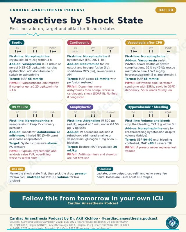
← Back to posts
← Back to posts

Myocardial O2 Supply vs Demand

View on Instagram

Understanding the post

Myocardial oxygen supply depends on coronary blood flow and the oxygen carried in arterial blood. Coronary flow is influenced by perfusion pressure, the time available for diastolic flow and resistance within the coronary circulation. Demand rises with heart rate, contractility and ventricular wall stress. This is a useful framework for understanding myocardial ischaemia. Kaplan’s Cardiac Anesthesia, 8th ed. (2023), chapter 6, printed pp. 154–158; Goodwill and colleagues, 2017.

The table’s basal metabolism row is also relevant: the heart uses some oxygen for basic cell maintenance even apart from the extra work of contraction. Kaplan, chapter 6, printed pp. 154–159; Goodwill and colleagues, 2017.

How to read the post

  • “The heart already extracts about 70%” mainly describes the resting LV. Resting LV extraction is commonly about 70–80%, leaving relatively little extra extraction available when demand rises. The normal RV extracts less and has more reserve. The same percentage does not apply to every chamber or clinical state. Goodwill, metabolic-control section; Kaplan, printed pp. 156–158.
  • More coronary flow provides the main increase in oxygen supply. Increased coronary flow is the principal response to higher LV demand. Arterial oxygen content also matters: haemoglobin concentration and oxygen saturation can change delivery even when flow is unchanged. Kaplan, printed pp. 154–158.
  • The effect of tachycardia depends on coronary reserve and the clinical setting. Faster rates raise demand and shorten diastole. A healthy coronary circulation can often compensate by increasing flow; ischaemia is more likely when reserve is limited, for example by coronary stenosis or ventricular hypertrophy. Tachycardia can also be a response to another problem, so the cause matters before treating the number. Kaplan, printed p. 156.
  • “Coronary diameter” includes more than a large artery’s lumen. Small resistance vessels and their ability to dilate are central to matching flow to demand. A normal-looking large coronary artery does not exclude a microvascular supply problem. Kaplan, printed pp. 155–156; Goodwill, coronary anatomy and metabolic control.

References

  1. Crystal GJ, Pagel PS. Coronary Physiology. In: Kaplan’s Cardiac Anesthesia, 8th ed. (2023), chapter 6. Printed pp. 154–158 cover flow regulation, oxygen extraction and supply–demand matching.
  2. Goodwill AG, Dick GM, Kiel AM, Tune JD. Regulation of Coronary Blood Flow. Compr Physiol. 2017;7:321–382. Mechanical determinants and metabolic control sections. Full text.

For education. Apply the cited sources in their clinical context and follow current local guidance; this material is not patient-specific advice.
← Back to posts
← Back to posts

Coronary Blood Flow: LV vs RV Timing

View on Instagram

Understanding the post

Left-ventricular coronary flow is predominantly diastolic because contraction compresses vessels within the ventricular wall, especially in the inner layer. The normal low-pressure RV has substantial flow during both systole and diastole. Pulmonary hypertension or RV pressure overload can make its flow more dependent on diastole. Kaplan’s Cardiac Anesthesia, 8th ed. (2023), chapter 6, printed pp. 154–156; Goodwill and colleagues, 2017.

How to read the post

  • “Systolic compression stops subendocardial flow” is a simplified description of the inner LV layer. Flow there is very low during systole and may briefly reverse. This does not mean all blood flow in the entire left coronary circulation stops during systole; the outer layers have a different pattern. Goodwill, mechanical-determinants section.
  • “LV coronary perfusion pressure is aortic diastolic pressure minus LVEDP” is an estimate, not a complete definition. It is a useful approximation for the pressure available to perfuse the inner LV wall during diastole. Actual coronary flow also depends on microvascular resistance, pressure within the myocardium and the available flow reserve. It should not be applied unchanged to the RV. Goodwill, determinants of coronary driving pressure; Kaplan, printed pp. 155–156.
  • “Tachycardia steals diastolic time” is correct, but does not always cause ischaemia. A healthy coronary circulation can compensate by increasing flow. Risk rises when reserve is reduced by coronary narrowing, hypertrophy or other disease, especially if perfusion pressure or oxygen content is also low. Kaplan, printed p. 156.

References

  1. Crystal GJ, Pagel PS. Coronary Physiology. In: Kaplan’s Cardiac Anesthesia, 8th ed. (2023), chapter 6, printed pp. 154–158. Includes LV/RV flow timing and the effect of pulmonary hypertension.
  2. Goodwill AG, Dick GM, Kiel AM, Tune JD. Regulation of Coronary Blood Flow. Compr Physiol. 2017;7:321–382. Mechanical determinants, transmural flow and coronary driving-pressure sections. Full text.

For education. Apply the cited sources in their clinical context and follow current local guidance; this material is not patient-specific advice.
← Back to posts
← Back to posts

Esmolol: Mechanism, Doses and Cautions

View on Instagram

Sources supporting this post

  • Baxter BREVIBLOC US label: BREVIBLOC prescribing information, Baxter March 2025. Supports selective beta-blocking action, red-cell enzyme breakdown, roughly 2-minute distribution and 9-minute elimination half-lives (§12), labelled doses (§2), concentrations (§3), contraindications (§4) and adverse effects (§§5–6). Preventive dosing and other uses outside the labelled indications need a separate clinical protocol.
  • BREVIBLOC prescribing information: original linked listing: Original reference named in the post, alongside the dated Baxter version above.
  • 2022 ACC/AHA Aortic Disease Guideline: Supports IV beta-blockers, including esmolol, for acute aortic syndromes unless contraindicated (§7.3.1); blood-pressure reduction must preserve organ perfusion.
  • 2014 ESC aortic-disease guideline: Original reference named in the post for the aortic-dissection box; retained alongside later guidance.
  • Stoelting’s Pharmacology: Original textbook reference named in the post.
  • Hensley’s Practical Approach to Cardiothoracic Anesthesia, 6th ed (2019), chapter 2, section III (PDF pp108–111): Supports lower heart rate, oxygen demand and contractility, longer diastole, and reduced dynamic outflow obstruction with beta-blockade.
  • Antiarrhythmic mechanisms of beta-blocker therapy (2019): Explains the beta-receptor/cAMP diagram, including calcium handling and the heart’s pacemaker channels.
  • IAEM hypercyanotic Tet-spell guideline (2024): Supports esmolol as a specialist rescue option for persistent hypercyanotic spells, not routine treatment of every fast pulse.
  • AHA 2024 HCM guideline clinical-update slides: Supports beta-blockade for obstructive HCM within a treatment plan that preserves filling and blood pressure.
  • 2024 ESC peripheral arterial and aortic disease guideline: Current ESC targets and treatment order for acute aortic syndromes.

Understanding the post

  • The “Pre-laryngoscopy/sternotomy bolus 0.5–1.5 mg/kg” entry describes preventive use, rather than a labelled preventive regimen. The US label covers treatment of fast heart rate or high blood pressure around surgery and does not recommend using esmolol to prevent those events. Preventive practice needs a separate clinical protocol. Baxter BREVIBLOC US label (§1.2 and §2.2).
  • The concentration warning in the post does not mean every preparation stronger than 10 mg/mL is forbidden. Both 10 and 20 mg/mL premixed products are labelled. Check the actual product and infusion site; leakage outside the vein can cause serious tissue injury. Baxter BREVIBLOC US label (§3 and §5.7).
  • Low blood pressure is common, may be severe, and usually improves within 30 minutes after reducing or stopping treatment. The post’s “gone in 10–20 min” therefore overstates how reliably hypotension resolves. Recovery is not guaranteed within a fixed time. Baxter BREVIBLOC US label (§5.1 and §6.1).
  • “Wears off in minutes” fits the label: substantial recovery from beta-blocking effects is seen in 10–20 minutes after stopping. That is different from promising that low blood pressure has resolved. Baxter BREVIBLOC US label (§12.2 versus §5.1).
  • “SVT · AF · ischaemia · TOF spell / Fast, reversible rate control” groups different problems together. A Tet spell is a paediatric emergency: beta-blockers can reduce the obstruction to blood leaving the right ventricle. Esmolol belongs in a monitored specialist rescue plan, not simply a routine rate-control pathway. IAEM hypercyanotic Tet-spell guideline (2024) (p11).
  • “LVOTO / SAM, HOCM” describes selected patients with dynamic obstruction, not all patients with low blood pressure. Treatment must also preserve filling and blood pressure; beta-blockade can worsen shock from other causes. AHA 2024 HCM guideline clinical-update slides (slide22); BREVIBLOC label (§§4,5.3).
  • “Sux block ↑ (~5→8 min)” means esmolol can prolong succinylcholine muscle relaxation. Do not treat those minutes as a reliable recovery time for an individual patient. BREVIBLOC label (§7).
  • The “70 kg: load 35 mg, start 3.5 mg/min = 21 mL/h of 10 mg/mL” calculation is correct for the illustrated 500-microgram/kg load and 50-microgram/kg/min infusion. Loading is optional in the label’s SVT pathway, and doses must be adjusted to response. “Not with bicarbonate” reflects an incompatibility warning, not a prohibition on treating a separate clinical problem. BREVIBLOC label (§§2.1,2.4).

Guideline update

For esmolol dosing and product safety, use the named prescribing information: Baxter March 2025. Its prevention-versus-treatment distinction, available concentrations and recovery warnings are product-label guidance, not a new resuscitation recommendation. Baxter BREVIBLOC US label (§§1.2,3,5.1,12.2). The 2022 aortic guideline specifies a heart-rate target of 60–80 beats/min and systolic pressure below 120 mmHg, or the lowest pressure that maintains organ blood supply, for acute aortic syndromes. These are guideline-specific goals, not universal esmolol targets. ACC/AHA 2022, §7.3.1.

The older ESC reference and its pressure target remain above. The ACC/AHA heart-rate range is a guideline-specific comparison, not proof that a target of 60 or less is an error. The 2024 HCM guidance adds context for the obstruction box: treat acute low blood pressure with attention to filling and vascular support, with beta-blockade where appropriate. AHA 2024 HCM guideline clinical-update slides (slide22).

ESC 2024 updates the older ESC reference: for acute aortic syndromes, aim for heart rate 60/min or less and systolic pressure below 120 mmHg, while preserving blood supply to organs. Higher pressures may be needed with impaired organ blood flow, spinal ischaemia or brain injury. It names IV labetalol or esmolol first, adding a vasodilator if needed. The post’s heart-rate target remains consistent with ESC; ACC/AHA 2022 instead specifies 60–80/min. These are distinct, guideline-specific targets. ESC 2024, §9.3.1.4.1, table 45 and figure 31.

For education. Apply the cited sources in their clinical context and follow current local guidance; this material is not patient-specific advice.
← Back to posts
← Back to posts

Aortic Root Anatomy

View on Instagram

Sources supporting this post

How to read the post

Coronary height in TAVI planning

The post flags an ostial height <10 mm above the annulus. A low coronary opening is an important warning, but 10 mm is not a universal safe/unsafe boundary. CT planning must also assess the sinuses, sinotubular junction, leaflet and outflow-tract calcification, the intended valve and how these structures interact. A height above 10 mm does not exclude obstruction. TAVI means transcatheter aortic valve implantation. ESC/EACTS 2025, §8.2.3 and procedural selection

The short-axis cusp pattern

The three closed semilunar cusps form the familiar “Mercedes-Benz” Y in the TEE ME AV SAX view. TEE ME AV SAX means the mid-oesophageal short-axis aortic-valve view. In a standard view, the noncoronary cusp lies beside the atrial septum and the right coronary cusp is anterior; the actual anatomical relationships are more reliable than screen position alone. ASE/SCA 2013, pp945–946

The conduction system and interleaflet triangles

The RCC–NCC entry highlights the membranous septum and His bundle. The key operative relationship is the right/noncoronary interleaflet triangle, the membranous septum and the adjacent conduction axis. Injury here can cause atrioventricular block. The left/noncoronary region is continuous with the fibrous aorto-mitral curtain and anterior mitral leaflet. The abbreviations mean right, noncoronary and left coronary cusps. Charitos and Sievers, interleaflet triangles

Adult root dimensions

The table presents adult TTE population means ± SD. The numbers match the cited 2015 table. In millimetres, men/women: annulus 26±3 / 23±2; sinuses 34±3 / 30±3; sinotubular junction 29±3 / 26±3; proximal ascending aorta 30±4 / 27±4. These are population means and standard deviations, not individual upper limits, TAVI sizing rules or thresholds for surgery. An individual measurement is interpreted in relation to age and body size. ASE/EACVI 2015, Table 14

Annulus measurement and the other root dimensions

The annulus entry specifies hinge-to-hinge measurement in mid-systole, inner edge to inner edge. This measurement convention applies to the annulus. The same 2015 guideline measures the other listed root dimensions at end-diastole using the leading-edge convention. Comparison with the reference values requires consistent technique and timing. The “annulus” here is the virtual basal ring through the leaflet attachment nadirs, not a separate circular fibrous structure. ASE/EACVI 2015, p32; Charitos and Sievers, the annulus

Aortic valve area versus root diameter

The post quotes a normal AVA of 3–4 cm². Aortic valve area is different from the root diameters in the table. This is an approximate teaching range; Kaplan gives 2.6–3.5 cm². It should not be used for prosthesis sizing. Kaplan, p610

How the root components fit together

The virtual basal ring, crown-shaped leaflet attachments, three interleaflet triangles, sinuses of Valsalva and sinotubular junction are real components. They overlap in height rather than forming separate flat layers in a stack. The usual right and left coronary openings arise in their named sinuses; the noncoronary sinus normally has no coronary opening. Anatomical variants also occur. Charitos and Sievers, root anatomy and sinuses; ASE/SCA 2013, p945

TEE angles and anatomical orientation

The short-axis view is labelled approximately 30–60°. The view angles are approximate. The atrial septum helps identify the noncoronary cusp; the right coronary cusp is anterior toward the right-ventricular outflow region. In the long-axis view, usually around 120–140°, the anterior cusp is right coronary; the posterior structure varies with the imaging plane. Adjacent anatomy and multiple views establish orientation; the stylized illustration is not a fixed screen-orientation map. ASE/SCA 2013, standard views and pp945–946

The noncoronary sinus and adjacent atria

The NCC entry describes its relationship to the interatrial septum and both atria. This is a useful neighbouring-structure description: the noncoronary sinus lies beside the paraseptal regions of both atria. Its right/anterior side is related to right atrial tissue and its leftward side to left atrial tissue. The pulmonary root is adjacent to the right and left coronary side of the aortic root. The card’s “LCC towards the PA” is a rough anatomical cue, not a universal screen position; PA means pulmonary artery. Taylor et al., §3.2 Anatomy; AATS, Pulmonary Valve section

Guideline update: The 2015 population table remains the source of these particular reference values. Its description of three-dimensional echo and CT as emerging sizing methods should be read alongside ESC/EACTS 2025, §8.2.3, which identifies cardiac CT as the preferred tool for TAVI anatomical planning. This develops the CT-based approach already present in ESC/EACTS 2021, §5.1.3; it does not turn normal population means or a single coronary-height cutoff into a procedural decision rule.

For education. Apply the cited sources in their clinical context and follow current local guidance; this material is not patient-specific advice.
← Back to posts
← Back to posts

Pulmonary Hypertension: Definition, WHO Groups and Haemodynamic Cut-offs

View on Instagram

Understanding the post

The haemodynamic thresholds shown in the post match the 2022 ESC/ERS guideline. They are measurements made at rest by right-heart catheterisation:

  • Pulmonary hypertension: mean PAP >20 mmHg.
  • Pre-capillary PH: mean PAP >20, PAWP ≤15 mmHg and PVR >2 Wood units.
  • Isolated post-capillary PH: mean PAP >20, PAWP >15 mmHg and PVR ≤2 Wood units.
  • Combined post- and pre-capillary PH: mean PAP >20, PAWP >15 mmHg and PVR >2 Wood units.

The mean-PAP requirement applies to every subtype, even where the card does not repeat it. PAWP is pulmonary arterial wedge pressure; PVR is pulmonary vascular resistance. 2022 ESC/ERS guideline, section 3.1 and Table 5.

How to read the post

  • “Group 2” is not limited to isolated post-capillary PH. PH caused by left-heart disease can have either an isolated post-capillary pattern or a combined post- and pre-capillary pattern. The combined form therefore also belongs within group 2, despite the card’s narrower parenthetical description. 2022 guideline, sections 3.1 and 8.1.
  • “4 CTEPH” is useful shorthand but incomplete. Group 4 includes chronic thromboembolic PH and other pulmonary-artery obstructions. The five groups are: 1, pulmonary arterial hypertension; 2, left-heart disease; 3, lung disease/hypoxia; 4, pulmonary-artery obstructions; and 5, unclear or multiple contributing mechanisms. 2022 guideline, Table 6.
  • A pre-capillary pattern is not automatically group 1 PAH. Lung disease and chronic thromboembolic disease can produce the same pattern. Clinical classification requires the cause, not only the pressure numbers. Some high-flow patients have mean PAP >20 with both normal PVR and a low wedge pressure and do not fit either pre- or post-capillary definitions. 2022 guideline, section 3.1.
  • These are diagnostic boundaries, not automatic drug-treatment thresholds. Echocardiography estimates the probability of PH; it does not replace properly performed catheter measurements when a definitive haemodynamic diagnosis is needed. Treatment depends on the PH group, severity and the whole clinical picture. 2022 guideline, sections 5.1.5 and 5.1.12; 2024 World Symposium statement.
  • The “vasoreactive on iNO” numbers are correct, but the purpose of the test matters. A positive acute response is a fall in mean PAP of at least 10 mmHg to 40 mmHg or less, with cardiac output unchanged or increased. Testing to identify people suitable for high-dose calcium-channel blockers is recommended for idiopathic, heritable or drug-associated PAH, in a specialist PH centre. It is not a routine test for every PH group. Other specialist uses, such as transplant or congenital-shunt assessment, are separate decisions. 2022 ESC/ERS guideline, section 5.1.12.2 and Recommendation Table 1.

References

Source named in the post

  1. Humbert M, et al. 2022 ESC/ERS Guidelines for the diagnosis and treatment of pulmonary hypertension. Eur Heart J. 2022;43:3618–3731. Sections 3.1–3.2; Tables 5–6; right-heart catheterisation and vasoreactivity sections 5.1.12.1–5.1.12.2. Guideline.

Later updates

  1. Kovacs G, et al. Definition, classification and diagnosis of pulmonary hypertension. Eur Respir J. 2024;64:2401324. Seventh World Symposium statement. Full text.
  2. Kovacs G, Zeder K, Humbert M, et al. European Respiratory Society clinical practice guidelines update for the treatment of pulmonary arterial hypertension. Eur Respir J. 2026;2601178. Guideline.

Guideline update

The 2024 World Symposium statement keeps the 2022 pressure and resistance cut-offs and the five main groups. It refines subgroups, including long-term calcium-channel-blocker responders within idiopathic PAH and more disease-based subdivisions of lung-related PH. It also emphasises that a mildly abnormal pressure or resistance does not by itself justify PAH drugs. 2024 statement.

The 2026 ERS update concerns treatment of adult group 1 PAH, not a new definition of every type of PH. It recommends adding sotatercept for patients already taking PAH drugs who remain at intermediate-low, intermediate-high or high risk during follow-up, with specialist-centre monitoring. It also supports follow-up catheterisation in those risk groups when the result is expected to change treatment. These recommendations must not be applied to group 2 PH or to anyone merely crossing the post’s diagnostic threshold. 2026 guideline, scope and PICO recommendations.

For education. Apply the cited sources in their clinical context and follow current local guidance; this material is not patient-specific advice.
← Back to posts
← Back to posts

Fick Principle: Calculating Cardiac Output

View on Instagram

Sources supporting this post

  • Kaplan, 8th ed. (2023): Table 9.5 p.301; pp.305–306; chapter 2 pp.38–39. Supports oxygen-content/Fick calculations, measured oxygen consumption, mixed-venous sampling, other reasons for low SvO₂ and shunt-specific measurements.
  • Narang et al. Am J Cardiol 2012;109:594–598. Directly examines the assumed 125 mL/min/m² × body-surface-area estimate in adults and its error compared with measured oxygen use.
  • Hensley, 6th ed. (2019), chapter 10, §II.C.1, PDF p.476. Supports the card’s specific 1.34 and 0.003 oxygen-content coefficients, with Hb in g/dL, saturation as a fraction and PaO₂ in mmHg.

Understanding the post

  • “A widening arterial–venous O₂ difference means falling cardiac output” holds only if oxygen consumption is unchanged. Likewise, a lower mixed-venous oxygen saturation can reflect lower output, greater oxygen use, lower haemoglobin or lower arterial saturation. It does not diagnose low output by itself. Kaplan, 8th ed., oxygen transport and Fick sections, pp.305–306.
  • “CO = VO₂ ÷ (CaO₂ − CvO₂)” is the Fick relationship, but the units must match. If VO₂ is mL/min and oxygen content is mL/dL, cardiac output in L/min is VO₂ ÷ [10 × (CaO₂ − CvO₂)]. Use true mixed-venous sampling for this calculation. Kaplan, 8th ed., Table 9.5 p.301 and Fick section p.306.
  • “Assumed VO₂ ≈ 125 mL/min/m²” is an estimate rather than a measured value. Multiply by body surface area to estimate total VO₂ before calculating cardiac output; without that step, the result is indexed to body size. In one 75-adult study, estimates differed from measured VO₂ by more than 25% in one fifth of participants. It is not a universally validated constant. Narang et al. 2012, methods and results.
  • “Inaccurate with shunts or high FiO₂ error” should not imply that the Fick method cannot be used when a shunt is present. It needs appropriate sampling and separate assessment of pulmonary and systemic flow. High inspired oxygen introduces additional measurement and dissolved-oxygen considerations. Kaplan, 8th ed., chapter 2, pp.38–39.

For education. Apply the cited sources in their clinical context and follow current local guidance; this material is not patient-specific advice.

← Back to posts
← Back to posts

Severe Chronic AR: Anaesthetic Goals and Bypass Pitfalls

View on Instagram

Sources supporting this post

How to read the post

Markers of severe chronic regurgitation

The post lists vena contracta >0.6 cm, PHT <200 ms and holodiastolic flow reversal in the descending aorta. These are recognized severe-regurgitation markers. Vena contracta is the narrowest jet width; pressure half-time (PHT) describes pressure equalization; holodiastolic reversal means backward flow throughout diastole. Their interpretation includes valve anatomy, quantitative regurgitation and ventricular size/function. Pressure half-time is affected by ventricular filling pressure and compliance and is not a stand-alone diagnosis. ASE/SCMR 2017, Tables 10–11

Understanding “full, fast and forward”

The mnemonic is accompanied by HR 80–100, maintained preload and lower SVR. This mnemonic refers to severe chronic aortic regurgitation (AR). The goals are adequate filling, preserved forward output and controlled afterload, with avoidance of bradycardia. “Full” does not mean overloading a congested ventricle, and 80–100 is not a compulsory heart rate. Systemic vascular resistance (SVR), perfusion pressure, ventricular function and myocardial oxygen balance must guide adjustments. Kaplan explicitly recommends individualised management and notes that faster heart rate does not invariably reduce regurgitant fraction. Kaplan, pp620–621

Why significant AR limits balloon-pump use

IABP is contraindicated in moderate-to-severe AR. This is supported by the AATS teaching chapter: balloon inflation raises diastolic aortic pressure and can worsen regurgitation. IABP means intra-aortic balloon pump. The manufacturer manual separately lists severe aortic insufficiency. The caption’s “never” refers to significant unrepaired regurgitation, not every degree of leakage or a successfully corrected valve. AATS TSRA Primer, Contraindications; CARDIOSAVE Rescue, p xii

Choosing the cardioplegia route

The post highlights coronary-ostial or retrograde delivery when antegrade root delivery is ineffective. Root cardioplegia may be ineffective in significant AR. Regurgitation can divert solution into and distend the left ventricle, compromising coronary delivery. Direct coronary-ostial and/or retrograde delivery should be planned with adequate decompression and monitoring. Retrograde delivery alone can under-protect the right ventricle. Kaplan, p622; Hensley, Chapter 23, cardioplegia; CPB guideline, pp26–27

How root cardioplegia reaches the coronaries

The phrase “will not reach the coronaries through an incompetent valve” needs an anatomical distinction. Cardioplegia placed in the aortic root normally reaches the coronary openings directly; it does not need to cross the aortic valve. The problem with an incompetent valve is loss of solution into the ventricle and inadequate root pressure, not an anatomical requirement to pass through the valve. Hensley, Chapter 23, cardioplegia delivery; Kaplan, p622

Distension and decompression on bypass

The early-venting warning concerns a slow or fibrillating heart before cross-clamping. Left-ventricular distension is a risk when starting bypass if effective ejection is lost while aortic regurgitation continues. Slow or fibrillating rhythm can increase that risk, but “distends fast” is a warning, not a universal time course. Decompression requires planning and monitoring with the surgical/perfusion team. Venting does not replace effective cardioplegia delivery. Kaplan, p622; Hensley, Chapter 23, §V.E

Heart rate and regurgitant volume

The bradycardia warning reflects the longer diastolic interval. A longer filling interval can increase regurgitant volume per beat, supporting avoidance of clinically important bradycardia. Regurgitant volume and forward output also depend on valve-orifice size, pressure differences and ventricular function; simply increasing heart rate does not guarantee an improved regurgitant fraction or perfusion. Kaplan, pp620–621

Guideline update: The 2024 CPB guideline, published in 2025 supports adapting cardioplegia distribution and emphasizes the limitations of retrograde-only right-ventricular protection. ESC/EACTS 2025, Recommendation Table 3 addresses definitive valve treatment, not a universal anaesthetic heart-rate target: surgery remains recommended for symptomatic severe AR regardless of left-ventricular function. Compared with 2021, it upgrades repair in selected patients at experienced centres to Class IIa B and provides a Class IIb B option for TAVI in symptomatic patients ineligible for surgery with suitable anatomy.

For education. Apply the cited sources in their clinical context and follow current local guidance; this material is not patient-specific advice.

← Back to posts

Privacy and optional analytics

Cardiac Anaesthesia Podcast uses Google Analytics 4 on this reference library only if you choose to accept analytics. Rejecting analytics does not restrict access to the library.

When enabled, Google Analytics receives a random browser identifier, the public reference pages you view, reference links you click, time the page is visible and active, and a limited traffic source. It also processes browser, device and approximate-location information. Google says it does not log or store individual IP addresses in Analytics 4.

We do not send your name, email address, newsletter signup details, form contents or arbitrary URL parameters to Analytics. Advertising features, Google Signals and ad personalisation are disabled. Analytics does not identify a specific person, and visit counts are estimates affected by consent choices and browser blocking.

Analytics cookies and your consent choice expire after 180 days. You can change your choice at any time using “Privacy & analytics”. Rejecting analytics stops future collection in this browser and removes this site's Analytics cookies; it does not erase measurements already collected. Browser Do Not Track and Global Privacy Control signals keep analytics off.

The optional newsletter form is provided by Substack and is separate from this analytics choice. Hosting providers may process technical requests needed to serve this site. See Google's privacy policy, how Google uses information from partner sites, Substack's privacy policy and Vercel's privacy policy.